Published online Dec 26, 2020. doi: 10.12998/wjcc.v8.i24.6425
Peer-review started: August 2, 2020
First decision: September 30, 2020
Revised: October 10, 2020
Accepted: October 26, 2020
Article in press: October 26, 2020
Published online: December 26, 2020
Processing time: 139 Days and 13.2 Hours
Most melanomas identified in the stomach are metastatic. The primary gastric melanoma (PGM) is extremely rare. As such, clinical reports of PGM are scarce in the literature, lending to the challenge of diagnosis and treatment.
A 31-year-old woman presented with a 1-mo history of dysphagia but no symptoms of abdominal pain, abdominal distension, nausea, vomiting, hematemesis, or melena. The patient reported an unintentional weight loss of 6 kg within that time. History-taking revealed no previous medical conditions or surgical events. Abdominal computed tomography at a local hospital had suggested gastric tumor. Endoscopic examination in our hospital found a large, irregular, black mass. Subsequent laparoscopic exploration found the tumor on the side of the stomach fundus penetrating through the serosa, and enlarged lymph nodes (groups 1, 3, 7, and 9) fused into a mass, surrounding the peripheral artery and inseparable. Postoperative immunohistochemistry suggested gastric malignant melanoma. Positron emission tomography-computed tomography confirmed PGM. Treatment with programmed cell death protein 1 antagonist (toripalimab) plus chemotherapy (paclitaxel) was initiated but discontinued upon tumor bleeding. At the last telephone follow-up, the patient reported poor general condition but was alive.
Although unresolved and ongoing, this rare case of PGM expands the overall knowledge about this rare tumor’s diagnosis and management.
Core Tip: Primary gastric melanoma (PGM) is a rare malignant tumor, and there is a lack of clinical data in the literature. We describe a case of PGM presenting with dysphagia and unintentional weight loss, which was confirmed by immunohisto-chemistry and positron emission tomography-computed tomography. We also review the literature, summarizing and discussing the limited knowledge on PGM pathogenesis, clinical manifestations, diagnosis, treatment and prognosis.
- Citation: Long GJ, Ou WT, Lin L, Zhou CJ. Primary gastric melanoma in a young woman: A case report. World J Clin Cases 2020; 8(24): 6425-6431
- URL: https://www.wjgnet.com/2307-8960/full/v8/i24/6425.htm
- DOI: https://dx.doi.org/10.12998/wjcc.v8.i24.6425
Melanomas are malignant tumors that develop from melanocytes. Although they usually occur in the skin, oropharynx, and eyes, rare cases have been reported in the digestive tract, involving the esophagus, stomach, and intestines[1]. Melanoma of the gastrointestinal tract is generally considered to be metastatic, and it has been reported that up to 4% of patients with cutaneous melanoma have clinical gastrointestinal tract involvement antemortem and up to 60% at autopsy[2].
The concept of primary gastrointestinal melanoma (PGIM) is controversial, and it remains unknown whether the absence of primary skin lesions is merely due to spontaneous regression. Nevertheless, reports of cases of PGIMs without primary skin lesions are increasing in the literature[1,3,4], supporting the theory of PGIMs being a distinctive entity. Cheung et al[5] reviewed 659 PGIMs that had been reported from 1973 to 2004, and calculated an annual incidence of approximately 0.47 cases per million in 2000. They noted that the incidence of PGIM tumors was greatest in the oral-nasopharynx (32.8%), anal canal (31.4%), and rectum (22.2%), with rarer instances in the esophagus (5.9%), stomach (2.7%), small bowel (2.3%), gallbladder (1.4%), and large bowel (0.9%). Correspondingly, knowledge of the pathogenic course and clinical management of PGIM in the low-incidence sites, such as stomach (i.e., primary gastric melanoma, PGM), is lacking.
Here, we report a case of PGM to raise awareness of these rare tumors and enhance the overall knowledge for improving diagnosis and treatment of these tumors.
A 31-year-old woman was admitted to our hospital on December 19, 2019, with a chief complaint of dysphagia.
The patient reported a 1-mo history of dysphagia but denied experiencing any instances of abdominal pain, abdominal distension, nausea, vomiting, hematemesis, or melena. However, the patient reported an unintentional weight loss of 6 kg over the past month.
The patient’s past medical and surgical histories were negative.
The patient’s personal and family histories were negative.
Cardiopulmonary and abdominal physical examinations showed no abnormalities.
Serum levels of the carcinoembryonic antigen, cancer antigen-199 and cancer antigen-153 tumor markers were within normal ranges. Liver and kidney function markers and electrolytes were all within normal ranges.
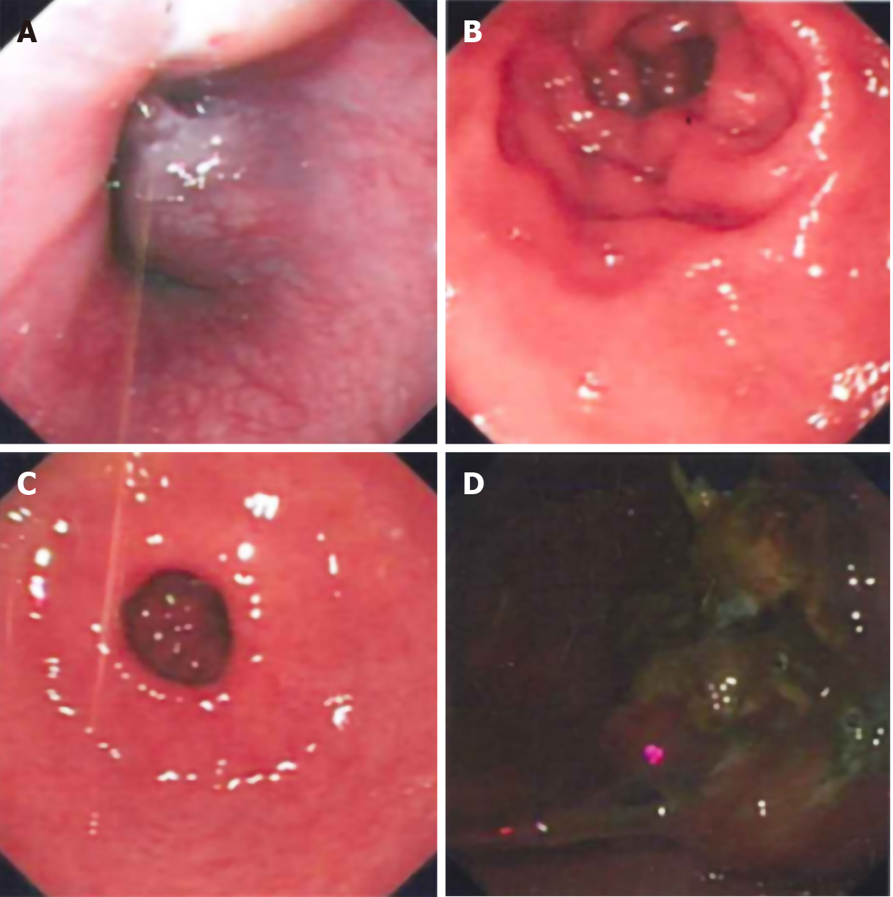
A computed tomography (CT) scan performed at a local hospital (before the patient was admitted to our hospital) had suggested a gastric tumor. Endoscopic examination at our hospital showed a large, irregular, black mass extending from the posterior wall of the cardia to the posterior wall and left lateral wall of the gastric fundus. The mass had unclear boundaries and was prone to bleeding when touched (Figure 1).
Based on the patient’s previous medical history and current positron emission tomography/computed tomography (PET-CT) findings, there was no additional malignant melanoma at any other site. Therefore, a clinical diagnosis of PGM was made.
Following standard preoperative preparation, the patient underwent laparoscopic exploration on day 3 after admission. The tumor was visualized at the side of the stomach fundus and found penetrating through the serosa. No metastases were observed in the liver, omentum, or abdominal wall. However, enlarged lymph nodes (in groups 1, 3, 7, and 9) were found to be fused into a mass, surrounding the peripheral artery; the mass was inseparable. The intraoperative findings suggested a diagnosis of gastric lymphoma, and the operation was performed after obtaining a few tissue specimens for pathological examination.
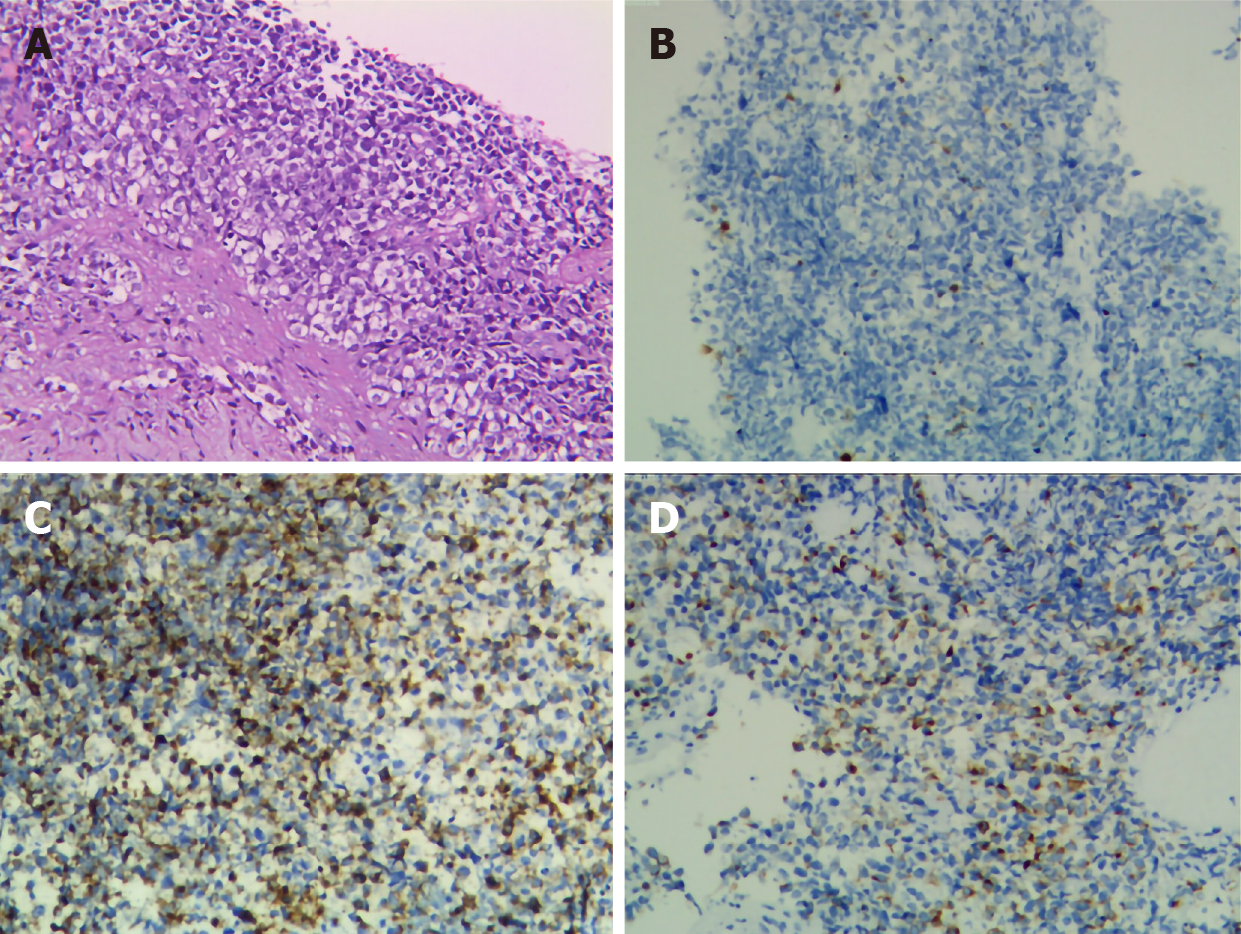
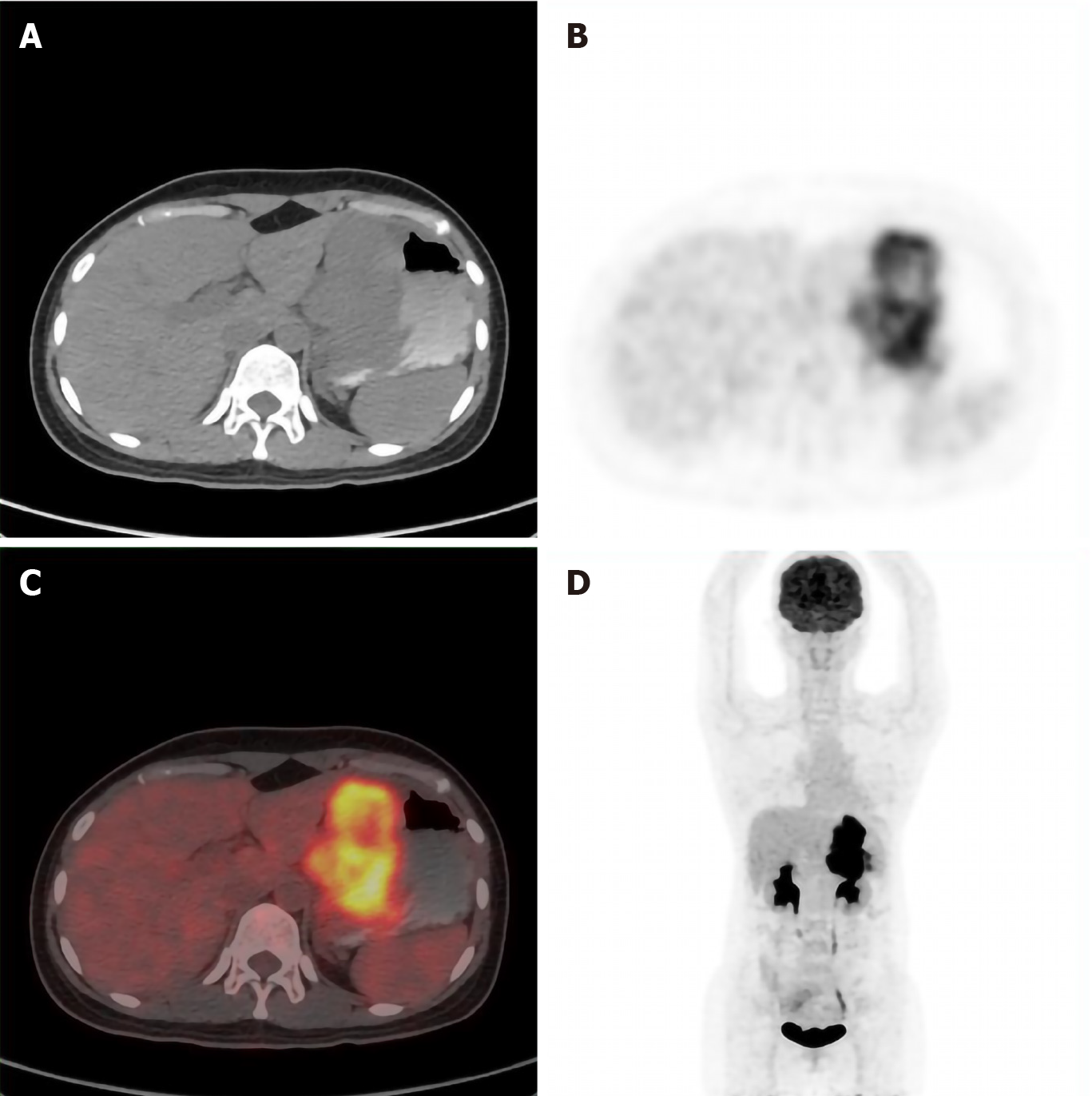
The intraoperative and endoscopic pathology findings suggested malignant melanoma of the stomach. Immunohistochemical analysis of the specimens showed a staining profile of CD79a (-), CK (-), cyclinD1 (+), HMB45 (+), ki-67 (60%+), LCA (-), MDM2 (+), melan-A (+), P16 (-), and S-100 (+) (Figure 2). We inquired the patient’s medical history again and confirmed that the patient had no history of cutaneous melanoma and melanoma resection. To confirm the metastasis of the tumor, the patient underwent PET-CT, which showed the gastric wall to be remarkably thickened and the glucose metabolism to be substantially increased. The gastric cancer (T4) tissue was found to have invaded the pancreatic tail and surrounding soft tissue; in addition, the perigastric omentum and mesentery showed signs of invasion or metastasis from the tumor. No other lesions were observed (Figure 3).
After full communication with the patient's family, the treatment strategy of programmed cell death protein 1 (PD-1) antagonist (toripalimab) + chemotherapy (paclitaxel) was initiated.
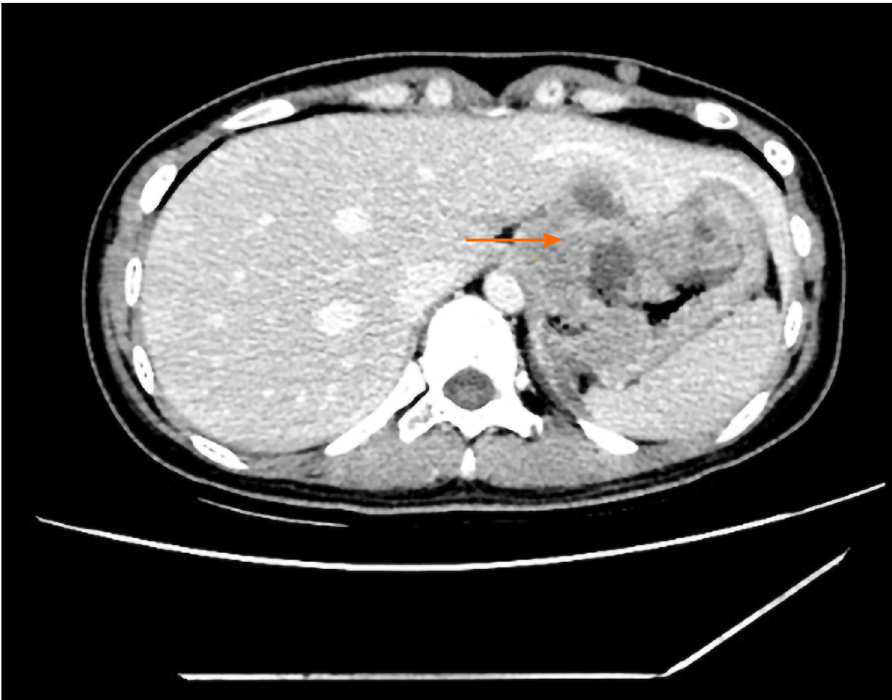
Gastrointestinal bleeding occurred in the fourth cycle of treatment, which was relieved by conservative management. Nevertheless, the patient and her family refused to continue the treatment. Systemic CT scan after the stage 4 chemotherapy (Figure 4) indicated that there was no significant change in tumor size and no metastasis to other new organs. The last telephone follow-up was on July 1, 2020. The patient was alive, but still experiencing symptoms of gastrointestinal bleeding and poor general condition.
PGM is a rare malignant tumor, which should meet two criteria for diagnosis: (1) immunohistochemistry of the tumor being consistent with the immunohistochemistry of melanoma, and positivity for S-100, HMB45 and melan-A; and (2) no history of skin melanoma or skin melanoma resection. However, it is difficult to distinguish metastatic melanoma with spontaneous regression of the primary site from PGM[6]. We inferred that metastatic melanoma with spontaneous regression of the primary lesion would be multifocal or that metastatic lesions would also occur in other organs. In our case, however, the PET-CT scan found lesions in the stomach only, and the patient denied any related skin history. The final diagnosis of PGM was thus made.
The pathogenesis of PGM remains poorly understood. While some studies have found melanocytes in the anal canal and esophagus[7,8], no studies to date have found melanocytes in the stomach. So, where do the malignant melanocytes come from? Two mechanisms have been proposed. Firstly, melanocytes from other sites, such as the anal canal and esophagus, would migrate into the stomach and become malignant in a new tissue environment[3,9]. Secondly, neural crest derivatives, such as amine-precursor uptake and decarboxylation cells, may gain or retain the ability to dedifferentiate into melanocytes and subsequently undergo malignant transformation[10]. It is generally believed that ultraviolet radiation is the main cause of skin malignant melanoma, but some sites of melanoma have never been exposed to ultraviolet rays, such as the digestive tract. Therefore, it is not unreasonable to consider that other factors, such as genetics, immune factors or viral infection, may also play a role in the development of malignant melanoma, especially those not exposed to ultraviolet radiation. We hypothesized that the skin releases a cytokine into the blood circulation due to the effect of sunlight on the exposed area, affecting the melanocytes in the non-exposed areas of the body and resulting in malignant transformation; this theory will need to be confirmed by further studies.
The clinical manifestations of PGM are similar to other common gastric tumors and lack specificity. In our case, dysphagia and wasting were the main clinical manifestations. For the case reported by Wang et al[1], the patient presented with recurrent chest tightness and chest pain as the initial symptoms. Both of the cases reported by Augustyn et al[3] and Bolzacchini et al[4] were hospitalized in an emergency with gastrointestinal bleeding. Therefore, PGM is occult at onset and lacks specific and obvious symptoms and signs in the early stage. B-ultrasonography, gastrointestinal angiography, and CT only showed gastric mass without obvious specificity.
Melanoma produces a paramagnetic substance called melanin; melanin-containing melanoma is indicated by hyperintensity on T1WI and hypointensity on T2WI in magnetic resonance imaging (MRI). PGM is a malignant tumor with abundant blood supply. On diffusion-weighted imaging, the tumor demonstrates a considerably high signal intensity. Thus, MRI is very helpful in the diagnosis of PGM and is even considered as the gold-standard preoperative assessment technique[1]. However, MRI is expensive and rarely used in physical examination. Early diagnosis of such tumors requires endoscopy and tissue biopsy. The manifestations of PGM under endoscopy are mostly isolated eminence lesions, or multiple small lesions, with or without melanin pigmentation, which bleed easily upon contact. Immunohistochemistry mainly shows positivity for HMB45 and S-100. In our case, black coloration was observed on the tumor surface during endoscopy, but the pigmentation was not considered in the diagnosis. During the surgical exploration, numerous enlarged lymph nodes were seen and determined to be fused into a mass. The diagnosis of gastric malignant lymphoma was considered. Gastric melanoma was not confirmed until immunohistochemical results were reported. This clinical course to diagnosis may serve as a profile to alert healthcare teams to the possibility of this type of tumor upon detection of gastric tumor pigmentation during endoscopy.
PGM is a mucosal melanoma, which is more aggressive and associated with a worse prognosis than cutaneous melanoma. This may be due to the general delay in diagnosis, the inherently more aggressive melanoma behavior, or earlier dissemination via the lymphatic and vascular supply of the gastrointestinal tract[11]. In 2009, the American Joint Committee on Cancer introduced a new classification for melanoma of the upper aerodigestive tract[12]. In this classification, T1 and T2 as well as Stage I and Stage II have been removed due to the aggressive nature of mucosal melanomas. Thus, our case was classified as Stage IVA (T4aN1M0).
Because the disease is rare, no standard treatment based on staging has been proposed. For early PGM, surgical resection is still recommended and the range of resection should be no less than that of gastric cancer. In the case reported by Wang et al[13], postoperative adjuvant chemoradiotherapy was not given after tumor resection, and the patient developed metastasis at 1 mo after surgery, and died of multiple metastases in the abdominal cavity at 2 mo after surgery. Therefore, the authors strongly recommended postoperative adjuvant chemoradiotherapy. However, it is currently believed that melanoma is not sensitive to radiotherapy and chemotherapy, and targeted therapy and immunotherapy are more commonly used[4]. Our case was treated with a PD-1 inhibitor (toripalimab) combined with paclitaxel and the tumor did not continue to grow nor was there any additional metastasis. Unfortunately, the patient eventually developed tumor bleeding and was forced to suspend treatment.
Among all the PGIM, PGM has the shortest median survival (5 mo)[5]. The treatment for PGM is still a comprehensive strategy based on surgery. The immune mechanism may play an important role in the spontaneous regression of malignant melanoma. Therefore, in the future, finding new immune drugs and new gene targets will be an important direction for improving the treatment of this rare tumor type.
We here report a rare case of PGM. We also review the related literature to summarize and consider the overall current knowledge on the diagnosis, incidence, pathogenesis, clinical manifestations, treatment, and prognosis of PGM.
Manuscript source: Unsolicited manuscript
Specialty type: Medicine, research and experimental
Country/Territory of origin: China
Peer-review report’s scientific quality classification
Grade A (Excellent): 0
Grade B (Very good): B
Grade C (Good): 0
Grade D (Fair): 0
Grade E (Poor): 0
P-Reviewer: Yong D S-Editor: Gao CC L-Editor: MedE-Ma JY P-Editor: Ma YJ
| 1. | Wang J, Yang F, Ao WQ, Liu C, Zhang WM, Xu FY. Primary gastric melanoma: A case report with imaging findings and 5-year follow-up. World J Gastroenterol. 2019;25:6571-6578. [RCA] [PubMed] [DOI] [Full Text] [Full Text (PDF)] [Cited by in CrossRef: 9] [Cited by in RCA: 13] [Article Influence: 2.2] [Reference Citation Analysis (3)] |
| 2. | Patel JK, Didolkar MS, Pickren JW, Moore RH. Metastatic pattern of malignant melanoma. A study of 216 autopsy cases. Am J Surg. 1978;135:807-810. [RCA] [PubMed] [DOI] [Full Text] [Cited by in Crossref: 452] [Cited by in RCA: 429] [Article Influence: 9.1] [Reference Citation Analysis (0)] |
| 3. | Augustyn A, de Leon ED, Yopp AC. Primary gastric melanoma: case report of a rare malignancy. Rare Tumors. 2015;7:5683. [RCA] [PubMed] [DOI] [Full Text] [Full Text (PDF)] [Cited by in Crossref: 13] [Cited by in RCA: 14] [Article Influence: 1.4] [Reference Citation Analysis (0)] |
| 4. | Bolzacchini E, Marcon I, Bernasconi G, Pinotti G. Primary melanoma of the stomach treated by BRAF inhibitor and immunotherapy. Dig Liver Dis. 2016;48:974. [RCA] [PubMed] [DOI] [Full Text] [Cited by in Crossref: 5] [Cited by in RCA: 5] [Article Influence: 0.6] [Reference Citation Analysis (0)] |
| 5. | Cheung MC, Perez EA, Molina MA, Jin X, Gutierrez JC, Franceschi D, Livingstone AS, Koniaris LG. Defining the role of surgery for primary gastrointestinal tract melanoma. J Gastrointest Surg. 2008;12:731-738. [RCA] [PubMed] [DOI] [Full Text] [Cited by in Crossref: 99] [Cited by in RCA: 93] [Article Influence: 5.5] [Reference Citation Analysis (0)] |
| 6. | Bulkley GB, Cohen MH, Banks PM, Char DH, Ketcham AS. Long-term spontaneous regression of malignant melanoma with visceral metastases. Report of a case with immunologic profile. Cancer. 1975;36:485-494. [RCA] [PubMed] [DOI] [Full Text] [Cited by in RCA: 1] [Reference Citation Analysis (0)] |
| 7. | DE LA PAVA S, NIGOGOSYAN G, PICKREN JW, CABRERA A. Melanosis of the esophagus. Cancer. 1963;16:48-50. [RCA] [PubMed] [DOI] [Full Text] [Cited by in RCA: 2] [Reference Citation Analysis (0)] |
| 8. | Clemmensen OJ, Fenger C. Melanocytes in the anal canal epithelium. Histopathology. 1991;18:237-241. [RCA] [PubMed] [DOI] [Full Text] [Cited by in Crossref: 56] [Cited by in RCA: 51] [Article Influence: 1.5] [Reference Citation Analysis (0)] |
| 9. | Horowitz M, Nobrega MM. Primary anal melanoma associated with melanosis of the upper gastrointestinal tract. Endoscopy. 1998;30:662-665. [RCA] [PubMed] [DOI] [Full Text] [Cited by in Crossref: 18] [Cited by in RCA: 17] [Article Influence: 0.6] [Reference Citation Analysis (0)] |
| 10. | Krausz MM, Ariel I, Behar AJ. Primary malignant melanoma of the small intestine and the APUD cell concept. J Surg Oncol. 1978;10:283-288. [RCA] [PubMed] [DOI] [Full Text] [Cited by in Crossref: 33] [Cited by in RCA: 37] [Article Influence: 0.8] [Reference Citation Analysis (0)] |
| 11. | Castro C, Khan Y, Awasum M, Belostocki K, Rosenblum G, Belilos E, Carsons S. Case report: primary gastric melanoma in a patient with dermatomyositis. Am J Med Sci. 2008;336:282-284. [RCA] [PubMed] [DOI] [Full Text] [Cited by in Crossref: 14] [Cited by in RCA: 16] [Article Influence: 0.9] [Reference Citation Analysis (0)] |
| 12. | Edge SB, Byrd DR, Carducci MA, Compton CC, Fritz AG, Greene F, Trotti A. Edge SB, Byrd DR, Carducci MA, Compton CC, Fritz AG, Greene F, Trotti A. American Joint Comission on Cancer (AJCC): Cancer Staging Manual. 7th ed. New York: Springer, 2009. |
| 13. | Wang L, Zong L, Nakazato H, Wang WY, Li CF, Shi YF, Zhang GC, Tang T. Primary advanced esophago-gastric melanoma: A rare case. World J Gastroenterol. 2016;22:3296-3301. [RCA] [PubMed] [DOI] [Full Text] [Full Text (PDF)] [Cited by in CrossRef: 13] [Cited by in RCA: 12] [Article Influence: 1.3] [Reference Citation Analysis (0)] |


