Published online Dec 26, 2020. doi: 10.12998/wjcc.v8.i24.6346
Peer-review started: July 1, 2020
First decision: August 8, 2020
Revised: August 14, 2020
Accepted: November 12, 2020
Article in press: November 12, 2020
Published online: December 26, 2020
Processing time: 171 Days and 7.6 Hours
Colonic duplication is a rare congenital anomaly. Many types of heterotopic tissue were identified within the wall of duplication. However, studies of ectopic immature renal tissue (EIRT) involving colon duplication in an adult have yet to be reported.
A 23-year-old woman visited our hospital with symptoms of recurrent abdominal pain and chronic constipation. Image analysis via abdomino-pelvic computed tomography, Gastrografin contrast study, and colonoscopy showed a blind and dilated bowel loop filled with fecal material located on the mesenteric side of the sigmoid colon. We established a diagnosis of sigmoid colon duplication and decided to perform a laparoscopic investigation. Segmental resection of the sigmoid colon with duplication was done. Microscopically, the duplicated segment showed all three layers of the bowel wall and EIRT in the wall of the duplication. The postoperative period was uneventful and the patient was discharged nine days after the surgery without complications. She has been doing well 12 mo after the follow-up period.
A comprehensive histopathologic examination for ectopic tissues or tumors is mandatory after resection of colon duplication.
Core Tip: Many types of heterotopic tissue were identified within the wall of duplication. We report an adult case of sigmoid colon duplication with ectopic immature renal tissue (EIRT). EIRT is a metanephric remnant arrested in an extra-renal site due to migratory defect and rarely results in extra-renal Wilms’ tumors. Detection of EIRT warrants a proper histological analysis for differential diagnosis between benign EIRT and a true Wilms’ tumor. This case highlights the importance of thorough histopathologic examination of ectopic tissues or tumors in colon duplications.
- Citation: Namgung H. Sigmoid colon duplication with ectopic immature renal tissue in an adult: A case report. World J Clin Cases 2020; 8(24): 6346-6352
- URL: https://www.wjgnet.com/2307-8960/full/v8/i24/6346.htm
- DOI: https://dx.doi.org/10.12998/wjcc.v8.i24.6346
Several studies have reported different types of heterotopic tissue within duplications. The common types of ectopic tissue include gastric mucosal, squamous, and pancreatic tissues[1]. Ectopic immature renal tissue (EIRT) is a metanephric remnant arrested in an extra-renal site due to abnormal migration[2]. We report a case of sigmoid colon duplication with EIRT. To our knowledge, this is the first report of EIRT occurring within the wall of a colonic duplication in an adult.
A 23-year-old woman visited our hospital with symptoms of recurrent abdominal pain and chronic constipation.
The patient’s history reveals multiple hospitalizations during childhood for similar symptoms without a clear diagnosis.
The patient had a free previous medical history.
No personal and family history was identified.
The physical examination was unremarkable except for tenderness in the right lower quadrant.
All laboratory tests were in the normal range.
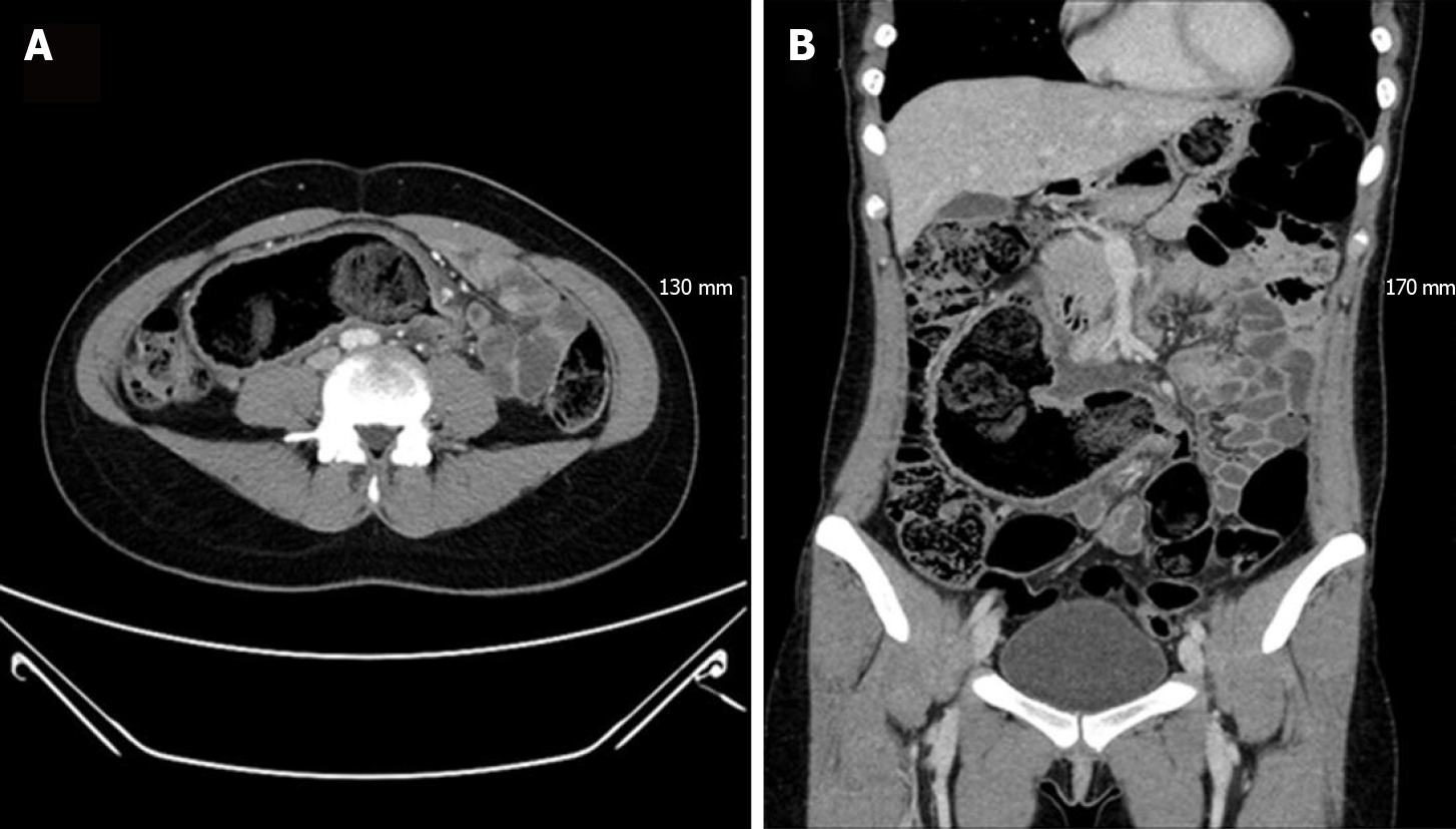
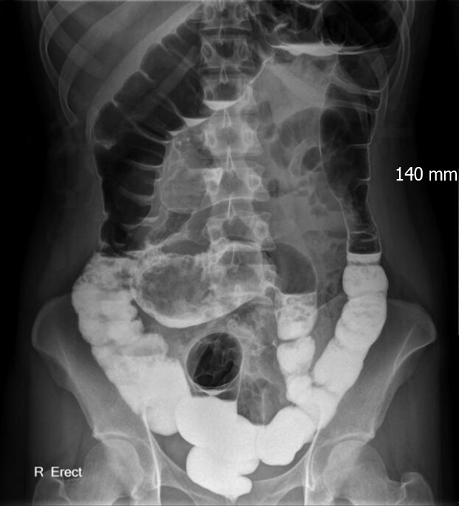
Abdomino-pelvic computed tomography (CT) showed a blind, dilated bowel loop filled with fecal material, directed to the right upper quadrant (RUQ) of the abdomen. This bowel loop communicated with the sigmoid colon and was located on the mesenteric side (Figure 1). The Gastrografin contrast study revealed a Y-shaped structure formed by the sigmoid colon and the duplicated colonic segment (Figure 2).
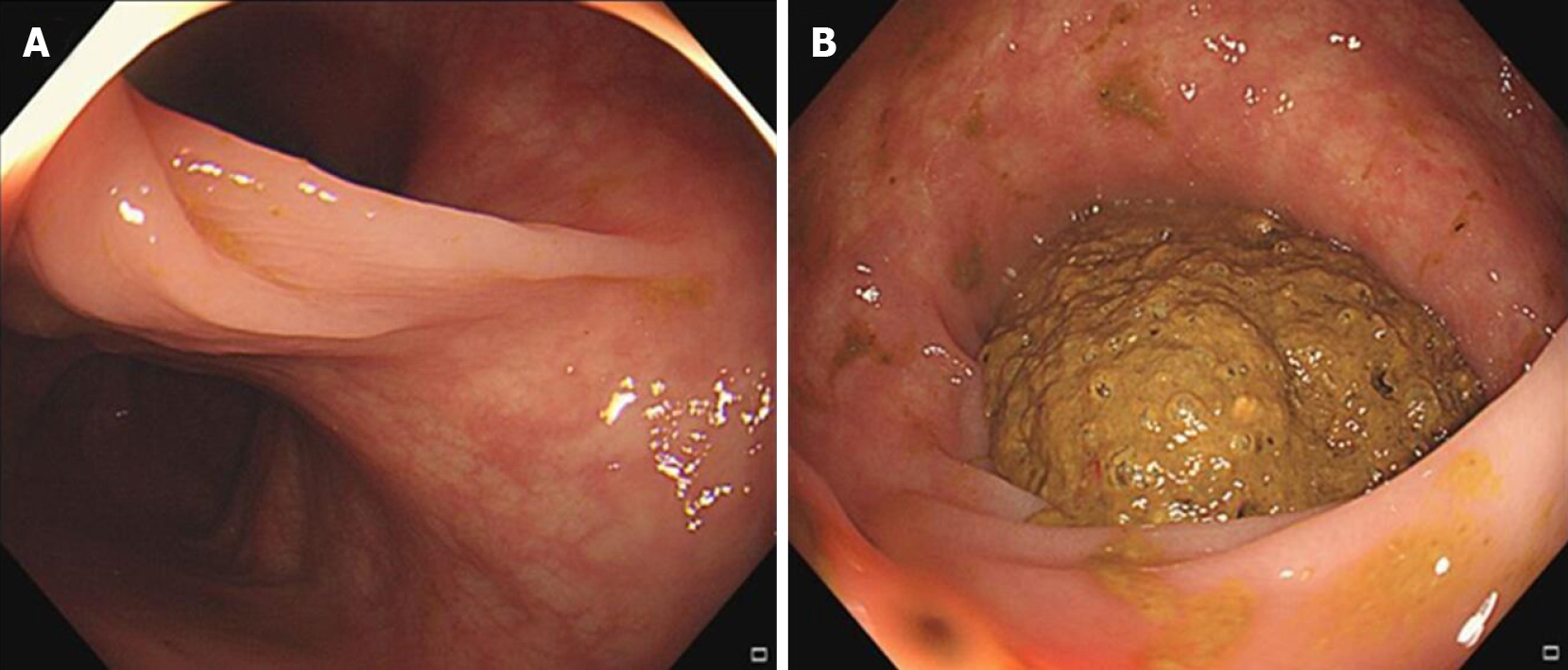
Colonoscopy showed bifurcation of the colonic lumen at the sigmoid colon and the duplicated segment was filled with huge fecalomas (Figure 3).
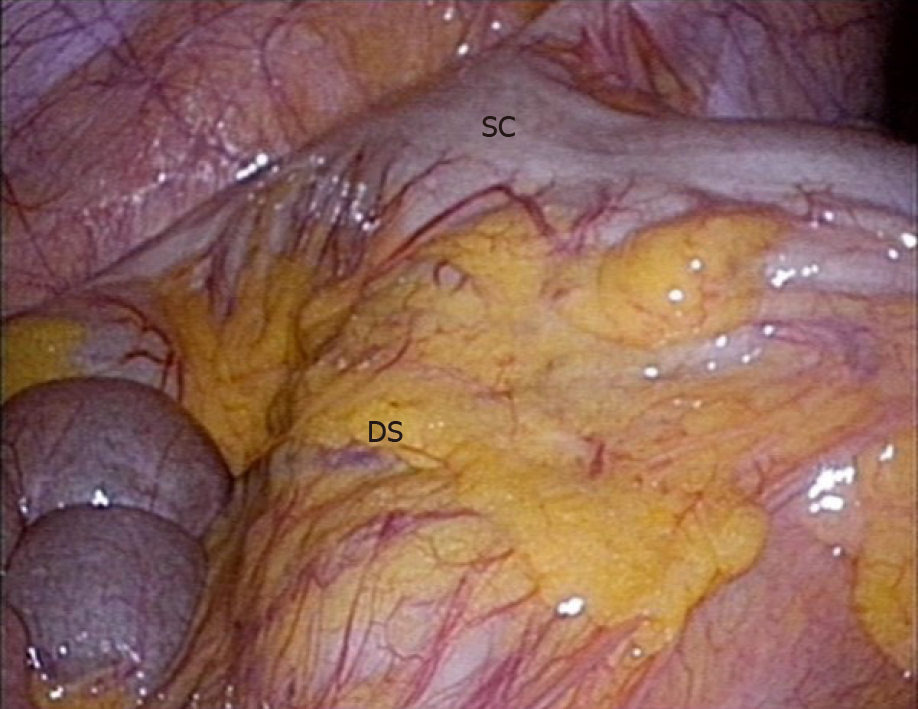
We established a diagnosis of sigmoid colon duplication based on these findings and decided to perform a laparoscopic examination. An approximately 30-cm-long, tubular bowel segment originating in the mesenteric side of the sigmoid colon was identified (Figure 4). This bowel segment was located under the mesocolon. It extended to the RUQ of the abdomen and ended near the duodenum. The surgery was converted to open surgery due to adhesion. Segmental resection of the sigmoid colon with duplication was performed.
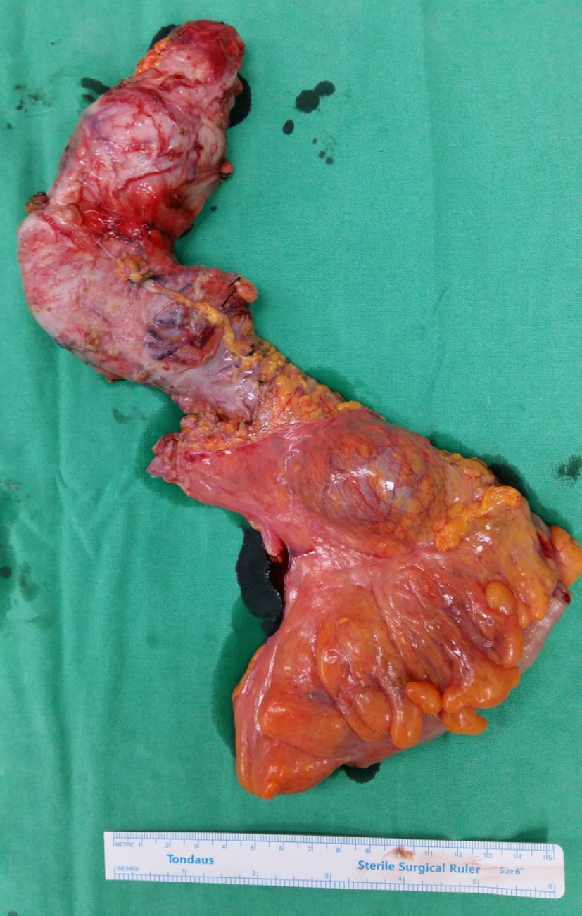
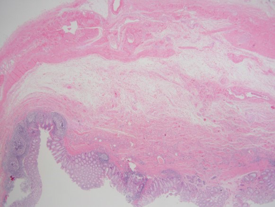
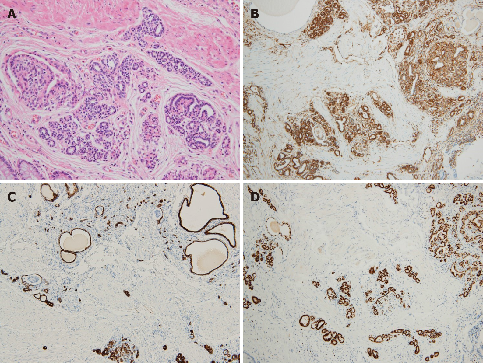
Grossly, the duplicated segment, measuring 34 cm in length, was connected to the native sigmoid colon on the mesenteric side (Figure 5). Microscopically, the duplicated segment revealed all three layers of the bowel wall with scattered heterotopic tissue (Figure 6). Heterotopic tissue composed of fetal glomeruli and scattered tubules was detected under higher magnification, with immunoreactivity against vimentin, CK7, and PAX8. A diagnosis of EIRT associated with colonic duplication was made (Figure 7).
The final diagnosis was sigmoid colon duplication with benign EIRT.
Segmental resection of the sigmoid colon with duplication was performed.
The postoperative period was uneventful and the patient was discharged nine days after the surgery without complications. She has been doing well and was satisfied with the outcome 12 mo after the follow-up.
Alimentary tract duplication is a very rare congenital malformation that occurs most commonly in the small bowel[3]. Colonic duplications account for only 6%-7% of all duplications, with the cecum the most common site[4]. Various theories have been proposed, but the etiology of colonic duplication has not been established. This anomaly is often diagnosed in childhood, but some may go undiagnosed until adulthood[5-7]. A combination of abdominal pain and intestinal obstruction symptoms is the most common clinical manifestation of colonic duplications. Patients with colonic duplication are often accompanied by vertebral and genitourinary anomalies[3,8]. However, the patient in this case report did not have any other anomalies except colonic duplication.
A preoperative diagnosis of colonic duplication is often difficult[1,4]. General imaging modalities, such as plain abdominal radiography or ultrasonography, provide limited information. The diagnosis is best established with CT imaging or contrast enema. Although a large diverticulum may appear similar to tubular type colonic duplication, haustral marking on contrast enema may suggest duplication, as in this case.
Colonic duplication characteristically arises from the mesenteric border of the colon and may have direct communication[1]. It has multiple bowel wall layers, including a smooth muscle coat and an epithelial mucosal lining. There have been reports of many types of heterotopic tissue identified within the duplications[1,3]. The common types of ectopic tissue include gastric mucosal, squamous, and pancreatic tissue. Rarely, malignant change can occur in a colonic duplication[9]. EIRT was found in the wall of the duplication in this case. EIRT is a metanephric remnant arrested in an extra-renal site due to a migratory defect and rarely can give rise to extra-renal Wilms tumors[2]. EIRT was composed of fetal glomeruli and scattered tubules. EIRT is rarely reported and most cases are associated with teratoma. There has been report of the presence of EIRT within the wall of a colonic duplication in an 8-mo-old male child[2], and this is the first report of EIRT found in the colonic duplication in an adult, to our knowledge. Whenever EITR is found, a proper histological interpretation is mandatory for a differential diagnosis between benign EIRT and a true Wilms tumor[2,10]. Because this case was not associated with teratoma and did not show any malignant features such as cellular atypia or nuclear pleomorphism, we plan to follow-up without further treatment. Surgical resection is the treatment of choice for symptomatic and asymptomatic colonic duplications to prevent complications and a tendency for malignant degeneration[1,4]. Because duplications always share blood supply with the native colon and malignant changes can occur in the conjunction area, the extent of resection should include the duplication and a short segment of normal colon[3].
Many types of heterotopic tissue and tumor were identified within the wall of colon duplication. EIRT was found in the wall of the duplication in this case. Treatment plan is modified based on histological findings. Therefore, a comprehensive histopathologic examination for ectopic tissues or tumors is mandatory after resection of colon duplication.
Manuscript source: Unsolicited manuscript
Specialty type: Surgery
Country/Territory of origin: South Korea
Peer-review report’s scientific quality classification
Grade A (Excellent): 0
Grade B (Very good): 0
Grade C (Good): C, C
Grade D (Fair): 0
Grade E (Poor): 0
P-Reviewer: Al-Shouk AAAM, Yu B S-Editor: Wang JL L-Editor: A P-Editor: Liu JH
| 1. | Jeziorczak PM, Warner BW. Enteric Duplication. Clin Colon Rectal Surg. 2018;31:127-131. [RCA] [PubMed] [DOI] [Full Text] [Cited by in Crossref: 19] [Cited by in RCA: 21] [Article Influence: 3.0] [Reference Citation Analysis (0)] |
| 2. | Mitra S, Singla N, Singh Sandhu G, Bal A. Ectopic Immature Renal Tissue Associated with Lipomeningomyelocele and Enteric Duplication Cyst: A Report of Two Cases. Fetal Pediatr Pathol. 2016;35:98-103. [RCA] [PubMed] [DOI] [Full Text] [Cited by in Crossref: 5] [Cited by in RCA: 5] [Article Influence: 0.6] [Reference Citation Analysis (0)] |
| 3. | Ildstad ST, Tollerud DJ, Weiss RG, Ryan DP, McGowan MA, Martin LW. Duplications of the alimentary tract. Clinical characteristics, preferred treatment, and associated malformations. Ann Surg. 1988;208:184-189. [RCA] [PubMed] [DOI] [Full Text] [Cited by in Crossref: 145] [Cited by in RCA: 144] [Article Influence: 3.9] [Reference Citation Analysis (0)] |
| 4. | Mourra N, Chafai N, Bessoud B, Reveri V, Werbrouck A, Tiret E. Colorectal duplication in adults: report of seven cases and review of the literature. J Clin Pathol. 2010;63:1080-1083. [RCA] [PubMed] [DOI] [Full Text] [Cited by in Crossref: 31] [Cited by in RCA: 42] [Article Influence: 2.8] [Reference Citation Analysis (0)] |
| 5. | Cheng KC, Ko SF, Lee KC. Colonic duplication presenting as a huge abdominal mass in an adult female. Int J Colorectal Dis. 2019;34:1995-1998. [RCA] [PubMed] [DOI] [Full Text] [Cited by in Crossref: 3] [Cited by in RCA: 4] [Article Influence: 0.7] [Reference Citation Analysis (0)] |
| 6. | Al-Jaroof AH, Al-Zayer F, Meshikhes AW. A case of sigmoid colon duplication in an adult woman. BMJ Case Rep. 2014;2014. [RCA] [PubMed] [DOI] [Full Text] [Cited by in Crossref: 8] [Cited by in RCA: 9] [Article Influence: 0.8] [Reference Citation Analysis (1)] |
| 7. | Kiu V, Liang JT. Laparoscopic resection of Y-shaped tubular duplication of the sigmoid colon: report of a case. Dis Colon Rectum. 2010;53:949-952. [RCA] [PubMed] [DOI] [Full Text] [Cited by in Crossref: 5] [Cited by in RCA: 5] [Article Influence: 0.3] [Reference Citation Analysis (0)] |
| 8. | Jung HI, Lee HU, Ahn TS, Lee JE, Lee HY, Mun ST, Baek MJ, Bae SH. Complete tubular duplication of colon in an adult: a rare cause of colovaginal fistula. Ann Surg Treat Res. 2016;91:207-211. [RCA] [PubMed] [DOI] [Full Text] [Full Text (PDF)] [Cited by in Crossref: 6] [Cited by in RCA: 7] [Article Influence: 0.8] [Reference Citation Analysis (0)] |
| 9. | Kang M, An J, Chung DH, Cho HY. Adenocarcinoma arising in a colonic duplication cyst: a case report and review of the literature. Korean J Pathol. 2014;48:62-65. [RCA] [PubMed] [DOI] [Full Text] [Full Text (PDF)] [Cited by in Crossref: 6] [Cited by in RCA: 8] [Article Influence: 0.7] [Reference Citation Analysis (0)] |
| 10. | Coli A, Angrisani B, Chiarello G, Massimi L, Novello M, Lauriola L. Ectopic immature renal tissue: clues for diagnosis and management. Int J Clin Exp Pathol. 2012;5:977-981. [PubMed] |


