Published online Dec 6, 2020. doi: 10.12998/wjcc.v8.i23.6009
Peer-review started: April 13, 2020
First decision: June 18, 2020
Revised: July 27, 2020
Accepted: October 12, 2020
Article in press: October 12, 2020
Published online: December 6, 2020
Processing time: 235 Days and 8.9 Hours
Gastrointestinal stromal tumors (GISTs) are mesenchymal tissue tumors originating from Cajal cells, presenting diverse clinical manifestations due to the different sizes, locations, and growth patterns of the lesions. Duodenum is an uncommon site of GISTs, more with gastrointestinal obstruction and bleeding as the first symptoms. Ectopic duodenal varix, as a rare varix occurring outside the gastroesophageal region, is the main type of heterotopic varices and an unusual cause of gas-trointestinal hemorrhage. The etiology is mainly seen in liver cirrhosis, portal hypertension, vasculitis, portal vein embolism and obstruction caused by various factors. Reports of duodenal stromal tumor combined with ectopic variceal hemorrhage are rarely seen; however, when it occurs, the situation can sometimes be urgent and life-threatening, especially when traditional endoscopy and imaging fail to detect the lesion timely.
We report a 52-year-old female patient who had no obvious inducement to develop black stool. Gastroscopy in a local hospital revealed that the duodenal horizontal ectopic varices were ruptured and bleeding. After metal clamping hemostasis, she still had gastrointestinal bleeding and was transferred to our hospital. Gastroscopy showed that active bleeding was still seen in the horizontal part of duodenum, and suspicious submucosal eminence was seen in the bleeding part. Abdominal computed tomography showed a huge stromal tumor of duodenum, specimens were pathologically confirmed after surgery. After a 3-mo follow-up, no gastrointestinal hemorrhage and complications occurred.
Ectopic variceal hemorrhage is rare but sometimes fatal. It may be combined with stromal tumor, which can be diagnosed by multiple methods. Endoscopic and surgical treatment are effective.
Core Tip: Giant duodenal stromal tumor with ectopic variceal hemorrhage as the initial symptom is extremely rare in the clinic. For gastrointestinal hemorrhage caused by rare sites and causes, meticulous and skillful endoscopic examination technology can prevent focus from missing, while multiple methods (computed tomography, endoscopy, etc.) are of great value in clarifying the etiology and reducing missed diagnosis.
- Citation: Li DH, Liu XY, Xu LB. Duodenal giant stromal tumor combined with ectopic varicose hemorrhage: A case report. World J Clin Cases 2020; 8(23): 6009-6015
- URL: https://www.wjgnet.com/2307-8960/full/v8/i23/6009.htm
- DOI: https://dx.doi.org/10.12998/wjcc.v8.i23.6009
Duodenal stromal tumor accounts for only 3%-5% of gastrointestinal stromal tumors[1]. Most of the existing studies on this rare disease are case-based and more detailed research is objectively needed. Duodenal stromal tumor can be asymptomatic or present with diverse manifestations such as abdominal mass, abdominal distension, abdominalgia, jaundice and gastrointestinal hemorrhage according to the different location, size, and growth patterns of tumors. Gastrointestinal hemorrhage is the most common clinical feature, which is induced by tumor expansion growth[2]. Generally, it can be found through endoscopy, imaging or surgery. At present, the treatment of duodenal stromal tumor is mainly surgical operation, and targeted therapy is carried out according to pathological diagnosis results[3]. Duodenal variceal hemorrhage accounts for 2%-5% of all variceal hemorrhage[4], which is a rare cause of gastrointestinal hemorrhage. We present a case of gastrointestinal hemorrhage caused by duodenal ectopic varices, which led to the discovery of giant duodenal stromal tumor. Repeated endoscopic hemostasis failed, and the bleeding eventually stopped only after surgical resection of stromal tumors. The patient recovered smoothly and was discharged from hospital.
The 52-year-old female patient was admitted to our hospital because of repeated black stool for more than 1 wk. There is nothing special about her family history and past history.
The patient underwent endoscopy twice due to melena, and metal clip was used to stop bleeding after initially found the ruptured hemorrhage of ectopic varices in duodenal horizontal part. Gastrointestinal hemorrhage recurred later, with suspected submucosal eminence at the bleeding site. Abdominal computed tomography (CT) revealed huge duodenal stromal tumor, which was then surgically removed and confirmed pathologically.
She had no gastrointestinal inflammation, ulcer or solid tumor before.
Other history could cause gastrointestinal hemorrhage, such as medication and alcoholism, were excluded. Her family history has nothing notable.
She had an anemic appearance, a flat and soft abdomen without tenderness, rebound pain or muscle tension.
The results showed positive stool occult blood test and severe anemia (hemoglobin 45 g/L). Albumin was assessed at 33.3 g/L, fibrinogen was 4.53 g/L, and no significant abnormality was found in tumor markers or other routine examinations.
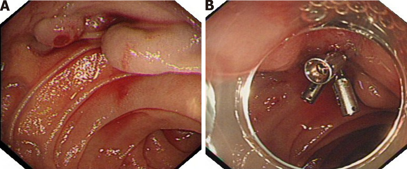
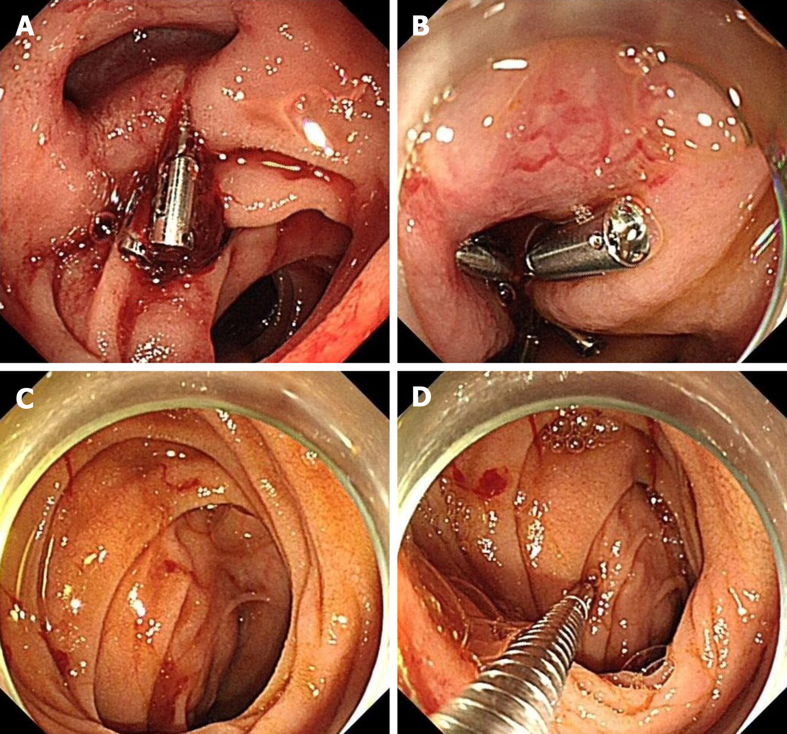
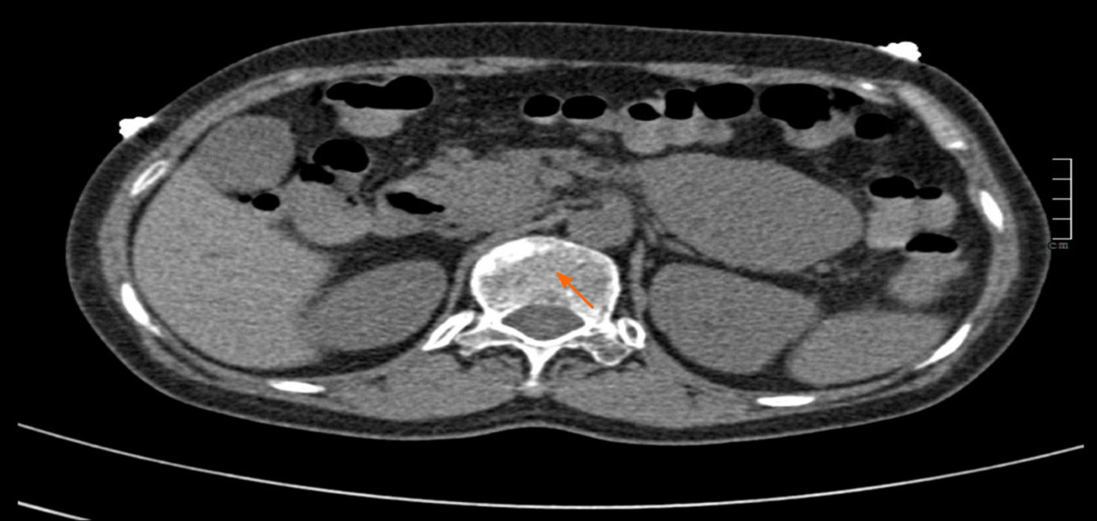
The first endoscopy was performed in the outer hospital, revealed that multiple ectopic varices in the horizontal part of duodenum, with a diameter of about 0.5 cm to 0.8 cm. Rupture accompanied by active hemorrhage could be seen on the surface of varices, and three metal clips were used for hemostasis (Figure 1). Gastroscopy in our hospital showed that there was residual metal clip in the horizontal part of duodenum and bleeding was still active locally. We applied two metal clips to stop bleeding. In addition, suspicious submucosal bulges were found in varicose vein areas, which was hard when touched by biopsy forceps and lacked obvious mucosal glide motion (Figure 2). Abdominal CT revealed lumpy soft tissue density shadow in the horizontal part of duodenum, with a size of about 7.0 cm × 4.8 cm × 5.7 cm (Figure 3).
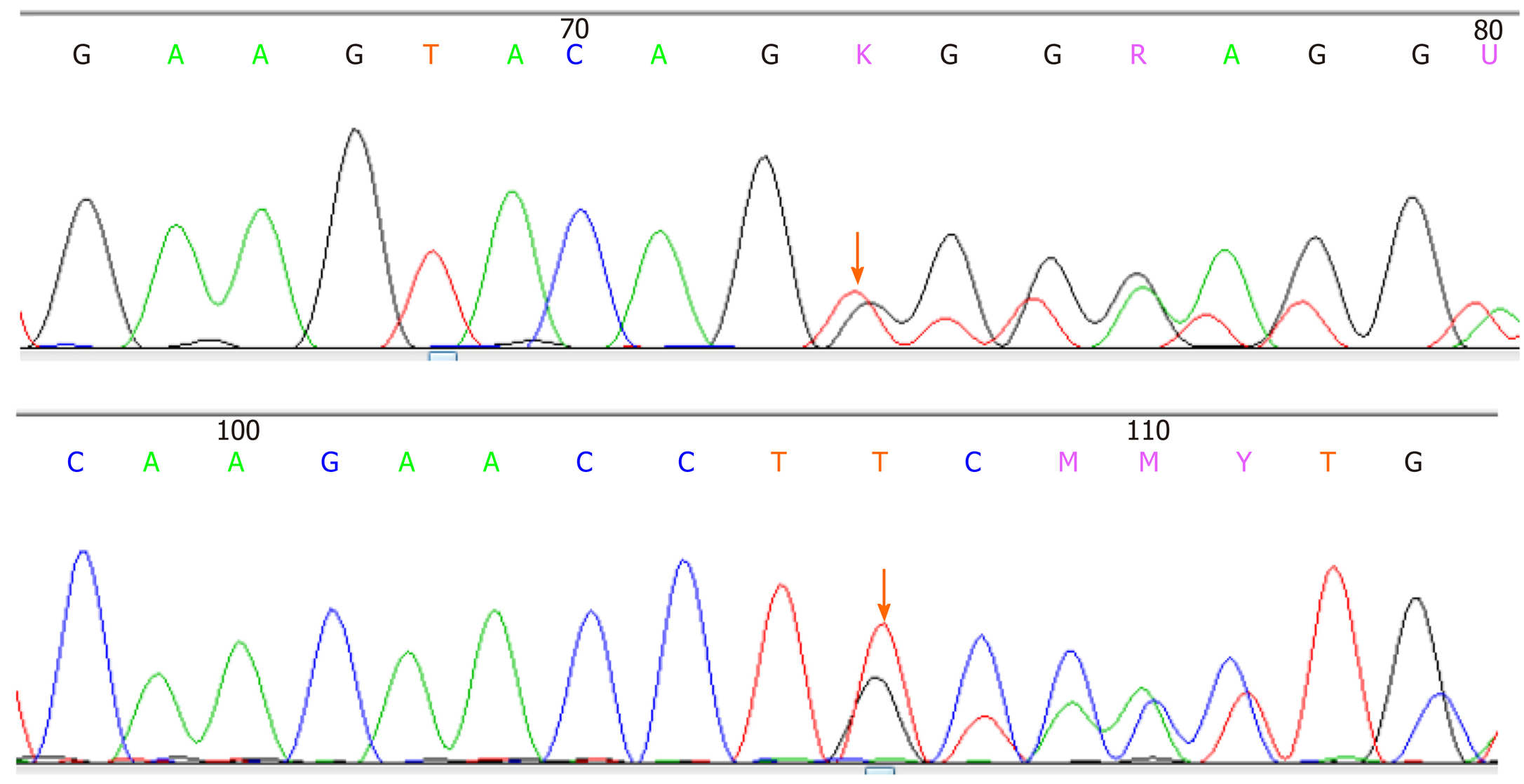
Pathologically, the postoperative specimens were confirmed to be duodenal horizontal stromal tumors with moderate risk, tumor size was about 7 cm × 7 cm × 5 cm, mitosis had 1/50 high power field (Figure 4A and B), There are curved and dilated blood vessels in submucosa, some of which are congested, which is in accordance with the pathological manifestations of varicose veins, but some of them are blocked by compression (Figure 4C). The immunohistochemical results are as follows: Cytokeratin (-), vimentin (+), CD34+, diffuse CD117+ and discovered on gastrointestinal stromal tumor-1 (+), S100-, smooth muscle actin (+), desmin+, caldesmin-, Ki-67 less than 1% (Figure 5). Gene testing found that C-KIT gene Exon-11 had c.1669_1674 deltggag (p.w557_k558del) mutation, while platelet-derived growth factor receptor alpha and related exons had no mutation (Figure 6).
Synthesizing endoscopy, imaging and pathology, we finally diagnosed the patient as gastrointestinal hemorrhage caused by duodenal giant stromal tumor combined with ectopic varicose rupture.
The patient suffered from massive gastrointestinal hemorrhage after she had completed two times of endoscopic metal clamp hemostasis, urgent exploratory laparotomy was consequently carried out, abdominal adhesion lysis and resection of tumor at the initial part of duodenal level were performed. Anti-inflammatory, proton pump inhibitor, and symptomatic support treatment were given after operation, and imatinib mesylate capsules was taken according to the gene test results.
No gastrointestinal bleeding occurred after the patient was hospitalized for 5 d, and hemoglobin recovered to 114 g/L. At 3 mo postoperative follow-up, the patient showed that the patient recovered smoothly and had no typical discomfort or complications.
Duodenal stromal tumor is a rare tumor originating from mesenchymal tissue. The common clinical manifestations include abdominal pain, abdominal mass, and gastrointestinal hemorrhage. Among them, gastrointestinal hemorrhage is mainly black stool, which is caused by tumor growth invading mucosal layer and forming ulcer[5]. Previous reports of duodenal stromal tumor combined with variceal hemorrhage can hardly be seen. Ectopic varices as a rare cause of gastrointestinal hemorrhage, is varices occurring outside the stomach and esophagus. The most common site of ectopic varices is duodenum, which is usually caused by embolism and obstruction of branches of portal vein and retroperitoneal vena cava due to liver cirrhosis, portal hypertension, extrahepatic portal vein occlusion, and vascular malformation[6]. Some regional factors (e.g., such as local vascular occlusion, thrombosis, diversion after surgery, inflammation) are also responsible for the formation of duodenal varicose veins and secondary hemorrhage[7,8]. Considering that the patient we reported had no relevant medical history and clinical manifestations of hepatitis, liver cirrhosis or elevated portal pressure, taking into account the evidence of endoscopy, abdominal CT, and pathological examination, we speculate that the ectopic varicose veins in this patient are caused by the compression of giant stromal tumors, especially when many tortuous and dilated blood vessels in submucosa can be seen in pathological sections. It can be seen that some vascular cavities are compressed and occluded due to the compression of stromal tumors, which reveals the formation mechanism of the disease. Unfortunately, the patient underwent emergency surgery and failed to perform abdominal enhanced CT examination to further clarify the origin of ectopic varicose veins and the portal system due to her sudden and fatal gastrointestinal bleeding. In this case, the patient presented with black stool as the first symptom and found ectopic varicose veins in the horizontal part of the duodenum during gastroscopy in other hospital, but the huge bulge around the bleeding site was ignored. The revelation to us is that for patients with acute gastrointestinal hemorrhage, when the hemorrhage focus is found, attention should be paid to the observation around the bleeding site, especially in rare parts and hemorrhage caused by unusually causes, such as duodenum, its unique anatomical characteristics (small intestinal cavity, numerous plica and large curvature) lead to the lesions being ignored to some extent, consequently, meticulous observation is particularly essential to avoid omission. We suggest slowly withdrawing the endoscope and flexibly rotate the small knob when observing the duodenum, to obtain the maximum observation field of vision, especially in the bent part of intestinal lumen, to avoid missing lesions or misdiagnosis as much as possible. Due to the sudden life-threatening gastrointestinal hemorrhage of the patient, we performed emergency surgery and completely removed the tumor. The final pathological diagnosis was stromal tumor in the horizontal part of duodenum with moderate risk. For endoscopic treatment of ectopic varicose veins, we can choose intravenous injection of sclerosing agent, tissue glue, or clamping varicose veins with metal clips according to the operation level of endoscopists and patients' conditions, among which clamping with metal clips for unexplained ectopic veins is considered to be the safe and effective treatment[9]. Surgery and molecular targeted therapy are currently the main treatment methods for duodenal stromal tumors[10], and the prognosis depends on tumor size, mitosis image and postoperative risk classification[11].
Giant duodenal stromal tumor combined with ectopic varices with rupture and hemorrhage as the initial symptom is extremely rare. We emphasize great efforts should be made to observe the periphery of hemorrhage focus, especially gastrointestinal hemorrhage in rare parts, skilled operation also contributes to reduce missed diagnosis during endoscopic observation for special parts, timely application of multi-means inspection method is of great significance to clarify the cause and reduce missed diagnosis. This report has won the informed consent of patient and has no conflicts of interest.
Appreciation is expressed to the Department of Pathology and Imaging of Affiliated Hospital of Guizhou Medical University, who provided the original examination pictures and opinions for this case.
Manuscript source: Unsolicited manuscript
Specialty type: Gastroenterology and hepatology
Country/Territory of origin: China
Peer-review report’s scientific quality classification
Grade A (Excellent): 0
Grade B (Very good): B
Grade C (Good): C, C
Grade D (Fair): 0
Grade E (Poor): 0
P-Reviewer: Chiu KW, Kikuchi H S-Editor: Liu M L-Editor: Filipodia P-Editor: Zhang YL
| 1. | Schottenfeld D, Beebe-Dimmer JL, Vigneau FD. The epidemiology and pathogenesis of neoplasia in the small intestine. Ann Epidemiol. 2009;19:58-69. [RCA] [PubMed] [DOI] [Full Text] [Full Text (PDF)] [Cited by in Crossref: 232] [Cited by in RCA: 203] [Article Influence: 12.7] [Reference Citation Analysis (0)] |
| 2. | Liu Z, Zheng G, Liu J, Liu S, Xu G, Wang Q, Guo M, Lian X, Zhang H, Feng F. Clinicopathological features, surgical strategy and prognosis of duodenal gastrointestinal stromal tumors: a series of 300 patients. BMC Cancer. 2018;18:563. [RCA] [PubMed] [DOI] [Full Text] [Full Text (PDF)] [Cited by in Crossref: 21] [Cited by in RCA: 26] [Article Influence: 3.7] [Reference Citation Analysis (0)] |
| 3. | Vitiello GA, Medina BD, Zeng S, Bowler TG, Zhang JQ, Loo JK, Param NJ, Liu M, Moral AJ, Zhao JN, Rossi F, Antonescu CR, Balachandran VP, Cross JR, DeMatteo RP. Mitochondrial Inhibition Augments the Efficacy of Imatinib by Resetting the Metabolic Phenotype of Gastrointestinal Stromal Tumor. Clin Cancer Res. 2018;24:972-984. [RCA] [PubMed] [DOI] [Full Text] [Cited by in Crossref: 31] [Cited by in RCA: 43] [Article Influence: 5.4] [Reference Citation Analysis (0)] |
| 4. | Norton ID, Andrews JC, Kamath PS. Management of ectopic varices. Hepatology. 1998;28:1154-1158. [RCA] [PubMed] [DOI] [Full Text] [Cited by in Crossref: 239] [Cited by in RCA: 254] [Article Influence: 9.4] [Reference Citation Analysis (0)] |
| 5. | Shen C, Chen H, Yin Y, Chen J, Han L, Zhang B, Chen Z, Chen J. Duodenal gastrointestinal stromal tumors: clinicopathological characteristics, surgery, and long-term outcome. BMC Surg. 2015;15:98. [RCA] [PubMed] [DOI] [Full Text] [Full Text (PDF)] [Cited by in Crossref: 28] [Cited by in RCA: 31] [Article Influence: 3.1] [Reference Citation Analysis (0)] |
| 6. | Hashizume M, Tanoue K, Ohta M, Ueno K, Sugimachi K, Kashiwagi M, Sueishi K. Vascular anatomy of duodenal varices: angiographic and histopathological assessments. Am J Gastroenterol. 1993;88:1942-1945. [PubMed] |
| 7. | Du S, Mao Y, Sang X, Lu X, Yang Z, Zhong S, Huang J. Bleeding ectopics. Lancet. 2010;375:2192. [RCA] [PubMed] [DOI] [Full Text] [Cited by in Crossref: 2] [Cited by in RCA: 2] [Article Influence: 0.1] [Reference Citation Analysis (0)] |
| 8. | Hwang SW, Sohn JH, Kim TY, Kim JY, Yhi J, Kwak DS, Kim HS, Song SY. Long-term successful treatment of massive distal duodenal variceal bleeding with balloon-occluded retrograde transvenous obliteration. Korean J Gastroenterol. 2014;63:248-252. [RCA] [PubMed] [DOI] [Full Text] [Cited by in Crossref: 7] [Cited by in RCA: 9] [Article Influence: 0.9] [Reference Citation Analysis (0)] |
| 9. | Malik A, Junglee N, Khan A, Sutton J, Gasem J, Ahmed W. Duodenal varices successfully treated with cyanoacrylate injection therapy. BMJ Case Rep. 2011;2011:bcr0220113913. [RCA] [PubMed] [DOI] [Full Text] [Cited by in Crossref: 10] [Cited by in RCA: 12] [Article Influence: 0.9] [Reference Citation Analysis (0)] |
| 10. | Lee SY, Goh BK, Sadot E, Rajeev R, Balachandran VP, Gönen M, Kingham TP, Allen PJ, D'Angelica MI, Jarnagin WR, Coit D, Wong WK, Ong HS, Chung AY, DeMatteo RP. Surgical Strategy and Outcomes in Duodenal Gastrointestinal Stromal Tumor. Ann Surg Oncol. 2017;24:202-210. [RCA] [PubMed] [DOI] [Full Text] [Cited by in Crossref: 40] [Cited by in RCA: 45] [Article Influence: 5.0] [Reference Citation Analysis (0)] |
| 11. | Zhang Q, Shou CH, Yu JR, Yang WL, Liu XS, Yu H, Gao Y, Shen QY, Zhao ZC. Prognostic characteristics of duodenal gastrointestinal stromal tumours. Br J Surg. 2015;102:959-964. [RCA] [PubMed] [DOI] [Full Text] [Full Text (PDF)] [Cited by in Crossref: 15] [Cited by in RCA: 16] [Article Influence: 1.6] [Reference Citation Analysis (0)] |


