Published online Nov 26, 2020. doi: 10.12998/wjcc.v8.i22.5701
Peer-review started: August 2, 2020
First decision: September 24, 2020
Revised: October 8, 2020
Accepted: October 26, 2020
Article in press: October 26, 2020
Published online: November 26, 2020
Processing time: 114 Days and 19.9 Hours
Bouveret syndrome, also known as gallstone ileus, is a rare form of gastric outlet obstruction accounting for 1%-3% of cases. This condition is most often reported in females. The diagnosis can be challenging and is often missed due to atypical presentations, which occasionally mimic gastric outlet obstruction symptoms such as nausea, vomiting, loss of appetite and hematemesis. The symptoms vary with stone size. Larger stones are managed with a surgical approach, but this carries increased morbidity and mortality. Over the past decade, the endoscopic approach has emerged as an alternative mode of treatment, but it is generally unsuccessful in the management of larger-sized stones. A literature review revealed cases of successful endoscopic treatment requiring multiple sessions for stone sizes measuring up to about 4.5 cm. Here we present a unique case of an elderly patient with Bouveret syndrome with a 5 cm stone mimicking a gastric mass and causing gastric outlet obstruction, who was successfully managed in a single session using a complete endoscopic approach with laser lithotripsy.
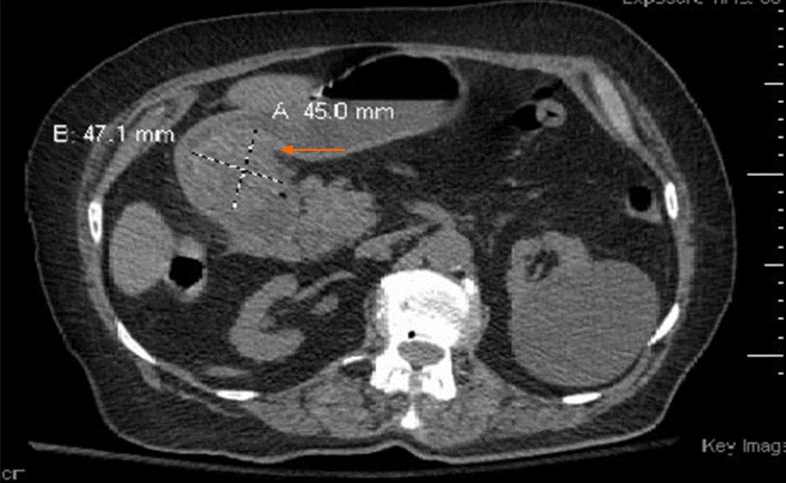
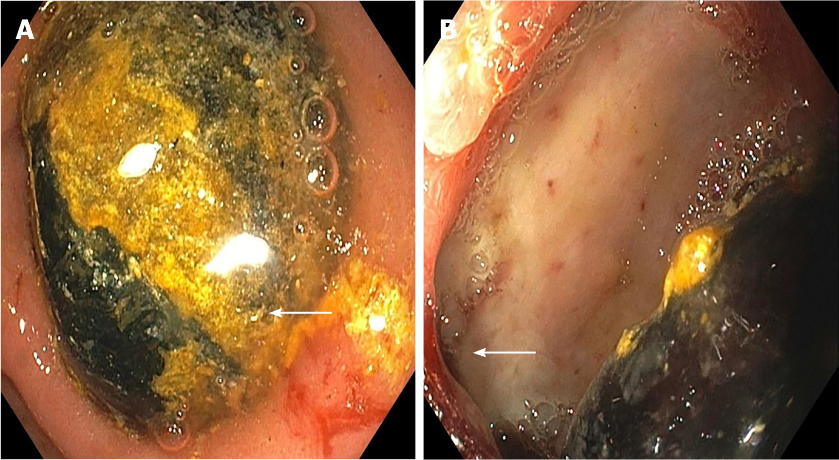
An 85-year-old female patient presented with 1-month history of intermittent abdominal pain, vomiting, decreased appetite and weight loss. An abdominal computed tomography showed a 4.5 cm × 4.7 cm partially calcified mass at the gastric pylorus causing gastric outlet obstruction. Endoscopy showed an ulcerated fistulous opening and a large 5 cm impacted gallstone in the duodenal bulb. Endoscopic nets and baskets were used in an attempt to remove the stone, but this approach was unsuccessful. Given her advanced age, poor physical condition and underlying comorbidities, she was deemed to be high-risk for surgery. Thus, a minimally invasive approach using endoscopic laser lithotripsy was attempted and successfully treated the stone. Post-procedure, the patient experienced complete resolution of her symptoms with no complications and was able to tolerate her diet. She was subsequently discharged home at 48 h, with an uneventful recovery.
In our paper we describe Bouveret syndrome and highlight its management with a novel endoscopic approach of laser lithotripsy in addition to various other endoscopic approaches available to date and its success rates.
Core Tip: An elderly female presented with features of gastric outlet obstruction found to have a large gall stone of 5 cm on endoscopy, successfully treated with complete endoscopic approach with laser lithotripsy.
- Citation: Parvataneni S, Khara HS, Diehl DL. Bouveret syndrome masquerading as a gastric mass-unmasked with endoscopic luminal laser lithotripsy: A case report. World J Clin Cases 2020; 8(22): 5701-5706
- URL: https://www.wjgnet.com/2307-8960/full/v8/i22/5701.htm
- DOI: https://dx.doi.org/10.12998/wjcc.v8.i22.5701
Gastric outlet obstruction is a condition characterized by mechanical impedance of gastric contents emptying into the proximal duodenum. Bouveret syndrome is a rare form of gastric outlet obstruction caused by gallstone impaction in the duodenum via a fistulous tract. First described by Beaussier in 1770, the syndrome was described again in two case reports published by Bouveret[1] in 1896, after which the syndrome was given his name[1]. This condition is most commonly seen in elderly patients, with a reported median age of 74 year, and occurs predominantly in females, with a female-to-male ratio of 1.82[2]. The diagnosis can be challenging and is often missed due to its nonspecific and atypical presentation that mimics the symptoms of gastric outlet obstruction, such as nausea, vomiting, weight loss, decreased appetite, and hematemesis, as seen with the present case[3]. We present a rare case of a patient who presented with signs of malignancy and was discovered to have a stone on endoscopy. The stone was treated successfully with a complete endoscopic resolution using laser lithotripsy.
An 85-year-old female presented to emergency room with vomiting and abdominal pain.
An elderly female patient presented with symptoms of intermittent abdominal pain around epigastric and umbilical area. The pain was dull aching 5/10 intensity that had been present for 1 mo and gradually worsened over the last 2 wk. The abdominal pain was non-radiating, exacerbated by food intake and had no associated relieving factors. Vomiting intermittently for the last 2 wk and increased in intensity over the past 48 h; it was non-blood stained and non-foul smelling. The patient had a history of loss of appetite and weight loss of about 15 Lbs. over the 1-mo duration. Her last bowel movement was 2 d prior to admission, with a normal consistency but black tarry in color. Review of systems unremarkable.
Past medical history included hypertension, cerebrovascular accident, diabetes, and chronic kidney disease stage 3. Family history did not reveal anything of significance to her present illness.
Family history did not reveal anything of significance to her present illness.
Heart rate of 110 beats per minute, blood pressure of systolic 80’s, temperature 36.4 °C, respiratory rate of 18. The patient abdomen was mildly distended, with tenderness in the mid epigastric region, no organomegaly, and normal bowel sounds.
Blood tests revealed elevated troponin of 86, electrocardiogram showed ST segment depressions likely demand ischemia from the acute illness. Basic metabolic panel showed creatinine of 3.1 (baseline was 1.4), potassium of 3.2, alkaline phosphatase of 177, Alanine transaminase of 53, aspartate transaminase of 53, and lactic acid of 2.3. On complete blood count, her white cell count of 20.78 neutrophilic leukocytosis, and hemoglobin of 9.8.
An abdominal computed tomography (CT) scan showed a 4.5 cm × 4.7 cm partially calcified mass at the gastric pylorus causing gastric outlet obstruction (Figure 1).
Endoscopy showed an ulcerated fistulous opening and a large 5 cm impacted gallstone in the duodenal bulb (Figure 2). Endoscopic nets and baskets were used in an attempt to remove the stone, but this approach was unsuccessful.
Given the endoscopic picture patient was diagnosed to have gastric outlet obstruction secondary to a large impacted gall stone which defines Bouveret syndrome, with a large duodenal ulcer.
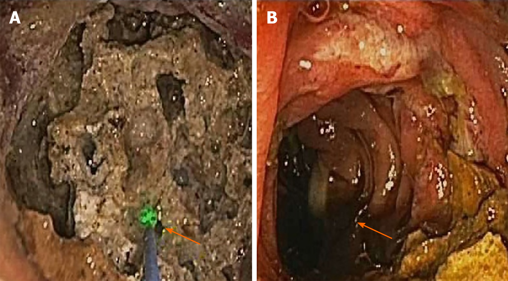
Given her advanced age, poor physical condition and underlying comorbidities, she was deemed to be high-risk for surgery. Thus, a minimally invasive approach using endoscopic laser lithotripsy was attempted. Holmium laser therapy was performed using an upper endoscope in the duodenal bulb with 9 Watts, 1000 pulses and 10 Joules; and after an extensive session of lithotripsy, the stone was eventually fragmented and removed piecemeal (Figure 3).
Post-procedure, the patient experienced complete resolution of her symptoms with no complications and was able to tolerate her diet. She was subsequently discharged home at 48 h, with an uneventful recovery.
Bouveret syndrome is a rare form of gallstone ileus accounting for approximately 1%-3% of cases[4]. In approximately 85% of these patients, gallstones are eliminated either by vomiting or in the feces; in the remaining 15% gallstones measuring more than 2.5 cm become impacted in the descending order in terminal ileum, proximal ileum, jejunum, colon, and rarely in the duodenum and stomach[5-7] . Imaging such as X-ray, CT scan, and ultrasound are used for diagnosis. X-ray is diagnostic in only 21% of cases, whereas CT scan has higher sensitivity for diagnosis. The classic sign seen on radiograph is the Riggler triad of bowel obstruction, pneumobilia and ectopic gallstone[8-10].
Treatment includes non-surgical and surgical options. In the past, surgical approaches using open enterolithotomy and cholecystectomy with fistula closure were considered first-line treatment, but surgery was associated with a morbidity and mortality of 37.5% and 11%, respectively[11]. The first successful reports describing endoscopic visualization and extraction of gallstones were published in three cases by Bedogni et al[12] in 1985. Later, endoscopic nets and lithotripsy techniques were developed for gallstone extraction. Endoscopic nets are a good alternative to the surgical approach in elderly patients in order to decrease the mortality risk, though nets are reported to be more effective for treating smaller-sized stones rather than larger ones[13]. This is evident in our case, in which the endoscopic net approach failed and lithotripsy was ultimately used for stone extraction.
Lithotripsy is a procedure used for stone destruction to facilitate easy passage and removal. It includes the use of mechanical, electrohydraulic, laser and extracorporeal shock wave techniques. Given the large stone size in our patient, we opted for laser lithotripsy. Laser lithotripsy was introduced in the mid-1980’s for the treatment of bile duct stones[14,15]. Holmium and neodymium yttrium aluminum garnet lasers are the Food and Drug Administration-approved laser treatments available in the market[16]. Laser lithotripsy has proven to be safe and effective in the treatment of complicated and refractory bile duct stones[17,18]. A literature review revealed case discussions in which laser lithotripsy was successfully used in the treatment of Bouveret syndrome. This approach was successful in patients with a maximal median size stone size of 4 cm (Interquartile range: 1.5) and was performed in a median of 1.5 sessions (Interquartile range: 1.5)[19].
To date, the present report is unique in describing a tailored endoscopic laser lithotripsy approach used to successfully treat a large stone with only one treatment session in an elderly patient with comorbidities. Bouveret syndrome is rare and should be considered in elderly patients with atypical presentations, and endoscopic approaches to fragmentation are a viable option that represent an alternative to surgery as first-line therapy.
Bouveret syndrome is a rare form of gastric outlet obstruction. Given the high incidence of morbidity and mortality associated with misdiagnosis and delayed treatment, it is important to keep Bouveret syndrome in the differential diagnosis of gastric outlet obstruction in elderly patients with atypical presentation. This case is unique because of its atypical presentation mimicking a malignant gastric outlet obstruction, which was successfully treated with advanced endoscopic intervention.
Manuscript source: Unsolicited manuscript
Specialty type: Medicine, research and experimental
Country/Territory of origin: United States
Peer-review report’s scientific quality classification
Grade A (Excellent): 0
Grade B (Very good): 0
Grade C (Good): C
Grade D (Fair): 0
Grade E (Poor): 0
P-Reviewer: Goral V S-Editor: Zhang L L-Editor: A P-Editor: Li JH
| 2. | Al-Habbal Y, Ng M, Bird D, McQuillan T, Al-Khaffaf H. Uncommon presentation of a common disease - Bouveret's syndrome: A case report and systematic literature review. World J Gastrointest Surg. 2017;9:25-36. [RCA] [PubMed] [DOI] [Full Text] [Full Text (PDF)] [Cited by in CrossRef: 18] [Cited by in RCA: 16] [Article Influence: 2.0] [Reference Citation Analysis (0)] |
| 3. | Cappell MS, Davis M. Characterization of Bouveret's syndrome: a comprehensive review of 128 cases. Am J Gastroenterol. 2006;101:2139-2146. [RCA] [PubMed] [DOI] [Full Text] [Cited by in Crossref: 161] [Cited by in RCA: 147] [Article Influence: 7.7] [Reference Citation Analysis (0)] |
| 4. | Heneghan HM, Martin ST, Ryan RS, Waldron R. Bouveret's syndrome--a rare presentation of gallstone ileus. Ir Med J. 2007;100:504-505. [PubMed] |
| 5. | Iñíguez A, Butte JM, Zúñiga JM, Crovari F, Llanos O. Bouveret syndrome: report of four cases. Rev Med Chil. 2008;136:163-168. [PubMed] |
| 6. | Brennan GB, Rosenberg RD, Arora S. Bouveret syndrome. Radiographics. 2004;24:1171-1175. [RCA] [PubMed] [DOI] [Full Text] [Cited by in Crossref: 57] [Cited by in RCA: 63] [Article Influence: 3.2] [Reference Citation Analysis (0)] |
| 7. | Mavroeidis VK, Matthioudakis DI, Economou NK, Karanikas ID. Bouveret syndrome-the rarest variant of gallstone ileus: a case report and literature review. Case Rep Surg. 2013;2013:839370. [RCA] [PubMed] [DOI] [Full Text] [Full Text (PDF)] [Cited by in Crossref: 31] [Cited by in RCA: 50] [Article Influence: 4.2] [Reference Citation Analysis (0)] |
| 8. | Trubek S, Bhama J K, Lamki N. Radiological findings in Bouveret's syndrome. Emerg Radiol. 2001;8:335-337. [RCA] [DOI] [Full Text] [Cited by in Crossref: 13] [Cited by in RCA: 5] [Article Influence: 0.2] [Reference Citation Analysis (0)] |
| 9. | Pickhardt PJ, Friedland JA, Hruza DS, Fisher AJ. Case report. CT, MR cholangiopancreatography, and endoscopy findings in Bouveret's syndrome. AJR Am J Roentgenol. 2003;180:1033-1035. [RCA] [PubMed] [DOI] [Full Text] [Cited by in Crossref: 51] [Cited by in RCA: 53] [Article Influence: 2.4] [Reference Citation Analysis (0)] |
| 10. | Yu CY, Lin CC, Shyu RY, Hsieh CB, Wu HS, Tyan YS, Hwang JI, Liou CH, Chang WC, Chen CY. Value of CT in the diagnosis and management of gallstone ileus. World J Gastroenterol. 2005;11:2142-2147. [RCA] [PubMed] [DOI] [Full Text] [Full Text (PDF)] [Cited by in CrossRef: 115] [Cited by in RCA: 126] [Article Influence: 6.3] [Reference Citation Analysis (2)] |
| 11. | Pavlidis TE, Atmatzidis KS, Papaziogas BT, Papaziogas TB. Management of gallstone ileus. J Hepatobiliary Pancreat Surg. 2003;10:299-302. [RCA] [PubMed] [DOI] [Full Text] [Cited by in Crossref: 47] [Cited by in RCA: 47] [Article Influence: 2.1] [Reference Citation Analysis (0)] |
| 12. | Bedogni G, Contini S, Meinero M, Pedrazzoli C, Piccinini GC. Pyloroduodenal obstruction due to a biliary stone (Bouveret's syndrome) managed by endoscopic extraction. Gastrointest Endosc. 1985;31:36-38. [RCA] [PubMed] [DOI] [Full Text] [Cited by in Crossref: 38] [Cited by in RCA: 37] [Article Influence: 0.9] [Reference Citation Analysis (1)] |
| 13. | Jindal A, Philips CA, Jamwal K, Sarin SK. Use of a Roth Net Platinum Universal Retriever for the endoscopic management of a large symptomatic gallstone causing Bouveret's syndrome. Endoscopy. 2016;48:E308. [RCA] [PubMed] [DOI] [Full Text] [Cited by in Crossref: 4] [Cited by in RCA: 4] [Article Influence: 0.4] [Reference Citation Analysis (0)] |
| 14. | Kozarek RA, Low DE, Ball TJ. Tunable dye laser lithotripsy: in vitro studies and in vivo treatment of choledocholithiasis. Gastrointest Endosc. 1988;34:418-421. [RCA] [PubMed] [DOI] [Full Text] [Cited by in Crossref: 44] [Cited by in RCA: 35] [Article Influence: 0.9] [Reference Citation Analysis (0)] |
| 15. | Cotton PB, Kozarek RA, Schapiro RH, Nishioka NS, Kelsey PB, Ball TJ, Putnam WS, Barkun A, Weinerth J. Endoscopic laser lithotripsy of large bile duct stones. Gastroenterology. 1990;99:1128-1133. [RCA] [PubMed] [DOI] [Full Text] [Cited by in Crossref: 99] [Cited by in RCA: 76] [Article Influence: 2.2] [Reference Citation Analysis (0)] |
| 16. | ASGE Technology Committee. , Watson RR, Parsi MA, Aslanian HR, Goodman AJ, Lichtenstein DR, Melson J, Navaneethan U, Pannala R, Sethi A, Sullivan SA, Thosani NC, Trikudanathan G, Trindade AJ, Maple JT. Biliary and pancreatic lithotripsy devices. VideoGIE. 2018;3:329-338. [RCA] [PubMed] [DOI] [Full Text] [Full Text (PDF)] [Cited by in Crossref: 30] [Cited by in RCA: 16] [Article Influence: 2.3] [Reference Citation Analysis (0)] |
| 17. | Lv S, Fang Z, Wang A, Yang J, Zhang W. Choledochoscopic Holmium Laser Lithotripsy for Difficult Bile Duct Stones. J Laparoendosc Adv Surg Tech A. 2017;27:24-27. [RCA] [PubMed] [DOI] [Full Text] [Cited by in Crossref: 6] [Cited by in RCA: 6] [Article Influence: 0.8] [Reference Citation Analysis (0)] |
| 18. | Schatloff O, Rimon U, Garniek A, Lindner U, Morag R, Mor Y, Ramon J, Winkler H. Percutaneous transhepatic lithotripsy with the holmium: YAG laser for the treatment of refractory biliary lithiasis. Surg Laparosc Endosc Percutan Tech. 2009;19:106-109. [RCA] [PubMed] [DOI] [Full Text] [Cited by in Crossref: 14] [Cited by in RCA: 14] [Article Influence: 0.9] [Reference Citation Analysis (0)] |
| 19. | Dumonceau JM, Devière J. Novel treatment options for Bouveret's syndrome: a comprehensive review of 61 cases of successful endoscopic treatment. Expert Rev Gastroenterol Hepatol. 2016;10:1245-1255. [RCA] [PubMed] [DOI] [Full Text] [Cited by in Crossref: 13] [Cited by in RCA: 13] [Article Influence: 1.4] [Reference Citation Analysis (0)] |


