Published online Jan 6, 2020. doi: 10.12998/wjcc.v8.i1.133
Peer-review started: October 15, 2019
First decision: November 13, 2019
Revised: November 18, 2019
Accepted: November 30, 2019
Article in press: November 30, 2019
Published online: January 6, 2020
Processing time: 83 Days and 6.1 Hours
Hyalinizing clear cell carcinoma (HCCC) is an uncommon tumor that originates in the salivary glands. This neoplasia constitutes less than 1% of minor salivary gland tumors.
A 67-year-old female visited the maxillofacial surgery department owing to a smooth, slightly yellowish protruding mass on the left side of the floor of the mouth, at the level of the molars; the tumor mass had a soft consistency on palpation and did not adhere to deep planes. The microscopical analysis of the excisional biopsy showed that the lesion was composed of sheets and cords of clear cells separated by thick eosinophilic bands of hyaline collagen. Normal glandular tissue was absent, periodic acid-Schiff with and without diastase stains, and immunohistochemical reactions were performed to confirm the diagnosis. This is the second case reported in the literature of HCCC arising in the floor of the mouth.
HCCC is a rare salivary gland tumor that has not been studied extensively. Its diagnosis is usually challenging, because clinically, it can be confused with a benign neoplasm.
Core tip: Hyalinizing clear cell carcinoma is a rare tumor that originates in the salivary glands. This neoplasia constitutes less than 1% of minor salivary gland tumors, the lesion is composed for clear cells that formed compact groups and cords that were separated by thick eosinophilic bands of collagen, with the appearance of hyaline. This carcinoma is a rare salivary gland tumor that has not been studied extensively. Its diagnosis is usually challenging, because clinically, it can be confused with a benign neoplasm.
- Citation: Donohue-Cornejo A, Paes de Almeida O, Sánchez-Romero C, Espinosa-Cristóbal LF, Reyes-López SY, Cuevas-González JC. Hyalinizing clear cell carcinoma-a rare entity in the oral cavity: A case report. World J Clin Cases 2020; 8(1): 133-139
- URL: https://www.wjgnet.com/2307-8960/full/v8/i1/133.htm
- DOI: https://dx.doi.org/10.12998/wjcc.v8.i1.133
Hyalinizing clear cell carcinoma (HCCC) is a rare tumor that originates in the salivary glands. Although this neoplasia constitutes less than 1% of minor salivary gland tumors, when this carcinoma presents, it has a predilection to develop in this type of gland. Due to its rarity, it has not been studied extensively. To this end, we present this case report, the clinical, histopathological, and immunohistochemical documentation of which can be useful for the clinician and pathologist when making the diagnosis, increasing our understanding and recognition of this carcinoma[1,2].
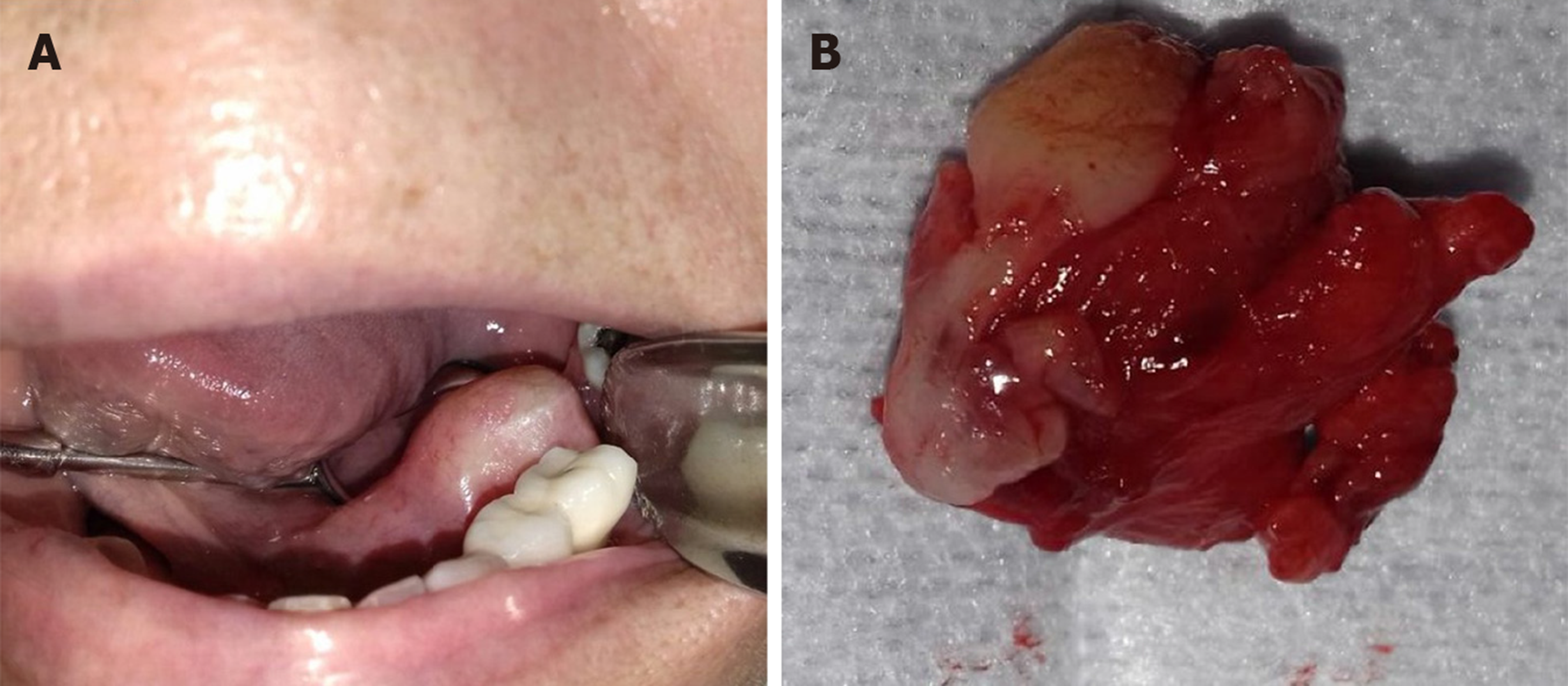
A 67-year-old female visited the maxillofacial surgery department due to a smooth, slightly yellowish protruding mass on the left side of the floor of the mouth, at the level of the molars (Figure 1A).
The tumor mass had a soft consistency on palpation and did not adhere to deep planes. The patient reported having noticed an increase in the volume of the mass for approximately 1 year asymptomatically.
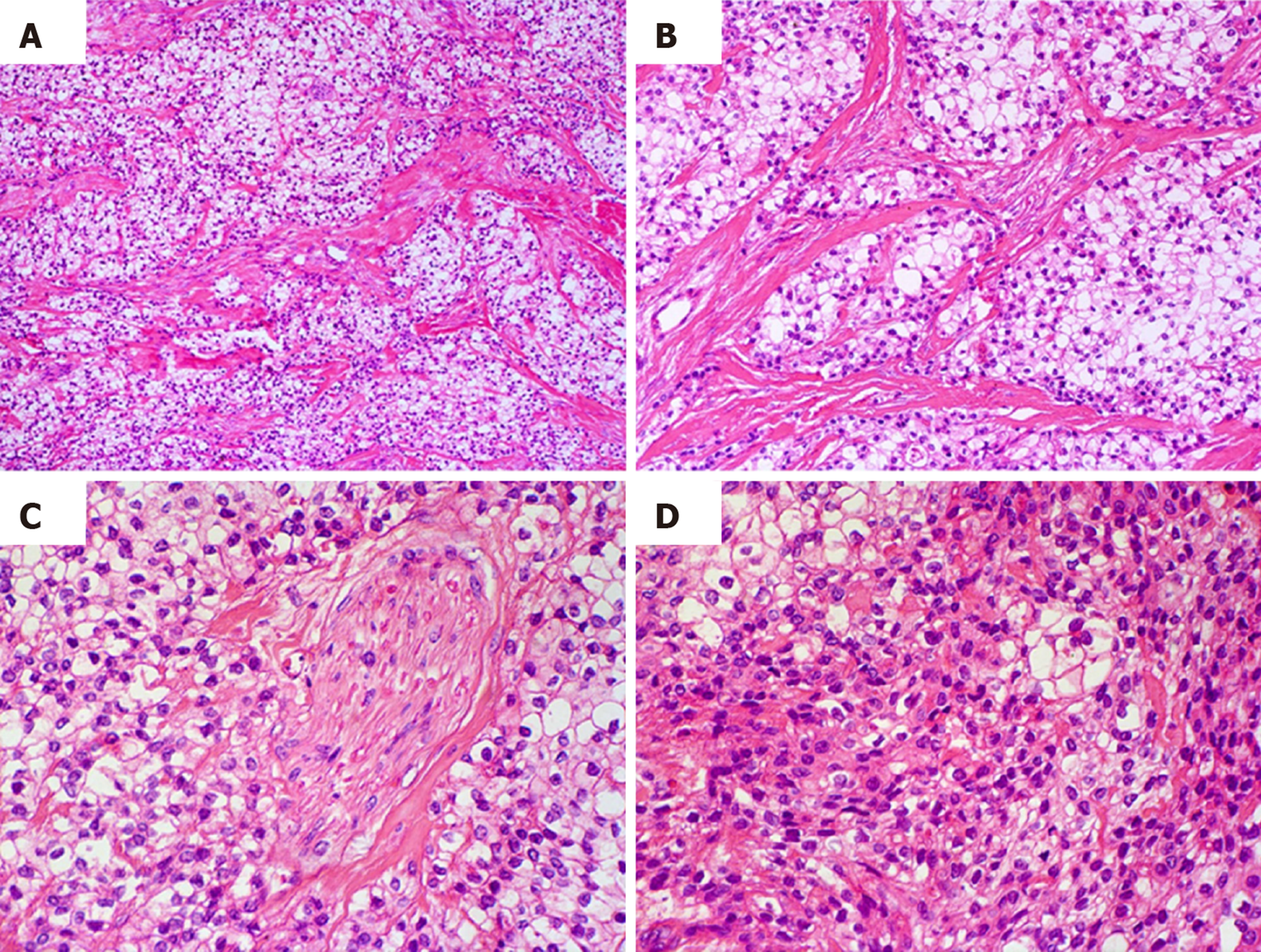
Based on the clinical features, the surgeon chose to perform an excisional biopsy. The tumor was well-demarcated from the surrounding tissues therefore, it was completely removed, measuring 5 cm × 4 cm × 4 cm (Figure 1B), and once the sample was processed and stained with hematoxylin and eosin (HE), a tumor lesion was observed, composed primarily of diffuse, proliferating clear cells that formed compact groups and cords that were separated by thick eosinophilic bands of collagen, with the appearance of hyaline (Figure 2A and B). Despite the predominance of clear cells, focal groups of tumor cells with eosinophilic cytoplasm were identified (Figure 2D), occasional mitoses and neural invasion were observed (Figure 2C).
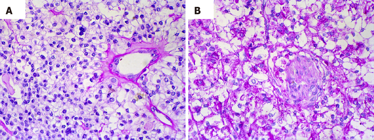
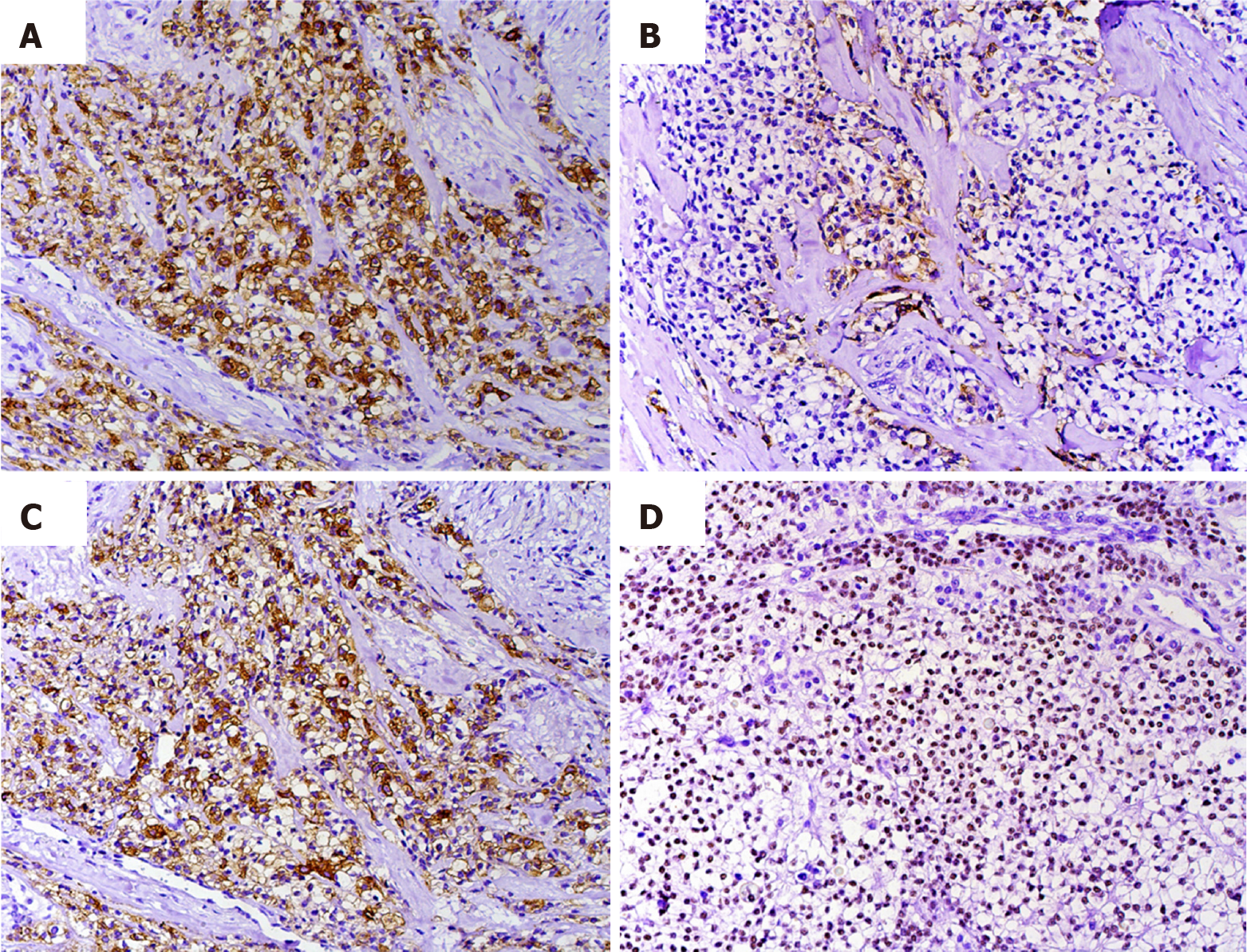
Periodic acid-Schiff (PAS) stains and immunohistochemical reactions were performed to confirm the diagnosis. HCCC is diastase sensitive due to the glycogen of the tumor cells, as showed by our case, it was negative for PAS with diastase (Figure 3A) and positive for PAS without diastase (Figure 3B). Antibodies against AE1-AE3, CK5, CK7, p63, and Ki-67 were positive. In contrast, there was no signal with CK14, CK19, or smooth muscle antibodies (SMAs) (Figures 3 and 4).
Considering the clinical, histopathological, histochemical, and immunohistochemical findings, a diagnosis of HCCC was reached. Once the diagnosis was established and the cell morphology was re-evaluated surgical borders with neoplastic cells were identified.
The patient underwent a second surgery, widen the margins, to ensure complete removal of the lesion.
Close long-term follow-up.
Several studies suggest that HCCC presents primarily as a painless lesion that most often affects females (2:1), between the sixth and seventh decades of life. It is considered a low-grade neoplasm that rarely recurs or metastasizes[3].
This case had clinical characteristics of a benign lesion with duration of a year, which was intact covering mucosa and well-demarcated from the surrounding tissues. When the computerized axial tomography was performed, a small isodense exophytic mass was described on the floor of the mouth, close to the left molar area without growth towards the deep planes. According to our research, this is the second case of HCCC located on the floor of the mouth (Table 1), the patient did not seek consultation until the volume increase was considerable.
| Ref. | Sex | Age (yr) | Location | Treatment | Prognosis |
| Bostanci et al[12], 2019 | F | 51 | Right upper gingiva | Maxillectomy | Tumor recurrence |
| Hwang et al[6], 2019 | F | 71 | Base of tongue | Surgical treatment and chemoradiation | Not referred |
| Gushi et al[9], 2017 | F | 31 | Maxillary alveolar mucosa | Excisional biopsy | Good |
| Moreno et al[13], 2014 | F | 47 | Base of tongue | Surgical treatment | Good |
| Lin et al[14], 2015 | F | 37 | Tongue | Surgical treatment | Good |
| Kim et al[15], 2013 | M | 54 | Mouth floor | Surgical treatment and radiation | Good |
| Gon et al[3], 2013 | F | 70 | Palate | Surgical treatment | Tumor recurrence |
| Saleh et al[16], 2012 | M | 31 | Soft Palate | Incisional biopsy | Bad, the patient declined any treatment |
| Roby et al[8], 2012 | F | 60 | Tongue | Surgical excision | Not referred |
| Baghirath et al[4], 2011 | F | 36 | Right upper gingiva | Surgical treatment | Not referred |
| Kauzman et al[17], 2011 | M | 53 | Right upper gingiva | Surgical excision and radiation treatment | Good |
| Chatelain et al[18], 2011 | M | 48 | Right upper gingiva | Maxillectomy, cervical lymph node dissection, and postoperative radiotherapy | No recurrence at 12 mo |
| Masilamani et al[19], 2011 | M | 73 | Base of tongue | Surgical excision | No recurrence at 12 mo |
Due to the clinical characteristics, such as its well-defined increase in volume, its similarity in color to the adjacent mucosa, and its asymptomatic nature and evolution, our clinical impression was that of a benign neoplasm. However, following the standard protocol of care, a biopsy was performed by histopathology and complementary approaches, which allowed us to reach a definitive diagnosis of HCCC.
Overall, 81.8% of cases of HCCC occur in the buccal cavity. The most frequent sites are the tongue, hard palate, floor of the mouth, and base of the tongue; HCCC is uncommon in the major salivary glands, nasopharynx, hypopharynx, and lacrimal gland[4]. In a review of the literature that we conducted in cases from 2011 to date, we found 13 published clinical cases located in oral cavity of which 8 (61%) affected the female sex and 5 (39%) the male (Table 1).
Histopathologically, HCCC comprises proliferating epithelial cells with a clear cytoplasm. In some cases, a subgroup of cells with eosinophilic cytoplasm are present, all of which are organized in trabeculae, cords, or solid nests that are surrounded by a stroma of hyaline fibrous tissue. This tumor usually presents with immuno-histochemical positivity for p63, CK5, CK7, CK14, and CK19 and negative S-100, CK 14, SMA, and PAX8 expression, in addition to negativity for PAS with diastase, which favors a diagnosis of HCCC and helps rule out other histopathological differential diagnoses, such as malignant myoepithelioma, epithelial-myoepithelial carcinoma, clear cell mucoepidermoid carcinoma, low-grade polymorphic adenocarcinoma, acinar cell carcinoma, clear cell oncocytoma, and renal metastatic carcinoma[5-9]. Thus, an immunohistochemical panel with the appropriate antibodies must be performed.
In our case, we observed immunolabeling with antibodies against AE1-AE3, CK5, CK7, p63, and Ki67, whereas SMA, CK14, and CD10 were negative. Thus, we recommend using these immunohistochemical markers to aid in the diagnosis of this neoplasia with the histopathological characteristics that we have described.
HCCC is considered a low-grade malignancy. However, the size of the tumor, the cellular atypia, and the number of mitoses is related to its prognosis, and as with any other carcinoma, to decrease the likelihood of recurrence, it must be removed completely[10]. In case series, up to 25% of evidence of metastasis has been reported[11]. Although the treatment depends on the clinical characteristics of the tumor, in 50% of cases, it is limited to surgery, versus surgery and radiotherapy in 25.7%, with a 5-year survival of 77.6%; the remainder corresponds to other treatment modalities or is unknown to due to the unavailability of data[1].
In our case, by histopathology, we noted that the frequency of atypia and mitosis was low and that the positivity to Ki-67 antibody was of 3%, which was consistent with a low-grade malignant neoplasm. Although some tumor cells had infiltrated nervous tissue, the presence of metastases was ruled out by imaging (positron emission tomography). Because the edges of the excisional biopsy were not free of tumor cells, the patient underwent a second surgery to eliminate the possibility of recurrence. After 2 years of follow-up, there are no data that suggest recurrence, but continuous and long-term follow-up is indicated to identify any eventual alterations in a timely manner.
HCCC is a rare salivary gland tumor that has not been studied extensively. We presented herein the second case reported in the literature affecting the floor of the mouth. Its diagnosis is usually challenging, because clinically, it can be confused with a benign neoplasm, whereas histologically, there are several differential diagnoses. Thus, auxiliary techniques, such as PAS staining and immunohistochemistry, are valuable tools in reaching the correct diagnosis of this tumor.
Manuscript source: Unsolicited manuscript
Specialty type: Medicine, research and experimental
Country of origin: Mexico
Peer-review report classification
Grade A (Excellent): 0
Grade B (Very good): B
Grade C (Good): 0
Grade D (Fair): D
Grade E (Poor): 0
P-Reviewer: Ikura Y, Ishizawa K S-Editor: Ma YJ L-Editor: A E-Editor: Liu JH
| 1. | Oliver J, Wu P, Chang C, Roden D, Wang B, Liu C, Hu K, Schreiber D, Givi B. Patterns of Care and Outcome of Clear Cell Carcinoma of the Head and Neck. Otolaryngol Head Neck Surg. 2019;161:98-104. [RCA] [PubMed] [DOI] [Full Text] [Cited by in Crossref: 7] [Cited by in RCA: 10] [Article Influence: 1.7] [Reference Citation Analysis (0)] |
| 2. | Icard B, Grider DJ, Aziz S, Rubio E. Primary tracheal hyalinizing clear cell carcinoma. Lung Cancer. 2018;125:100-102. [RCA] [PubMed] [DOI] [Full Text] [Cited by in Crossref: 6] [Cited by in RCA: 18] [Article Influence: 2.6] [Reference Citation Analysis (0)] |
| 3. | Gon S, Bhattacharyya A, Majumdar B, Das TK. Post-radiotherapy locoregional recurrence of hyalinizing clear cell carcinoma of palate. J Cancer Res Ther. 2013;9:281-283. [RCA] [PubMed] [DOI] [Full Text] [Cited by in Crossref: 11] [Cited by in RCA: 11] [Article Influence: 1.0] [Reference Citation Analysis (0)] |
| 4. | Baghirath PV, Kumar JV, Vinay BH. Hyalinizing clear cell carcinoma: A rare entity. J Oral Maxillofac Pathol. 2011;15:335-339. [RCA] [PubMed] [DOI] [Full Text] [Full Text (PDF)] [Cited by in Crossref: 14] [Cited by in RCA: 15] [Article Influence: 1.2] [Reference Citation Analysis (0)] |
| 5. | Yamanishi T, Kutsuma K, Masuyama K. A Case of Hyalinizing Clear Cell Carcinoma, So-Called Clear Cell Carcinoma, Not Otherwise Specified, of the Minor Salivary Glands of the Buccal Mucosa. Case Rep Otolaryngol. 2015;2015:471693. [RCA] [PubMed] [DOI] [Full Text] [Full Text (PDF)] [Cited by in Crossref: 6] [Cited by in RCA: 6] [Article Influence: 0.6] [Reference Citation Analysis (0)] |
| 6. | Hwang G, Goldenberg D, Warrick J, Slonimsky G. A Hyalinizing Clear Cell Carcinoma of the Base of Tongue. Ear Nose Throat J. 2019;145561319840513. [RCA] [PubMed] [DOI] [Full Text] [Cited by in Crossref: 4] [Cited by in RCA: 4] [Article Influence: 0.7] [Reference Citation Analysis (0)] |
| 7. | AlAli BM, Alyousef MJ, Kamel AS, Al Hamad MA, Al-Bar MH, Algowiez RM. Primary paranasal sinus hyalinizing clear cell carcinoma: a case report. Diagn Pathol. 2017;12:70. [RCA] [PubMed] [DOI] [Full Text] [Full Text (PDF)] [Cited by in Crossref: 11] [Cited by in RCA: 14] [Article Influence: 1.8] [Reference Citation Analysis (0)] |
| 8. | Roby BB, Pambuccian SE, Khariwala SS. Pathology quiz case 2. Hyalinizing clear cell carcinoma. Arch Otolaryngol Head Neck Surg. 2012;138:207. [RCA] [PubMed] [DOI] [Full Text] [Cited by in Crossref: 7] [Cited by in RCA: 7] [Article Influence: 0.5] [Reference Citation Analysis (0)] |
| 9. | Gushi E, Seki U, Orhan K. Hyalinizing Clear Cell Carcinoma of Maxilla. J Clin Diagn Res. 2017;11:ZL01-ZL02. [RCA] [PubMed] [DOI] [Full Text] [Cited by in Crossref: 2] [Cited by in RCA: 4] [Article Influence: 0.5] [Reference Citation Analysis (0)] |
| 10. | Zhao W, Yang L, Wang L, Zuo W, Yuan S, Yu J, Yu Q, Hu X, Wang S, Liu N, Zhang H, Wei Y. Primary clear cell carcinoma of nasal cavity: report of six cases and review of literature. Int J Clin Exp Med. 2014;7:5469-5476. [PubMed] |
| 11. | O'Sullivan-Mejia ED, Massey HD, Faquin WC, Powers CN. Hyalinizing clear cell carcinoma: report of eight cases and a review of literature. Head Neck Pathol. 2009;3:179-185. [RCA] [PubMed] [DOI] [Full Text] [Cited by in Crossref: 72] [Cited by in RCA: 63] [Article Influence: 3.9] [Reference Citation Analysis (0)] |
| 12. | Bostanci A, Ozbudak IH, Turhan M. Hyalinizing Clear Cell Carcinoma of the Maxilla. J Maxillofac Oral Surg. 2019;18:391-394. [RCA] [PubMed] [DOI] [Full Text] [Cited by in Crossref: 4] [Cited by in RCA: 4] [Article Influence: 0.6] [Reference Citation Analysis (0)] |
| 13. | Moreno-Zafra S, Rodríguez-Verdugo M, Hernandez-Lopez R. Carcinoma de celulas claras en la base de la lengua. Acta Otorrinolaringol Esp. 2014;65:133-34. [RCA] [DOI] [Full Text] [Cited by in Crossref: 3] [Cited by in RCA: 1] [Article Influence: 0.1] [Reference Citation Analysis (0)] |
| 14. | Lin JC, Liao JB, Fu HT, Chang TS, Wang JS. Salivary Gland Hyalinizing Clear Cell Carcinoma. J Pathol Transl Med. 2015;49:351-353. [RCA] [PubMed] [DOI] [Full Text] [Full Text (PDF)] [Cited by in Crossref: 4] [Cited by in RCA: 4] [Article Influence: 0.4] [Reference Citation Analysis (0)] |
| 15. | Kim DW, Park HJ, Cha IH, Yang DH, Kim HS, Nam W. An atypical case of rare salivary malignancy, hyalinizing clear cell carcinoma. J Korean Assoc Oral Maxillofac Surg. 2013;39:283-288. [RCA] [PubMed] [DOI] [Full Text] [Full Text (PDF)] [Cited by in Crossref: 9] [Cited by in RCA: 10] [Article Influence: 0.8] [Reference Citation Analysis (0)] |
| 16. | Saleh KA, Nurishmah MI, Firouzeh GN, Goh BS. Primary clear cell carcinoma of minor salivary gland of the soft palate: a case report. Med J Malaysia. 2012;67:335-336. [PubMed] |
| 17. | Kauzman A, Tabet JC, Stiharu TI. Hyalinizing clear cell carcinoma: a case report and review of the literature. Oral Surg Oral Med Oral Pathol Oral Radiol Endod. 2011;112:e26-e34. [RCA] [PubMed] [DOI] [Full Text] [Cited by in Crossref: 31] [Cited by in RCA: 32] [Article Influence: 2.3] [Reference Citation Analysis (0)] |
| 18. | Chatelain B, Curlier A, Euvrard E, Vitte F, Ricbourg B, Meyer C. [Hyalinizing clear cell carcinoma of the maxilla]. Rev Stomatol Chir Maxillofac. 2011;112:183-186. [RCA] [PubMed] [DOI] [Full Text] [Cited by in Crossref: 3] [Cited by in RCA: 3] [Article Influence: 0.2] [Reference Citation Analysis (0)] |
| 19. | Masilamani S, Rao S, Chirakkal P, Kumar AR. Hyalinizing clear cell carcinoma of the base of tongue: a distinct and rare entity. Indian J Pathol Microbiol. 2011;54:167-169. [RCA] [PubMed] [DOI] [Full Text] [Cited by in Crossref: 14] [Cited by in RCA: 15] [Article Influence: 1.1] [Reference Citation Analysis (0)] |


