Published online Jun 16, 2015. doi: 10.12998/wjcc.v3.i6.538
Peer-review started: October 27, 2014
First decision: January 20, 2015
Revised: February 3, 2015
Accepted: March 16, 2015
Article in press: March 18, 2015
Published online: June 16, 2015
Processing time: 235 Days and 17.5 Hours
Primary adenocarcinoma of the appendix is a rare malignancythat constitutes < 0.5% of all gastrointestinalneoplasms. Moreover, primary signet ring cell carcinomaof the appendix is an exceedingly rare entity. In the present report, we describe a rare case of primary signet ring cell carcinoma of the appendix with ovarian metastasesand unresectable peritoneal dissemination occurring in a 45-year-old female patient. She was clinically misdiagnosed as torsion of ovarian cyst. She underwent appendicectomy and unilateral salpingo-oophorectomy.Histopathology revealed signet ring cell carcinoma and a right hemicolectomy was done. She then received palliative systemic chemotherapy with 12 cycles of oxaliplatin, 5-fluorouracil, and leucovorin (FOLFOX-4). The patient is doing well till today on follow up without progression of disease 10 mo after beginning chemotherapy.
Core tip: Meticulous histopathological evaluation along with clinical correlation is strongly recommended in such unusual circumstances. The clinician and surgical pathologist should keep in mind this rare entity as a differential diagnosis.
- Citation: Kulkarni RV, Ingle SB, Siddiqui S. Primary signet ring cell carcinoma of the appendix: A rare case report. World J Clin Cases 2015; 3(6): 538-541
- URL: https://www.wjgnet.com/2307-8960/full/v3/i6/538.htm
- DOI: https://dx.doi.org/10.12998/wjcc.v3.i6.538
Primary adenocarcinoma of the appendix, first invented in 1882, and constitutes 0.12 cases per one million people per year[1]. Primary signet ring cell carcinoma is an extremely unusual event in surgical practice[2]. Clinically it simulates acute appendicitis and difficult to distinguish from it[3,4]. So, it is a difficult task to diagnose it on clinical grounds. Usually the diagnosis is confirmed on histopathology of a surgically-removed inflamed appendix[5].
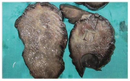
A 46-year-old female admitted in YCR Hospital Latur with persistent right lower quadrant abdominal pain. Baseline blood tests showed neutrophilic leukocytosis. Per abdominal examination revealed right abdominal distension. The patient was misdiagnosed as twisted ovarian cyst and emergency laparotomy was planned and performed. At laparotomy, the appendix appeared severely inflamed so the patient underwent an appendectomy and unilateral salphingo-oopherectomy. On gross examination, left large encapsulated, dark brown and smooth ovarian mass measuring 16 cm × 13 cm × 9.5 cm was found. The cut surface showed large hematoma with grey white visible areas. Attached fallopian tube measured 5 cm in length (Figure 1).
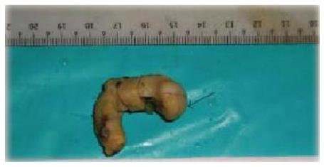
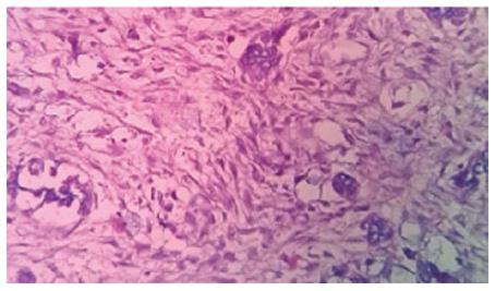
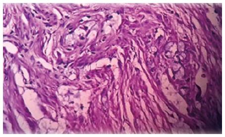
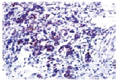
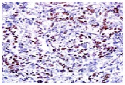
Appendix measured 6 cm in length. External surface was thickened and congested. Cut section showed obliterated lumen filled with gelatinous material (Figure 2). Microscopic examination of both left ovarian mass revealed signet ring cells showing an intracellular mucin to vacuolated cytoplasm shifting the nuclei towards periphery (Figure 3). Some extracellular mucin was seen infiltrating the stroma.Large areas of hemorrhage and necrosis are noted. In appendix the signet ring cells and extracellular mucin was seen invading the mucosa, submucosa and muscularis propia (Figure 4). Thus the case was finally diagnosed as infiltrating adenocarcinoma of the appendix with “signet ring cells” differentiation, with signs of infiltration of peri-appendicular fat. The tumor cells were immunopositive for CEA, cytokeratin 20 (Figure 5), MUC2, and CDX-2 (Figure 6). The patient underwent colonoscopy for the evaluation of synchronous disease, which was negative. Later computed tomography (CT) scan was done and an 8 cm × 5 cm lesion in right adnexal region was noted so the patient subsequently underwent right hemicolectomy with total abdominal hysterectomy and unilateral salphingo-oopherectomy and no residual carcinoma with negative lymph nodes was found.
Appendiceal adenocarcinoma is an unusual malignancy[1]. The reported prevalence rate is 0.3%[6]. Signet ring carcinoma constitutes only 4% of all neoplasms of appendix[2]. Malignant carcinoids are mainly found in younger age group (mean age, 38 years)[1]. The mean age of occurrence of mucinous adenocarcinoma is 60 years, while that of signet ring cell carcinoma is 62 years with male: female ratio 1:1[1].
According to International Classification of Diseases for Oncology appendiceal tumors are divided in to five classes: Mucinous adenocarcinoma, colonic type adenocarcinoma, goblet cell carcinoma, signet ring cell carcinoma and malignant carcinoid/adenocarcinoid[1,7]. Adenomas are the premalignant lesions[8,9]. The signet ring cell carcinomas are usually frequent in the stomach and intestine. In our case, the tumor cells were immunopositive for cytokeratin 20, CDX-2, MUC-2, and CEA. The CDX-2 marker is the key to confirm the final diagnosis[10].
Most of them are clinically low-grade tumors with indolent behavior. The overall 5-year survival rate is 20.5%. As per previous workers except for signet ring cell carcinoma and malignant characinoid, the histopathological variant does not affect the survival rate[11]. In fact the extent of the disease at the time of diagnosis is an important determinant of prognosis of patient. As per previous studies, the prognosis of patients with diffuse, peritoneal metastases is worse, with a 5-year survival rate of 6.7%-14%[12,13]. However, according to some workers signet ring cell carcinoma and poorly differentiated adenocarcinoma of the appendix had high propensity for development of metastasis with a 5-year survival rate of only 7%. So, signet ring cell carcinoma is considered as a separate tumor type in the appendix in view of its poor prognosis. Right hemicolectomy is the treatment of choice for all microscopic types of appendiceal carcinoma, even in cases with perforation. In case of metastasis treatment modalities are systemic chemotherapy along with intraoperative intraperitoneal chemotherapy, peritonectomy and cytoreductive surgery[1,5].
To conclude, meticulous histopathological examination of appendix is mandatory during exploratory laparotomy for ovarian masses and for diagnostic procedure. Exclusion of signet ring cell carcinoma from other carcinoma subtypes is of particular importance as it has an extremely poor prognosis and is usually diagnosed in advanced stages.
A 45-year-old woman presented to the Emergency Department of YCR Hospital Latur with persistent right lower quadrant abdominal pain.
Clinically diagnosed as twisted ovarian cyst.
Twisted ovarian cyst was the clinical differential diagnosis and signet ring carcinoma of either colon, stomach or appendix were the histological differential diagnosis.
Primary signet ring carcinoma of appendix with secondaries in the ovaries.
Computed tomography (CT) scan was done and an 8 cm × 5 cm lesion in right adnexal region was noted so the patient subsequently underwent right hemicolectomy with total abdominal hysterectomy and unilateral salphingo-oopherectomy.
Primary signet ring carcinoma of appendix with secondaries in the ovaries.
Right hemicolectomy.She then received palliative systemic chemotherapy with 12 cycles of oxaliplatin, 5-fluorouracil, and leucovorin (FOLFOX-4). The patient is doing well till today on follow up without progression of disease 10 mo after beginning chemotherapy.
Meticulous histopathological examination of appendix is mandatory during exploratory laparotomy for ovarian masses and for diagnostic procedure.
The authors have performed a good study, the manuscript is interesting.
P- Reviewer: Akbulut S, Virk JS S- Editor: Tian YL L- Editor: A E- Editor: Wu HL
| 1. | McCusker ME, Coté TR, Clegg LX, Sobin LH. Primary malignant neoplasms of the appendix: a population-based study from the surveillance, epidemiology and end-results program, 1973-1998. Cancer. 2002;94:3307-3312. [RCA] [PubMed] [DOI] [Full Text] [Cited by in Crossref: 386] [Cited by in RCA: 403] [Article Influence: 17.5] [Reference Citation Analysis (0)] |
| 2. | McGory ML, Maggard MA, Kang H, O’Connell JB, Ko CY. Malignancies of the appendix: beyond case series reports. Dis Colon Rectum. 2005;48:2264-2271. [RCA] [PubMed] [DOI] [Full Text] [Cited by in Crossref: 176] [Cited by in RCA: 175] [Article Influence: 8.8] [Reference Citation Analysis (0)] |
| 3. | Connor SJ, Hanna GB, Frizelle FA. Appendiceal tumors: retrospective clinicopathologic analysis of appendiceal tumors from 7,970 appendectomies. Dis Colon Rectum. 1998;41:75-80. [RCA] [PubMed] [DOI] [Full Text] [Cited by in Crossref: 402] [Cited by in RCA: 404] [Article Influence: 15.0] [Reference Citation Analysis (0)] |
| 4. | O’Donnell ME, Badger SA, Beattie GC, Carson J, Garstin WI. Malignant neoplasms of the appendix. Int J Colorectal Dis. 2007;22:1239-1248. [RCA] [PubMed] [DOI] [Full Text] [Cited by in Crossref: 61] [Cited by in RCA: 67] [Article Influence: 3.7] [Reference Citation Analysis (0)] |
| 5. | Ko YH, Jung CK, Oh SN, Kim TH, Won HS, Kang JH, Kim HJ, Kang WK, Oh ST, Hong YS. Primary signet ring cell carcinoma of the appendix: a rare case report and our 18-year experience. World J Gastroenterol. 2008;14:5763-5768. [RCA] [PubMed] [DOI] [Full Text] [Full Text (PDF)] [Cited by in CrossRef: 25] [Cited by in RCA: 25] [Article Influence: 1.5] [Reference Citation Analysis (0)] |
| 6. | Smeenk RM, van Velthuysen ML, Verwaal VJ, Zoetmulder FA. Appendiceal neoplasms and pseudomyxoma peritonei: a population based study. Eur J Surg Oncol. 2008;34:196-201. [RCA] [PubMed] [DOI] [Full Text] [Cited by in Crossref: 309] [Cited by in RCA: 366] [Article Influence: 20.3] [Reference Citation Analysis (4)] |
| 7. | Percy C, Van Holten V, Muir C, editors . International Classification of Diseases for Oncology (ICD-O). 2nd ed. Geneva: World Health Organization 1990; . |
| 8. | Higa E, Rosai J, Pizzimbono CA, Wise L. Mucosal hyperplasia, mucinous cystadenoma, and mucinous cystadenocarcinoma of the appendix. A re-evaluation of appendiceal “mucocele”. Cancer. 1973;32:1525-1541. [RCA] [PubMed] [DOI] [Full Text] [Cited by in RCA: 1] [Reference Citation Analysis (0)] |
| 9. | Djuranovic SP, Spuran MM, Kovacevic NV, Ugljesic MB, Kecmanovic DM, Micev MT. Mucinous cystadenoma of the appendix associated with adenocarcinoma of the sigmoid colon and hepatocellular carcinoma of the liver: report of a case. World J Gastroenterol. 2006;12:1975-1977. [PubMed] |
| 10. | Nonaka D, Kusamura S, Baratti D, Casali P, Younan R, Deraco M. CDX-2 expression in pseudomyxoma peritonei: a clinicopathological study of 42 cases. Histopathology. 2006;49:381-387. [RCA] [PubMed] [DOI] [Full Text] [Cited by in Crossref: 38] [Cited by in RCA: 35] [Article Influence: 1.8] [Reference Citation Analysis (0)] |
| 11. | Park IJ, Yu CS, Kim HC, Kim JC. Clinical features and prognostic factors in primary adenocarcinoma of the appendix. Korean J Gastroenterol. 2004;43:29-34. [PubMed] |
| 12. | Ronnett BM, Zahn CM, Kurman RJ, Kass ME, Sugarbaker PH, Shmookler BM. Disseminated peritoneal adenomucinosis and peritoneal mucinous carcinomatosis. A clinicopathologic analysis of 109 cases with emphasis on distinguishing pathologic features, site of origin, prognosis, and relationship to “pseudomyxoma peritonei”. Am J Surg Pathol. 1995;19:1390-1408. [RCA] [PubMed] [DOI] [Full Text] [Cited by in Crossref: 599] [Cited by in RCA: 545] [Article Influence: 18.2] [Reference Citation Analysis (0)] |
| 13. | Ronnett BM, Yan H, Kurman RJ, Shmookler BM, Wu L, Sugarbaker PH. Patients with pseudomyxomaperitonei associated with disseminated peritoneal adenomucinosis have a significantly more favorable prognosis than patients with peritoneal mucinous carcinomatosis. Cancer. 2001;92:85-91. [DOI] [Full Text] |


