Minireviews Open Access
Copyright ©2014 Baishideng Publishing Group Co., Limited. All rights reserved.
World J Clin Cases. Feb 16, 2014; 2(2): 27-31
Published online Feb 16, 2014. doi: 10.12998/wjcc.v2.i2.27
Unilateral peripheral neuropathic pain: The role of neurodiagnostic skin biopsy
Michelangelo Buonocore, Unit of Clinical Neurophysiology and Neurodiagnostic Skin Biopsy, Fondazione Salvatore Maugeri, Scientific Institute of Pavia, 27100 Pavia, Italy
Author contributions: Buonocore M contributed to the manuscript.
Correspondence to: Michelangelo Buonocore, MD, Clinical Neurophysiology Unit, “Salvatore Maugeri” Foundation-Scientific Institute of Pavia, Via Maugeri 10, 27100 Pavia, Italy. michelangelo.buonocore@fsm.it
Telephone: +39-382-592392 Fax: +39-382-592020
Received: October 3, 2013
Revised: December 9, 2013
Accepted: December 17, 2013
Published online: February 16, 2014
Processing time: 138 Days and 18.1 Hours

Abstract

According to the current definition of neuropathic pain (“pain arising as a direct consequence of a lesion or disease affecting the somatosensory system”), the demonstration of a lesion or disease involving the somatosensory system is mandatory for the diagnosis of definite neuropathic pain. Although several methods are currently available for this aim, none is suitable for every type of disease (or lesion). Neurodiagnostic skin biopsy (NSB) is a relatively new technique for the diagnosis of peripheral nerve lesions. It is an objective method, completely independent from the patient’s complaining, based on immunohistochemical staining techniques that allow measurement of the density of the epidermal nerve fibers, currently considered the free nerve endings of small diameter (A-delta and C) afferent fibers. NSB has the important property of being used to investigate the skin, allowing obtaining a diagnosis of small fiber axonal neuropathy of peripheral nerves supplying every body part covered by skin. This feature appears to be very important, particularly in cases of unilateral nerve lesions, because it allows going beyond the possibilities of neurophysiological tests which are available only for a limited number of peripheral nerves. All these characteristics make NSB a precious instrument for the diagnosis of peripheral unilateral neuropathic pain.

Key Words: Skin biopsy; Neuropathic pain; Diagnosis; Peripheral nerve lesion; Innervation

Core tip: The demonstration of a lesion or disease involving the somatosensory system is mandatory for the diagnosis of definite neuropathic pain. Unfortunately, none of the currently available methods is suitable for every type of nerve lesion. Neurodiagnostic skin biopsy (NSB) is an objective method to measure the density of epidermal sensory small fibers. In case of unilateral nerve lesions, it goes beyond the diagnostic possibilities of neurophysiological tests, allowing the diagnosis of axonal neuropathies of peripheral nerves supplying every body part covered by skin. For these reasons, NSB represents a precious tool for the diagnosis of peripheral unilateral neuropathic pain.


UNILATERAL NEUROPATHIC PAIN

In 2008, the special interest group for neuropathic pain of the International Association for the Study of Pain (IASP) proposed a new definition of neuropathic pain: “pain arising as a direct consequence of a lesion or disease affecting the somatosensory system”[1]. This new definition was accepted by the IASP and is now largely used all over the world. According to it, neuropathic pain conditions have several possible mechanisms which express different phenotypes and clinical presentations. On this basis, neuropathic pain has to be divided into at least central and peripheral neuropathic pain, depending on which part of the nervous system is involved. Peripheral neuropathic pain is sustained by peripheral nerve lesions that can have various types of presentation[2]. In particular, among the multiform pathophysiological expressions, one of the main differences is based on symmetrical or asymmetrical pathology sustaining the painful clinical syndromes. Nerve lesions associated with neuropathic pain can indeed be symmetrical, e.g., polyneuropathies, or asymmetrical, e.g., herpes zoster neuropathy. Although there are well-documented contralateral effects following a painful lesion[3], only asymmetrical nerve lesions sustain the so-called unilateral neuropathic pain.

Several forms of unilateral peripheral nerve lesions are possible[2,4]. They can be roughly divided into mononeuropathies, radiculopathies and plexopathies. The most common clinical presentation is mononeuropathy, i.e., a lesion involving only one peripheral nerve, such as the median nerve lesion in the carpal tunnel syndrome. Another common clinical presentation of unilateral peripheral nerve lesion is radiculopathy, a frequent consequence of an intervertebral disk herniation or other pathologies of the lumbar or cervical spine. Plexopathies are rarer than other unilateral nerve lesions, the most frequent being brachial plexopathy, commonly caused by trauma or disorders involving neighboring structures.

CLINICAL NEUROPHYSIOLOGICAL TESTS FOR THE DIAGNOSIS OF UNILATERAL PERIPHERAL NERVE LESIONS

In cases of suspected peripheral unilateral neuropathic pain, in order to demonstrate the presence of a peripheral nerve lesion sustaining the painful condition, the first diagnostic step following the clinical examination is usually the execution of neurophysiological tests, in particular electromyography (EMG) and electroneurography (ENG or nerve conduction studies)[5-8]. Unfortunately, those tests have two major limitations. The first is the impossibility of using them in every part of the body (e.g., the trunk) and the second is the fact that they investigate only large diameter fiber functions, part of the lemniscal system which is only one of the two tracts of the somatosensory system, the lesion (or disease) of which is mandatory for the diagnosis of neuropathic pain[1]. Another possible tool to test the function of peripheral large diameter/lemniscal fibers are somatosensory evoked potentials[7,9,10] that can be useful to study the proximal parts of the peripheral nervous system, but substantially share the same limitations of EMG and ENG. The small diameter fibers’ functions can be neurophysiologically investigated by Laser Evoked Potentials[10-12], although this test is still confined to specialized neurophysiological labs, is time consuming and currently is not used for routine diagnostic evaluation.

Finally, another test for evaluating small fiber afferent function is Quantitative Sensory Testing[13-15]. The controlled application of thermal stimuli indeed allows investigating the spinothalamic functions, both in its A-delta (cold stimuli) and C component (warm stimuli). The major limit of this method is the necessity for the patient’s cooperation. On the other hand, the most important advantage is the possibility of studying the entire body surface and above all to identify and measure hypersensitivity phenomena, such as thermal allodynia and hyperalgesia.

NEURODIAGNOSTIC SKIN BIOPSY

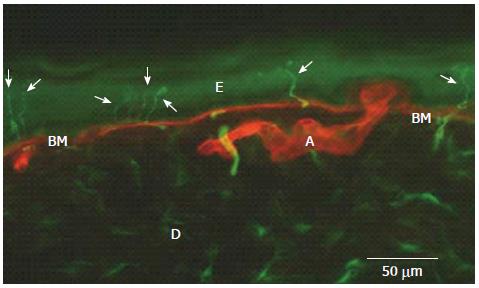
Neurodiagnostic skin biopsy (NSB) is a relatively new technique for the diagnosis of peripheral neuropathies[16-18]. It is an objective method based on a skin biopsy performed by a circular punch, usually 3 mm diameter, and on immunohistochemical staining techniques that allow identifying the epidermal nerve fibers. To this aim, both bright-field and immunofluorescence (Figure 1) can be used, allowing calculation of the Epidermal Nerve Fiber Density (ENFD)[19]. It is important to underline that epidermal nerve fibers are currently exclusively considered the free nerve endings of small diameter (A-delta and C) afferent fibers[20], a part of the spinothalamic tract physiologically conveying thermal and painful sensations from the periphery to the brain. Interestingly, among all the nerve fibers present in a peripheral nerve, the great majority are just small diameter fibers[21].

Figure 1
Figure 1 Epidermal nerve fibers (arrows) identified by immunofluorescence in the distal leg of a normal subject (in green, PGP 9. 5 staining of nerve fibers; in red, type IV collagen staining of basement membrane and blood vessels). E: Epidermis; D: Dermis; BM: Basement Membrane; A: Artery.
NSB FOR THE DIAGNOSIS OF PERIPHERAL NEUROPATHIES IN BODY PARTS IMPOSSIBLE TO INVESTIGATE BY CLINICAL NEUROPHYSIOLOGICAL TESTS

Due to the continuous advances in knowledge that have occurred in the last ten years, NSB is currently considered an important diagnostic tool for neurologists[22,23].

NSB has the important property of being used to investigate the skin, allowing obtaining a diagnosis of small fiber axonal neuropathy of peripheral nerves innervating every body part covered by skin[24,25].

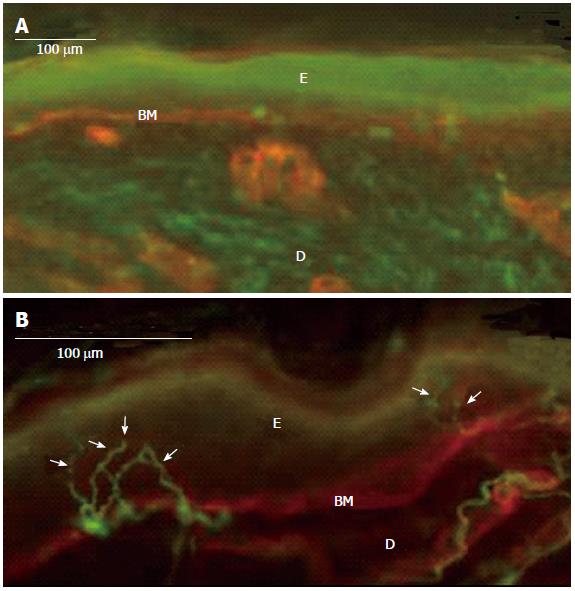
This feature appears to be very important because it allows going beyond the possibilities of neurophysiological tests which are available only for a limited number of peripheral nerves. It follows on that NSB allows reaching a diagnosis of neuropathy for “difficult” nerves, such as those of the trunk or occipital nerves frequently (and irregularly) involved in post-herpetic neuralgia[26]. An example of NSB clinical use is given in Figure 2 which shows a severe, unilateral decrease of ENFD in the neck skin of a patient with post-herpetic neuralgia.

Figure 2
Figure 2 Complete denervation of epidermis (and dermis) in an 82-year-old patient with a severe post-herpetic neuralgia (A), normal, contralateral, mirror skin innervation (13 fibers/mm) (B). Immunofluorescence method: In green, PGP 9.5 staining of nerve fibers; in red, type IV collagen staining of basement membrane and blood vessels; Arrows: Epidermal nerve fibers; E: Epidermis; D: Dermis; BM: Basement Membrane.

Another important property of NSB is the ability to identify lesions involving small branches of peripheral nerves which cannot be investigated by neurophysiological tests. This feature of NSB appears to be particularly important in post-traumatic peripheral nerve lesions where one lesion is different from another.

In this context, it is important to highlight a recent paper where NSB showed a significant asymmetry in a spinal cord injury patient complaining of a bilateral burning and pricking pain at the level of injury[27]. Interestingly, NSB not only allowed demonstration of the presence of two different mechanisms leading to identical symptoms, but also to justify a different efficacy of the same treatment in the two sides. Continuing to talk of possible pain mechanisms, in a very recent paper, NSB findings suggested skin hyperinnervation as a possible cause for the development of dynamic mechanical allodynia following finger amputation[28].

Another recent study confirmed that NSB can allow getting information from the skin of several parts of the body. In that study, the epidermal innervation was studied in burn patients with unilateral injuries, allowing to suggest a possible correlation between the residual cutaneous innervation and the development of chronic pain[29].

NSB can also be useful in other clinical contexts. For example, it can also be used in differentiating neuropathic from referred pain, as demonstrated in a very recent paper in patients with endometriosis and unilateral thigh pain[30]. Moreover, it has been used for assessing the involvement of the peripheral nervous system in a dermatological manifestation of neurological disease, such as a dyshidrotic eczema in a patient with ulnar neuropathy or a unilateral pruritus on the paretic side of a stroke patient[31]. Finally, NSB can also be used to exclude a neuropathic pathophysiology of a clinical pain, as demonstrated in Parry-Romberg syndrome, a rare painful condition characterized by progressive hemifacial atrophy and unilateral facial pain[32].

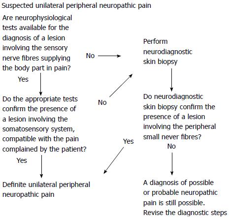
Considering the current evidence on NSB and the well-known specific diagnostic properties of neurophysiological tests, it is possible to suggest a diagnostic sequence that can be useful to confirm the diagnosis of unilateral peripheral neuropathic pain (Figure 3).

Figure 3
Figure 3 Suggested diagnostic sequences for the confirmation of a peripheral nerve lesion sustaining a definite, peripheral, unilateral neuropathic pain.
EPIDERMAL INNERVATION SYMMETRY RATIO

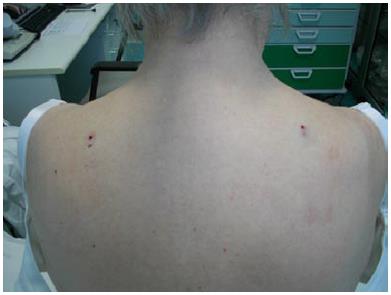
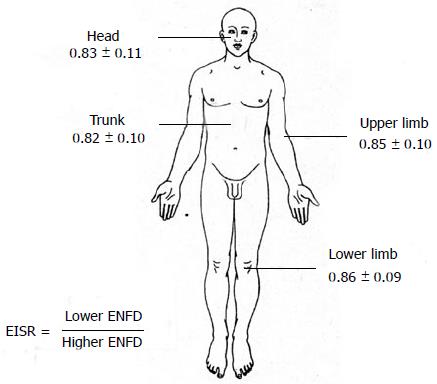
One of the main disadvantages of NSB is that it is very difficult to obtain robust normative data for any part of the body because of their different epidermal innervation patterns. This problem can be elegantly solved in cases of unilateral peripheral nerve lesions by comparing the neuropathic skin ENFD with the contralateral, normal side ENFD. To this aim, a bilateral biopsy is necessary (Figure 4). It is well known that the two sides of the body of a normal subject are only theoretically symmetrical and a significant asymmetry can be frequently found in several types of biological measures. NSB is not an exception. For this reason, a ratio (the Epidermal Innervation Symmetry Ratio) was developed to compare the epidermal innervation of two symmetrical, mirror parts of the body in normal subjects. Preliminary data were obtained from 133 normal subjects[33]. In particular, when comparing the ENFD of the right with the left side, the ratio showed a normal distribution (mean 1.02; median 1.01; standard deviation 0.21; asymmetry 1.86, kurtosis -0.97). Moreover, when confronting the lower with the higher (contralateral) ENFD, the ratio was quite constant and surprisingly reproducible, also considering different parts of the body (Figure 5).

Figure 4
Figure 4 Figure exemplifies the bilateral biopsy needed for comparing the epidermal nerve fiber density of two symmetrical, mirror skin parts.
Figure 5
Figure 5 Figure shows the values of epidermal innervation symmetry ratio obtained for different body parts in 133 normal subjects. ENFD: Epidermal Nerve Fiber Density.
LIMITATIONS

As with any other diagnostic method, the use of NSB for investigation of the peripheral nerve system has some limitations. First of all, it is important to mention that NSB is surely not useful for the diagnosis of entrapment neuropathies, particularly in their early phases when only large diameter fibers are involved. Another important limitation is that NSB is a time-consuming method; calculating the time for specimen processing and the manual counting of two blinded investigators and the time for obtaining a reliable medical report, it is usually not less than two weeks. Finally, NSB is currently confined to a small number of specialized diagnostic units.

CONCLUSION

The demonstration of a lesion or a disease involving the somatosensory system is mandatory for the diagnosis of definite neuropathic pain and objective diagnostic tools play an important role to reach the aim. To this end, several methods are currently available but none is suitable for every disease (or lesion). NSB can be an important diagnostic method for the demonstration of peripheral nervous system involvement, with a special reference to small fiber neuropathies and to peripheral nerves not evaluated by other tests. For these characteristics, NSB can be considered a precious instrument for the diagnosis of peripheral unilateral neuropathic pain.

ACKNOWLEDGMENTS

The author would like to thank the neuropathophysiology technicians Michela Canti and Rosa Bagnasco for their essential role in drafting the paper and Dr. Anna Maria Gatti for helping with the preparation of figures.

Footnotes

P- Reviewers: Alimehmeti R, Cho SY, Trohman RG S- Editor: Qi Y L- Editor: Roemmele A E- Editor: Wu HL

References
1.  Treede RD, Jensen TS, Campbell JN, Cruccu G, Dostrovsky JO, Griffin JW, Hansson P, Hughes R, Nurmikko T, Serra J. Neuropathic pain: redefinition and a grading system for clinical and research purposes. Neurology. 2008;70:1630-1635.  [PubMed]  [DOI]  [Cited in This Article: ]  [Cited by in Crossref: 1866]  [Cited by in F6Publishing: 1841]  [Article Influence: 102.3]  [Reference Citation Analysis (0)]
2.  Dick PJ, Thomas PK.  Peripheral neuropathy, 4th edition. Philadelphia: Elsevier Saunders 2005; .  [PubMed]  [DOI]  [Cited in This Article: ]
3.  Koltzenburg M, Wall PD, McMahon SB. Does the right side know what the left is doing? Trends Neurosci. 1999;22:122-127.  [PubMed]  [DOI]  [Cited in This Article: ]  [Cited by in Crossref: 354]  [Cited by in F6Publishing: 365]  [Article Influence: 14.0]  [Reference Citation Analysis (0)]
4.  Bromberg MB. An approach to the evaluation of peripheral neuropathies. Semin Neurol. 2010;30:350-355.  [PubMed]  [DOI]  [Cited in This Article: ]  [Cited by in Crossref: 6]  [Cited by in F6Publishing: 3]  [Article Influence: 0.2]  [Reference Citation Analysis (0)]
5.  Jillapalli D, Shefner JM. Electrodiagnosis in common mononeuropathies and plexopathies. Semin Neurol. 2005;25:196-203.  [PubMed]  [DOI]  [Cited in This Article: ]  [Cited by in Crossref: 12]  [Cited by in F6Publishing: 7]  [Article Influence: 0.4]  [Reference Citation Analysis (0)]
6.  Dumitru D Electrodiagnostic Medicine. Philadelphia: Hanley and Belfus 1995; .  [PubMed]  [DOI]  [Cited in This Article: ]
7.  Aminoff MJ Electrodiagnosis in clinical neurology, 4th edition. Philadelphia: Churchill livingstone 1999; .  [PubMed]  [DOI]  [Cited in This Article: ]
8.  Ross MA. Electrodiagnosis of peripheral neuropathy. Neurol Clin. 2012;30:529-549.  [PubMed]  [DOI]  [Cited in This Article: ]  [Cited by in Crossref: 9]  [Cited by in F6Publishing: 9]  [Article Influence: 0.7]  [Reference Citation Analysis (0)]
9.  Chiappa KH Evoked potential in clinical medicine, 3rd edition. Philadelphia: Lippincott-Raven 1997; .  [PubMed]  [DOI]  [Cited in This Article: ]
10.  Cruccu G, Aminoff MJ, Curio G, Guerit JM, Kakigi R, Mauguiere F, Rossini PM, Treede RD, Garcia-Larrea L. Recommendations for the clinical use of somatosensory-evoked potentials. Clin Neurophysiol. 2008;119:1705-1719.  [PubMed]  [DOI]  [Cited in This Article: ]  [Cited by in Crossref: 441]  [Cited by in F6Publishing: 444]  [Article Influence: 26.1]  [Reference Citation Analysis (0)]
11.  Bromm B, Treede RD. Laser-evoked cerebral potentials in the assessment of cutaneous pain sensitivity in normal subjects and patients. Rev Neurol (Paris). 1991;147:625-643.  [PubMed]  [DOI]  [Cited in This Article: ]
12.  Treede RD, Lorenz J, Baumgärtner U. Clinical usefulness of laser-evoked potentials. Neurophysiol Clin. 2003;33:303-314.  [PubMed]  [DOI]  [Cited in This Article: ]  [Cited by in Crossref: 297]  [Cited by in F6Publishing: 304]  [Article Influence: 14.5]  [Reference Citation Analysis (0)]
13.  Quantitative sensory testing: a consensus report from the Peripheral Neuropathy Association Neurology. 1993;43:1050-1052.  [PubMed]  [DOI]  [Cited in This Article: ]  [Cited by in Crossref: 95]  [Cited by in F6Publishing: 95]  [Article Influence: 3.0]  [Reference Citation Analysis (0)]
14.  Lindblom U. Analysis of Abnormal Touch, Pain, and Temperature Sensation in Patients. Touch, Temperature, and Pain in Health and Disease: Mechanisms and Assessments. Seattle: IASP Press 1994; 63-84.  [PubMed]  [DOI]  [Cited in This Article: ]
15.  Pfau DB, Geber C, Birklein F, Treede RD. Quantitative sensory testing of neuropathic pain patients: potential mechanistic and therapeutic implications. Curr Pain Headache Rep. 2012;16:199-206.  [PubMed]  [DOI]  [Cited in This Article: ]  [Cited by in Crossref: 55]  [Cited by in F6Publishing: 59]  [Article Influence: 4.5]  [Reference Citation Analysis (0)]
16.  Kennedy WR. Opportunities afforded by the study of unmyelinated nerves in skin and other organs. Muscle Nerve. 2004;29:756-767.  [PubMed]  [DOI]  [Cited in This Article: ]
17.  Lauria G, Hsieh ST, Johansson O, Kennedy WR, Leger JM, Mellgren SI, Nolano M, Merkies IS, Polydefkis M, Smith AG. European Federation of Neurological Societies/Peripheral Nerve Society Guideline on the use of skin biopsy in the diagnosis of small fiber neuropathy. Report of a joint task force of the European Federation of Neurological Societies and the Peripheral Nerve Society. Eur J Neurol. 2010;17:903-12, e44-9.  [PubMed]  [DOI]  [Cited in This Article: ]  [Cited by in Crossref: 490]  [Cited by in F6Publishing: 563]  [Article Influence: 40.2]  [Reference Citation Analysis (0)]
18.  Sommer C. Skin biopsy as a diagnostic tool. Curr Opin Neurol. 2008;21:563-568.  [PubMed]  [DOI]  [Cited in This Article: ]  [Cited by in Crossref: 30]  [Cited by in F6Publishing: 32]  [Article Influence: 1.9]  [Reference Citation Analysis (0)]
19.  Walk D. Role of skin biopsy in the diagnosis of peripheral neuropathic pain. Curr Pain Headache Rep. 2009;13:191-196.  [PubMed]  [DOI]  [Cited in This Article: ]  [Cited by in Crossref: 10]  [Cited by in F6Publishing: 10]  [Article Influence: 0.7]  [Reference Citation Analysis (0)]
20.  McCarthy BG, Hsieh ST, Stocks A, Hauer P, Macko C, Cornblath DR, Griffin JW, McArthur JC. Cutaneous innervation in sensory neuropathies: evaluation by skin biopsy. Neurology. 1995;45:1848-1855.  [PubMed]  [DOI]  [Cited in This Article: ]  [Cited by in Crossref: 351]  [Cited by in F6Publishing: 338]  [Article Influence: 11.3]  [Reference Citation Analysis (0)]
21.  Griffin JW, McArthur JC, Polydefkis M. Assessment of cutaneous innervation by skin biopsies. Curr Opin Neurol. 2001;14:655-659.  [PubMed]  [DOI]  [Cited in This Article: ]
22.  Lauria G, Lombardi R. Skin biopsy in painful and immune-mediated neuropathies. J Peripher Nerv Syst. 2012;17:38-45.  [PubMed]  [DOI]  [Cited in This Article: ]  [Cited by in Crossref: 23]  [Cited by in F6Publishing: 23]  [Article Influence: 1.8]  [Reference Citation Analysis (0)]
23.  Di Stefano G, La Cesa S, Biasiotta A, Leone C, Pepe A, Cruccu G, Truini A. Laboratory tools for assessing neuropathic pain. Neurol Sci. 2012;33:5-7.  [PubMed]  [DOI]  [Cited in This Article: ]  [Cited by in Crossref: 9]  [Cited by in F6Publishing: 9]  [Article Influence: 0.7]  [Reference Citation Analysis (0)]
24.  Lauria G, Lombardi R, Camozzi F, Devigili G. Skin biopsy for the diagnosis of peripheral neuropathy. Histopathology. 2009;54:273-285.  [PubMed]  [DOI]  [Cited in This Article: ]  [Cited by in Crossref: 86]  [Cited by in F6Publishing: 84]  [Article Influence: 4.9]  [Reference Citation Analysis (0)]
25.  Myers MI, Peltier AC. Uses of skin biopsy for sensory and autonomic nerve assessment. Curr Neurol Neurosci Rep. 2013;13:323.  [PubMed]  [DOI]  [Cited in This Article: ]  [Cited by in Crossref: 34]  [Cited by in F6Publishing: 32]  [Article Influence: 2.7]  [Reference Citation Analysis (0)]
26.  Buonocore M, Gatti AM, Amato G, Aloisi AM, Bonezzi C. Allodynic skin in post-herpetic neuralgia: histological correlates. J Cell Physiol. 2012;227:934-938.  [PubMed]  [DOI]  [Cited in This Article: ]  [Cited by in Crossref: 16]  [Cited by in F6Publishing: 16]  [Article Influence: 1.2]  [Reference Citation Analysis (0)]
27.  Westermann A, Krumova EK, Pennekamp W, Horch C, Baron R, Maier C. Different underlying pain mechanisms despite identical pain characteristics: a case report of a patient with spinal cord injury. Pain. 2012;153:1537-1540.  [PubMed]  [DOI]  [Cited in This Article: ]  [Cited by in Crossref: 36]  [Cited by in F6Publishing: 32]  [Article Influence: 2.5]  [Reference Citation Analysis (0)]
28.  Buonocore M, Gagliano MC, Bonezzi C. Dynamic mechanical allodynia following finger amputation: Unexpected skin hyperinnervation. World J Clin Cases. 2013;1:197-201.  [PubMed]  [DOI]  [Cited in This Article: ]  [Cited by in CrossRef: 3]  [Cited by in F6Publishing: 3]  [Article Influence: 0.3]  [Reference Citation Analysis (1)]
29.  Hamed K, Giles N, Anderson J, Phillips JK, Dawson LF, Drummond P, Wallace H, Wood FM, Rea SM, Fear MW. Changes in cutaneous innervation in patients with chronic pain after burns. Burns. 2011;37:631-637.  [PubMed]  [DOI]  [Cited in This Article: ]  [Cited by in Crossref: 39]  [Cited by in F6Publishing: 35]  [Article Influence: 2.5]  [Reference Citation Analysis (0)]
30.  Pacchiarotti A, Milazzo GN, Biasiotta A, Truini A, Antonini G, Frati P, Gentile V, Caserta D, Moscarini M. Pain in the upper anterior-lateral part of the thigh in women affected by endometriosis: study of sensitive neuropathy. Fertil Steril. 2013;100:122-126.  [PubMed]  [DOI]  [Cited in This Article: ]  [Cited by in Crossref: 12]  [Cited by in F6Publishing: 12]  [Article Influence: 1.0]  [Reference Citation Analysis (0)]
31.  Wallengren J, Tegner E, Sundler F. Cutaneous sensory nerve fibers are decreased in number after peripheral and central nerve damage. J Am Acad Dermatol. 2002;46:215-217.  [PubMed]  [DOI]  [Cited in This Article: ]  [Cited by in Crossref: 14]  [Cited by in F6Publishing: 14]  [Article Influence: 0.6]  [Reference Citation Analysis (0)]
32.  Falla M, Biasiotta A, Fabbrini G, Cruccu G, Truini A. Cutaneous innervation and trigeminal pathway function in a patient with facial pain associated with Parry-Romberg syndrome. J Headache Pain. 2012;13:497-499.  [PubMed]  [DOI]  [Cited in This Article: ]  [Cited by in Crossref: 7]  [Cited by in F6Publishing: 6]  [Article Influence: 0.5]  [Reference Citation Analysis (0)]
33.  Buonocore M, Gatti AM, Demartini L.  Epidermal innervation symmetry ratio (EISR) for the diagnosis of unilateral neuropathic pain (Abstract Book of the “4th International Congress on Neuropathic Pain”, 2013 May 23-26). Toronto: On USB key 2013; 740.  [PubMed]  [DOI]  [Cited in This Article: ]