Published online Feb 16, 2023. doi: 10.12998/wjcc.v11.i5.1182
Peer-review started: November 8, 2022
First decision: November 22, 2022
Revised: December 23, 2022
Accepted: January 16, 2023
Article in press: January 16,2023
Published online: February 16, 2023
Processing time: 97 Days and 21.4 Hours
Closed loop ileus caused by entrapment of bowel in a defect of the broad ligament is a rarity. Only a few cases have been reported in the literature.
We present the case of a 44-year-old, healthy patient with no prior history of abdominal surgery who developed a closed loop ileus due to an internal hernia secondary to a defect in the right broad ligament. She first presented to the emergency department with diarrhea and vomiting. As she had had no previous abdominal surgery, she was diagnosed with probable gastroenteritis and discharged. The patient subsequently returned to the emergency department due to a lack of improvement in her symptoms. Blood tests showed an elevated white blood cell count and a closed loop ileus was diagnosed on an abdominal computer tomography scan. Diagnostic laparoscopy revealed an internal hernia entrapped in a 2 cm large defect in the right broad ligament. The hernia was reduced and the ligament defect was closed using a running, barbed suture.
Bowel incarceration through an internal hernia may present with misleading symptoms and laparoscopy may reveal unexpected findings.
Core Tip: In young patients with negative history of abdominal surgery presenting at the emergency department with nausea and vomiting, the initial differential diagnosis should include ileus. If an ileus is suspected, computer tomography and laparoscopy are the diagnostic tools of choice. Internal hernias are rare, especially those through the broad ligament, but they should be considered to avoid complications such as bowel necrosis. Because of the rarity of the conditions, there are no studies or long-term data on the best treatment option, but most authors describe a direct defect closure.
- Citation: Zucal I, Nebiker CA. Closed loop ileus caused by a defect in the broad ligament: A case report. World J Clin Cases 2023; 11(5): 1182-1187
- URL: https://www.wjgnet.com/2307-8960/full/v11/i5/1182.htm
- DOI: https://dx.doi.org/10.12998/wjcc.v11.i5.1182
Internal hernias are responsible for up to 4% of bowel obstruction in the emergency setting[1,2]. Herniation of the bowel through the broad ligament has been reported to underlie 4%–7% of these cases[2-5]. A computer tomography (CT) scan is the diagnostic tool of choice, however, the cause of bowel obstruction is usually not identified. In this regard, diagnostic laparoscopy plays a crucial role. Not only the area of bowel entrapment can be identified, but the hernia can be reduced, and the defect surgically closed.
The initial clinical presentation may be misleading and non-specific, as affected women have typically not had previous abdominal surgery[5]. Here we present a case of a 44-year-old woman with closed loop ileus caused by a defect in the right broad ligament. The case was reported in accordance with the SCARE 2020 guidelines[6].
A 44-year-old female patient with sudden onset of abdominal pain was assigned to the surgical emergency department by the gynecological ward after exclusion of a gynecological pathology.
The pain was mainly localized in both lower abdominal quadrants and accompanied by nausea without vomiting.
History of past illness was negative.
She was otherwise healthy, had two children via vaginal delivery, and had no history of abdominal operations.
On clinical examination, pain was found on palpation of the right lower abdominal quadrant without signs of peritonism.
The blood tests showed an elevated white blood cell count (26 g/L) and normal C-reactive protein (CRP).
An ultrasound scan could not identify the appendix. However, distended bowel without peristalsis was seen in the right lower quadrant, possibly indicating a segmental obstruction. Because of a lack of previous abdominal operations, an ileus seemed unlikely and the patient was diagnosed with enteritis. She was scheduled for a control the next morning.
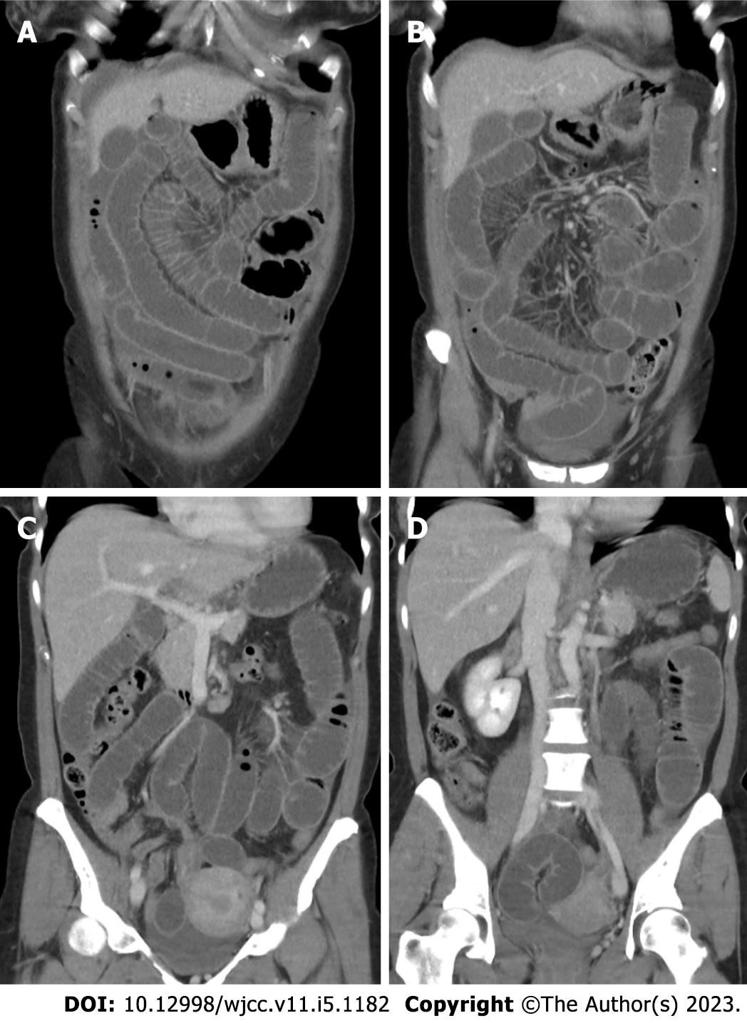
Within 12 h, the patient was brought back to the emergency department by ambulance and had developed additional vomiting and diarrhea. A diffuse pain on palpation of the lower abdominal quadrants was elicited and stool samples were collected for microbiological analysis. The white blood cell count had fallen to 14 g/L, and the CRP had risen to 20 mg/L. Again, the patient was discharged with the diagnosis of enteritis. On the next day, the patient came to the emergency department again with constant vomiting and new bloody diarrhea. A CT of the abdomen was performed, and a closed loop obstruction was postulated. Moreover, ascites was observed. The CT findings are shown in Figure 1. The patient was scheduled for an emergency laparoscopy which detected herniation of the ileum into a 2 cm defect in the right broad ligament.
An internal hernia through the right broad ligament.
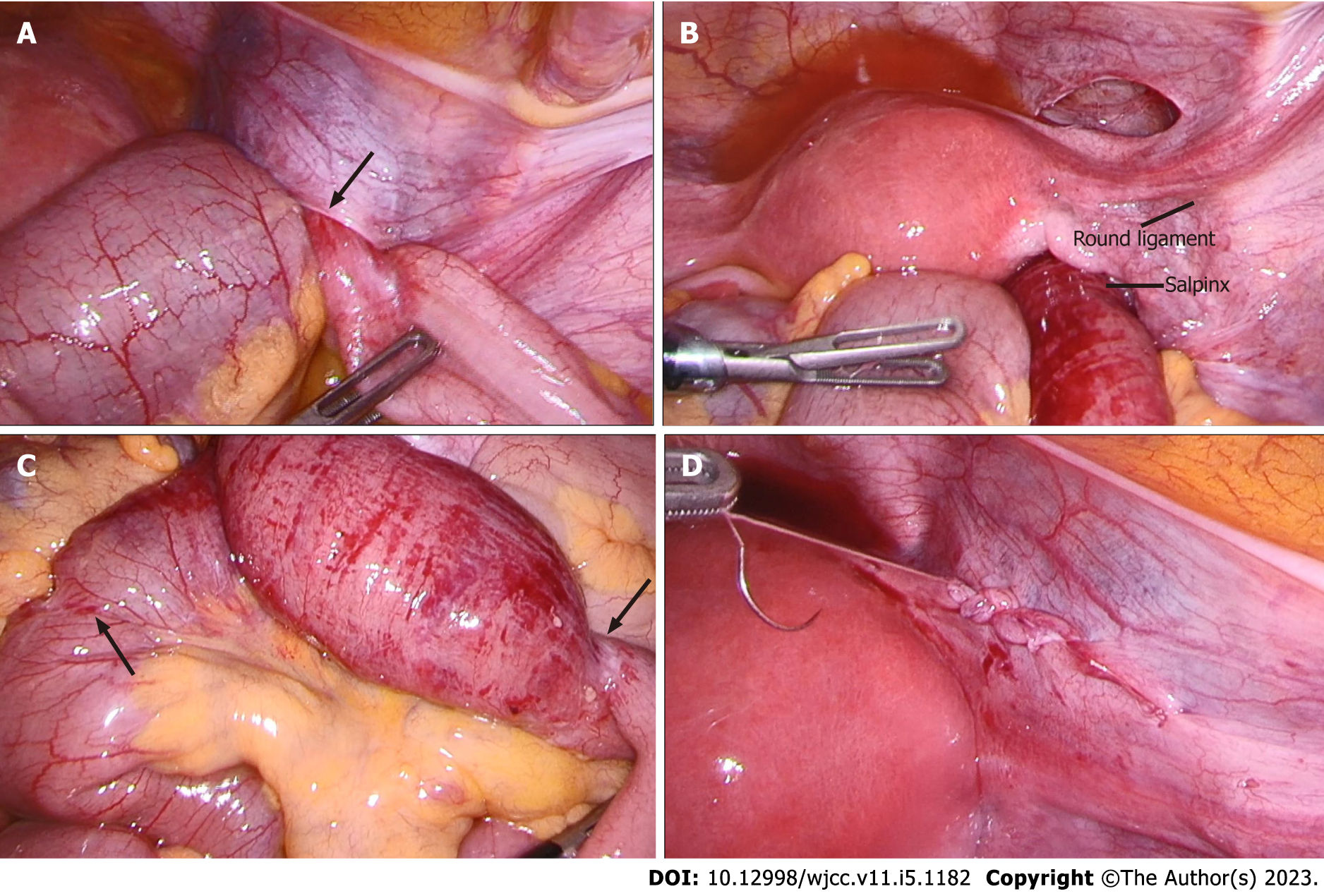
The bowel was successfully reduced and presented no signs of bowel ischemia. Two strangulation marks were identified but appeared to be without transmural necrosis. The defect in the right broad ligament was closed by using a barbed running suture. The intraoperative findings and defect closure are shown in Figure 2. The nasogastric tube could be removed the day after surgery. Food was well tolerated, and the patient was discharged on the third postoperative day.
After discharge, no further clinical follow-up was planned in the surgical outpatient clinic and the patient did not present again to the emergency department.
Bowel obstruction caused by herniation through the broad ligament is very rare and may occur in healthy patients with no prior history of abdominal surgery. Symptoms including nausea, vomiting and paradoxical diarrhea may therefore be attributed to enteritis, as happened in our case. In a case reported by Agrawal et al[7], a ruptured ovarian cyst was suspected before the exploratory laparotomy was performed and an internal hernia through the broad ligament was detected[7]. Such hernias usually manifest as closed loop obstruction on a CT scan[8,9], but the etiology is hard to detect. Thus, reaching the correct diagnosis may be delayed.
In the literature, only a few similar cases have been described. We were able to perform a laparoscopic hernia reduction and closure of the defect without complications. In other reported cases, the hernia had to be reduced via laparotomy[7-11]. Although the bowel was hyperemic, there were no signs of bowel ischemia, so no bowel resection had to be performed. In contrast, in other reported cases, strangulated bowel had to be resected[10]. In the case reported by Takahashi et al[9], the fallopian tube had to be removed due to necrosis[9]. Hashimoto et al[8] described recurrence of a broad ligament hernia in a 53-year-old woman 10 years after primary repair[8]. In a case reported by Rodrigues et al[12], asymptomatic, internal broad ligament hernia was an incidental finding in an exploratory laparoscopy to recover a lost intrauterine device[12].
According to Cilley et al's classification[13], we report a type I hernia as the defect was located caudal to the round ligament in the broad ligament. A type II hernia would have been located in the mesovarium and mesosalpinx above the round ligament, and a type III hernia has been described as a defect through the meso-ligamentum teres uteri[13]. Broad ligament defects and consecutive hernias can be congenital or acquired[14]. Congenital defects arise from spontaneous rupture of congenital cysts in the broad ligament and are usually bilateral, whereas acquired defects may be secondary to delivery trauma, pregnancy, surgery, or inflammatory disease[9,14]. Inspection of the contralateral broad ligament is important to avoid re-operation, however, guidelines on the optimal defect closure and long-term outcomes are missing.
A closed loop ileus due to an internal hernia in patients with a negative history of abdominal surgery may present with misleading symptoms. A CT scan is the diagnostic tool of choice to identify closed loop obstruction, but only laparoscopy can provide the correct diagnosis and treatment. Due to the rarity of broad ligament internal hernias, there is no consensus on the best surgical treatment. Observation of long-term outcomes of the reported cases with regards to hernia recurrence is needed.
Provenance and peer review: Unsolicited article; Externally peer reviewed.
Peer-review model: Single blind
Specialty type: Medicine, research and experimental
Country/Territory of origin: Switzerland
Peer-review report’s scientific quality classification
Grade A (Excellent): 0
Grade B (Very good): 0
Grade C (Good): C, C
Grade D (Fair): 0
Grade E (Poor): 0
P-Reviewer: Carannante F, Italy; Wang Y, China S-Editor: Li L L-Editor: A P-Editor: Li L
| 1. | Zemour J, Coueffe X, Fagot H. Herniation of the broad ligament… And the other side? Int J Surg Case Rep. 2019;65:354-357. [RCA] [PubMed] [DOI] [Full Text] [Full Text (PDF)] [Cited by in Crossref: 9] [Cited by in RCA: 6] [Article Influence: 1.0] [Reference Citation Analysis (0)] |
| 2. | Ghahremani GG. Internal abdominal hernias. Surg Clin North Am. 1984;64:393-406. [RCA] [PubMed] [DOI] [Full Text] [Cited by in Crossref: 113] [Cited by in RCA: 123] [Article Influence: 3.0] [Reference Citation Analysis (0)] |
| 3. | Martin LC, Merkle EM, Thompson WM. Review of internal hernias: radiographic and clinical findings. AJR Am J Roentgenol. 2006;186:703-717. [RCA] [PubMed] [DOI] [Full Text] [Cited by in Crossref: 360] [Cited by in RCA: 369] [Article Influence: 19.4] [Reference Citation Analysis (0)] |
| 4. | Fukuoka M, Tachibana S, Harada N, Saito H. Strangulated herniation through a defect in the broad ligament. Surgery. 2002;131:232-233. [RCA] [PubMed] [DOI] [Full Text] [Cited by in Crossref: 11] [Cited by in RCA: 12] [Article Influence: 0.5] [Reference Citation Analysis (0)] |
| 5. | Reyes N, Smith LE, Bruce D. Strangulated internal hernia due to defect in broad ligament: a case report. J Surg Case Rep. 2020;2020:rjaa487. [RCA] [PubMed] [DOI] [Full Text] [Full Text (PDF)] [Cited by in Crossref: 3] [Cited by in RCA: 5] [Article Influence: 1.0] [Reference Citation Analysis (0)] |
| 6. | Agha RA, Franchi T, Sohrabi C, Mathew G, Kerwan A; SCARE Group. The SCARE 2020 Guideline: Updating Consensus Surgical CAse REport (SCARE) Guidelines. Int J Surg. 2020;84:226-230. [RCA] [PubMed] [DOI] [Full Text] [Cited by in Crossref: 4265] [Cited by in RCA: 4711] [Article Influence: 942.2] [Reference Citation Analysis (0)] |
| 7. | Agrawal P, Grab JT IVs, Howe HR 3rd, Cross K. Ruptured Ovarian Cyst Masking Diagnosis of Hernia Through Broad Ligament of Uterus: A Case Report. J Investig Med High Impact Case Rep. 2022;10:23247096221100500. [RCA] [PubMed] [DOI] [Full Text] [Full Text (PDF)] [Cited by in Crossref: 1] [Reference Citation Analysis (0)] |
| 8. | Hashimoto Y, Kanda T, Chida T, Suda K. Recurrence hernia in the broad ligament of the uterus: a case report. Surg Case Rep. 2020;6:288. [RCA] [PubMed] [DOI] [Full Text] [Full Text (PDF)] [Cited by in Crossref: 2] [Cited by in RCA: 2] [Article Influence: 0.4] [Reference Citation Analysis (0)] |
| 9. | Takahashi M, Yoshimitsu M, Yano T, Idani H, Shiozaki S, Okajima M. Rare Contents of an Internal Hernia through a Defect of the Broad Ligament of the Uterus. Case Rep Surg. 2021;2021:5535162. [RCA] [PubMed] [DOI] [Full Text] [Full Text (PDF)] [Cited by in RCA: 3] [Reference Citation Analysis (0)] |
| 10. | Arif SH, Mohammed AA. Strangulated small-bowel internal hernia through a defect in the broad ligament of the uterus presenting as acute intestinal obstruction: A case report. Case Rep Womens Health. 2021;30:e00310. [RCA] [PubMed] [DOI] [Full Text] [Full Text (PDF)] [Reference Citation Analysis (0)] |
| 11. | Ohno S, Chikaishi W, Sugimoto T, Komori S, Kawai M. An incarcerated internal hernia of the sigmoid colon through a defect in the broad ligament: A case report. Int J Surg Case Rep. 2021;85:106169. [RCA] [PubMed] [DOI] [Full Text] [Full Text (PDF)] [Reference Citation Analysis (0)] |
| 12. | Rodrigues F, Sarmento I, Tiago P. Asymptomatic internal hernia through a defect of broad ligament: a surprising finding in a laparoscopic surgery to recover a lost levonorgestrel-releasing intrauterine system. BMJ Case Rep. 2015;2015. [RCA] [PubMed] [DOI] [Full Text] [Cited by in Crossref: 1] [Cited by in RCA: 2] [Article Influence: 0.2] [Reference Citation Analysis (0)] |
| 13. | Cilley R, Poterack K, Lemmer J, Dafoe D. Defects of the broad ligament of the uterus. Am J Gastroenterol. 1986;81:389-391. [PubMed] |


