Published online Nov 26, 2023. doi: 10.12998/wjcc.v11.i33.8089
Peer-review started: September 19, 2023
First decision: October 17, 2023
Revised: October 22, 2023
Accepted: November 14, 2023
Article in press: November 14, 2023
Published online: November 26, 2023
Processing time: 66 Days and 1.3 Hours
Dextroversion is defined as the presence of dextrocardia with situs solitus, dextro-loop ventricles, and normally related great arteries. Dextrocardia can pose tech
A 73-year-old woman with cardiac dextroversion suffered from a recurrence of atrial fibrillation after her radiofrequency catheter ablation and Despite the cessa
Dextroversion makes the implantation of leadless pacemakers more challenging, and appropriate adjustments in fluoroscope angles may be crucial for intracardiac operations. Additionally, when advancing delivery systems, attention should be paid to rotational direction during valve-crossing procedures; changes in the per
Core Tip: Dextroversion can be even more challenging given the distortion of normal anatomical relationships and the uncertainty of the accurate location and borders of the cardiac structures caused by the shift and rotation effected by the pathologic process. We present a complicated but successful case of implantation of a leadless pacemaker in a patient with cardiac dextroversion.
- Citation: Li N, Wang HX, Sun YH, Shu Y. Successful leadless pacemaker implantation in a patient with dextroversion of the heart: A case report. World J Clin Cases 2023; 11(33): 8089-8093
- URL: https://www.wjgnet.com/2307-8960/full/v11/i33/8089.htm
- DOI: https://dx.doi.org/10.12998/wjcc.v11.i33.8089
Abnormal heart structures complicate cardiac electrophysiology operative treatment, especially the implantation of devices like leadless pacemakers. It is primarily because the tools provided by manufacturers have not been sufficiently designed or tested for rare abnormal structures. Thus, implementing these delivery systems in intricately structured cases can be demanding or necessitate a level of innovation. The literature presents scarce accounts concerning the installation of leadless pacemakers in patients having dextrocardia[1-3]. Dextrocardia, a scarcely occurring inborn anomaly in the general population, is estimated to occur in 1 out of every 12000 live births, and it might be linked with substantial supplementary cardiac deformities[4]. The incidence of dextrocardia is evenly distributed between males and females at a ratio of 1:1. In cases of dextrocardia, the positioning of abdominal organs may be normal (situs solitus), reversed (situs inversus), or indeterminate (situs ambiguous or isomerism) in respective proportions of 32%-35%, 35%-39%, and 26%-28%[4,5]. Dextroversion is characterized by a right-sided cardiac placement with a rightward cardiac apex in the context of situs solitus. Unlike situs inversus, where the arrangement of the visceroatrial mirrors the typical layout, dextroversion features the conventional positioning of the tracheobronchial tree and abdominal organs[6].
The hominine embryonicheart originates from a rudimentary cardiac tube that has the sinus venosus, atrium, ventricle, bulbus cordis, and arterial trunks lined up in sequence. The end of veins and arteries are stationary. The atriums and veins return develop concurrently, thus anchoring the atria in place via the inflowing veins. The growth of the bulboventricular loop causes the cardiac tube to bend, creating morphological-biventricular chamber. This process does not impact position of the atria, which continues to correspond with the lacation of te internal organs[7,8]. During the initial phases of fetal development, situs solitus and the establishment of the dextro-loop take place, positioning the heart’s apex within the right hemithorax. In the initial four weeks of the newborn’s lifetime, the tip of heart transitions from the right thoracic cavity to the left half of the chest. Despite of the auricular positon, every dextro-bulboventricular loop ought to complete their advancement with the heart in the left half of the chest.
Dextroversion can be congenital as well as acquired. The former is due to defeat of the ultimate leftward shift of biventricular chambers duringthe process of embryogenesis. Although the morphologic right atrium and right ventricle are still located on the right side, they are positioned behind the corresponding left atrium. In this case, the right rotation of the heart was caused by mechanical morphological compression of the left diaphragm due to obvious elevation, which was considered to be related to phrenic nerve palsy after previous radiofrequency catheter ablation. Owing to the fact that the apex is towards the right with situs solitus, the cardiac shift in the thoracic cavity to the right causes changes in veinal junctions and also alters the dissecting associations among each vessel, the right heart system.This distortion, coupled with the variations among patients, makes it highly demanding to implement procedures of the heart. Furthermore, literatures provide scant reports on operation of leadless pacemaker in sufferers who have dextrocardia[2,3,9]. At present, this is the first case of leadless pacemaker implantation in a patient with dextroversion of the heart.
A 73-year-old woman was suffered from sinus pauses and symptomatic bradycardia (as low as 28 beats per minute) even after the cessation of antiarrhythmic drugs.
The patient experienced a relapse of atrial fibrillation and subsequently arrived at an external hospital exhibiting episodes of sinus pauses and symptomatic bradycardia (down to 28 beats per minute), even after ceasing the use of antiarrhythmic drugs.
The patient had a history of transient ischemic attack and severely symptomatic paroxysmal atrial fibrillation for 20 years with a history of radiofrequency catheter ablation and left atrial appendage closure in 2019. Initially, the left atrial appendage was occluded, succeeded by the implementation of pulmonary vein isolation (PVI). The placement of the Watchman device was successfully affirmed with no remaining flow. Subsequently, PVI was executed utilizing radiofrequency ablation. The blockage of both ingress and egress was substantiated in all veins]. And she suffered from dextroversion, which was proved by chest X-ray and Echocardiography.
This case had no specific personal or family history.
No abnormalities were detected in the physical examination.
All laboratory tests were normal.
A chest X-ray verified the presence of a cardiac shadow on the right side, with the apex of the heart orientated towards the right. However, there was no evidence of an expanded cardiac silhouette. The mediastinum was centrally positioned, with the liver’s shadow on the right aligning with situs solitus. Furthermore, the left hemidiaphragm displayed at a level higher than its right counterpart. Echocardiography indicated a dextro-loop configuration in ventricular morphology, accompanied by a right-oriented cardiac axis and a ventricular apex directed towards the right.
The patient had a history of transient ischemic attack and severely symptomatic paroxysmal atrial fibrillation for 20 years with a history of radiofrequency catheter ablation and left atrial appendage closure in 2019. Then she experienced a relapse of atrial fibrillation and had episodes of sinus pauses and symptomatic bradycardia (down to 28 beats per minute), even after ceasing the use of antiarrhythmic drugs.
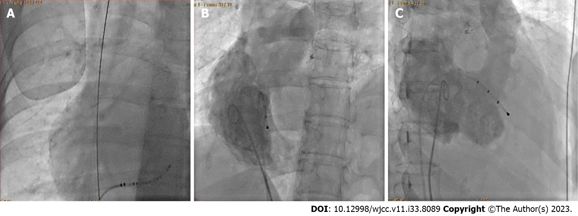
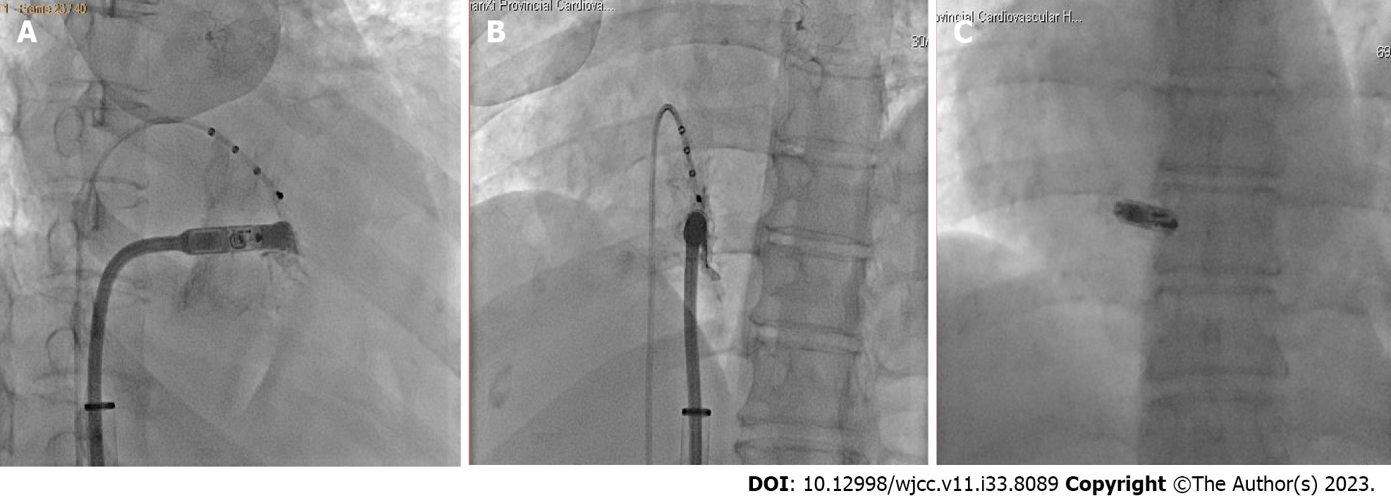
Right femoral venous access was secured under fluoroscopic guidance using a micro-puncture needle. A lengthy Amplatz stiff guidewire was threaded and pushed forward through the micro-puncture sheath, in anticipation of the Micra implantation. Fluoroscopic assessment of the guidewire in the thoracic area revealed the cordis image in the right half of chest (Figure 1A). Owing to the dextroversion, a quadripolar lead wire (F5QD252RT, Biosense Webster, CA, United States) was advanced to the heart to seek an optimal exposure angle (Figure 1B and C). Then, the right anterior oblique view of 45° and left anterior oblique view of 15° were applied by the electrophysiologist to give assistance in order to be more safer and precisein placement of the Medtronic Micra leadless pacemaker. Under fluoroscopic guidance, the pacemaker introducer sheath and delivery system were respectively positioned in the right atrium and right ventricle. Concurrently, the Micra was embedded in the right ventricle septum (Figure 2). Once the stability and electrical thresholds were confirmed, the Micrawas released from the catheter. The pacemaker was functioning properly, and its R wave is 8.0 mV, impedance is 840 ohms, and capture threshold is 0.8 V @0.24 ms.
In the instance, no prolonged complications were noted. The device function was appropriate at 3 mo, 6 mo and 12 mo following Micra leadless pacemaker placement.
In the conventional position of the heart, we usually use a right anterior oblique 30° and left anterior oblique 45° to determine the relative position between the delivery system and leadless pacemaker with the interventricular septum. However, due to the dextroversion in this patient, the fluoroscope angle needed to be adjusted accordingly based on anatomical distortion. In this case, a reference was made by placing a quadripolar lead wire in the right ventricular for pre-operation. This helped determine the optimal angle for exposing the delivery system and interventricular septum, which was a right anterior oblique 45° and left anterior oblique 15°. When crossing over the tricuspid valve, an additional counterclockwise rotation angle was required; while after the crossing-over, there was relatively less clockwise rotation compared to when in a normal cardiac position. Owing to the relatively sharp angular torque compared to conventional anatomical structures and the correspondingly reduced coaxiality generated in the delivery system rooting in the complicated anatomical structure, the severed tethers were discovered to be tight. Thus, utmost caution needed to be taken in removing the tethers in order to avoid negative impacts on pacemaker fixation during the release process.
Dextroversion makes the implantation of leadless pacemakers more challenging, and appropriate adjustments in fluoroscope angles may be crucial for intracardiac operations. Additionally, when advancing delivery systems, attention should be paid to rotational direction during valve-crossing procedures; changes in the perspective of posture angle between normal cardiac position and dextroversion can serve as references. This case study reports the successful implantation of a leadless pacemaker in a patient with dextroversion and provides invaluable clinical experience for the development of a relevant therapeutic regimen.
Provenance and peer review: Unsolicited article; Externally peer reviewed.
Peer-review model: Single blind
Specialty type: Cardiac and cardiovascular systems
Country/Territory of origin: China
Peer-review report’s scientific quality classification
Grade A (Excellent): 0
Grade B (Very good): B
Grade C (Good): 0
Grade D (Fair): 0
Grade E (Poor): 0
P-Reviewer: Sutton R, United Kingdom S-Editor: Liu JH L-Editor: A P-Editor: Liu JH
| 1. | Buxton AE, Morganroth J, Josephson ME, Perloff JK, Shelburne JC. Isolated dextroversion of the heart with asymmetric septal hypertrophy. Am Heart J. 1976;92:785-790. [RCA] [PubMed] [DOI] [Full Text] [Cited by in Crossref: 18] [Cited by in RCA: 16] [Article Influence: 0.3] [Reference Citation Analysis (0)] |
| 2. | Shenthar J, Rai MK, Walia R, Ghanta S, Sreekumar P, Reddy SS. Transvenous permanent pacemaker implantation in dextrocardia: technique, challenges, outcome, and a brief review of literature. Europace. 2014;16:1327-1333. [RCA] [PubMed] [DOI] [Full Text] [Cited by in Crossref: 12] [Cited by in RCA: 14] [Article Influence: 1.3] [Reference Citation Analysis (0)] |
| 3. | De Regibus V, Pardeo A, Artale P, Petretta A, Filannino P, Iacopino S. Leadless pacemaker implantation after transcatheter lead extraction in complex anatomy patient. Clin Case Rep. 2018;6:1106-1108. [RCA] [PubMed] [DOI] [Full Text] [Full Text (PDF)] [Cited by in Crossref: 11] [Cited by in RCA: 8] [Article Influence: 1.1] [Reference Citation Analysis (0)] |
| 4. | Bohun CM, Potts JE, Casey BM, Sandor GG. A population-based study of cardiac malformations and outcomes associated with dextrocardia. Am J Cardiol. 2007;100:305-309. [RCA] [PubMed] [DOI] [Full Text] [Cited by in Crossref: 93] [Cited by in RCA: 124] [Article Influence: 6.9] [Reference Citation Analysis (0)] |
| 5. | Garg N, Agarwal BL, Modi N, Radhakrishnan S, Sinha N. Dextrocardia: an analysis of cardiac structures in 125 patients. Int J Cardiol. 2003;88:143-55; discussion 155. [RCA] [PubMed] [DOI] [Full Text] [Cited by in Crossref: 59] [Cited by in RCA: 72] [Article Influence: 3.3] [Reference Citation Analysis (0)] |
| 6. | Tripathi S, Ajit Kumar VK. Comparison of Morphologic Findings in Patients with Dextrocardia with Situs Solitus vs Situs Inversus: a Retrospective Study. Pediatr Cardiol. 2019;40:302-309. [RCA] [PubMed] [DOI] [Full Text] [Cited by in Crossref: 8] [Cited by in RCA: 10] [Article Influence: 1.7] [Reference Citation Analysis (0)] |
| 7. | Vanpraagh R, Vanpraagh S, Vlad P, Keith JD. Anatomic types of congenital dextrocardia: Diagnostic and embryologic implications. Am J Cardiol. 1964;13:510-531. [RCA] [PubMed] [DOI] [Full Text] [Cited by in Crossref: 204] [Cited by in RCA: 176] [Article Influence: 2.9] [Reference Citation Analysis (0)] |
| 8. | Applegate KE, Goske MJ, Pierce G, Murphy D. Situs revisited: imaging of the heterotaxy syndrome. Radiographics. 1999;19:837-52; discussion 853. [RCA] [PubMed] [DOI] [Full Text] [Cited by in Crossref: 223] [Cited by in RCA: 178] [Article Influence: 6.8] [Reference Citation Analysis (0)] |
| 9. | Conti S, Sgarito G. Leadless pacemaker implantation in postpneumonectomy syndrome. HeartRhythm Case Rep. 2020;6:124-125. [RCA] [PubMed] [DOI] [Full Text] [Full Text (PDF)] [Cited by in Crossref: 3] [Cited by in RCA: 2] [Article Influence: 0.3] [Reference Citation Analysis (0)] |


