Published online Oct 6, 2023. doi: 10.12998/wjcc.v11.i28.6871
Peer-review started: June 17, 2023
First decision: August 24, 2023
Revised: September 2, 2023
Accepted: September 11, 2023
Article in press: September 11, 2023
Published online: October 6, 2023
Processing time: 100 Days and 3.7 Hours
We report a case with the displacement of an articular fracture fragment of the base of the second metacarpal from the ulnar to the volar side, treated via the dorsal approach. The dorsal approach can be a good option not only because it allows direct observation of ligament damage and fixation of bone fragments but also because the thin subcutaneous tissue makes the approach easier.
A 45-year-old man with a right hand injury visited the hospital. A small bone fragment was identified using plain radiography. Lateral radiography revealed the fragment as lying over the volar aspect of the carpometacarpal (CMC) joint. Computed tomography revealed that approximately one-third of the CMC joint surface of the second metacarpal was damaged. We provisionally diagnosed an intra-articular fracture with significant CMC joint instability and performed open reduction and internal fixation. We made a dorsal longitudinal incision over the CMC joint between the second and third metacarpals. The dorsal ligament of the third CMC joint was torn. We thought it had been dislocated to the volar side and spontaneously reduced to that position. There are only few reports of volar dis
Although past reports have used a palmar approach, the dorsal approach is a good option for these cases.
Core Tip: We report a case of displacement of an articular fracture fragment from the base of the second metacarpal from the ulnar to the volar side, treated via the dorsal approach. The dislocation mechanism was different from that in previously reported cases. The dorsal approach is a good option not only because it allows direct observation of ligament damage and fixation of bone fragments but also because the thin subcutaneous tissue makes the approach easier.
- Citation: Kurozumi T, Saito M, Odachi K, Masui F. Dorsal approach for isolated volar fracture-dislocation of the base of the second metacarpal: A case report. World J Clin Cases 2023; 11(28): 6871-6876
- URL: https://www.wjgnet.com/2307-8960/full/v11/i28/6871.htm
- DOI: https://dx.doi.org/10.12998/wjcc.v11.i28.6871
Although several reports have described dislocation of the carpometacarpal (CMC) joints, volar dislocation has been rarely reported[1-6]. Dislocations of the CMC joints of the second and third metacarpals are extremely rare because of the strong ligamentous attachments and stable bony structures[7-13]. In cases of dislocation of these joints, closed reduction is typically attempted[7,11,13]. However, if bone fragments on the articular surface are displaced and remain on the volar side, surgery is required. We report a case with the displacement of an articular fracture fragment of the base of the second metacarpal from the ulnar to the volar side that was treated via the dorsal approach.
A 45-year-old man sustained an injury to his right hand at night. The next morning, he noticed some abrasions and experienced pain and swelling in his hand.
The patient visited an initial hospital where a doctor identified a small bone fragment in his hand using plain radiography. The precise mechanism of the injury was unknown because the patient had been heavily drunk at the time of the accident and remembered nothing. The patient was referred to our hospital since the doctor could not identify the origin of the bone fragment.
Nothing of note.
Nothing of note.
The patient presented with severe swelling and tenderness over the CMC joint.
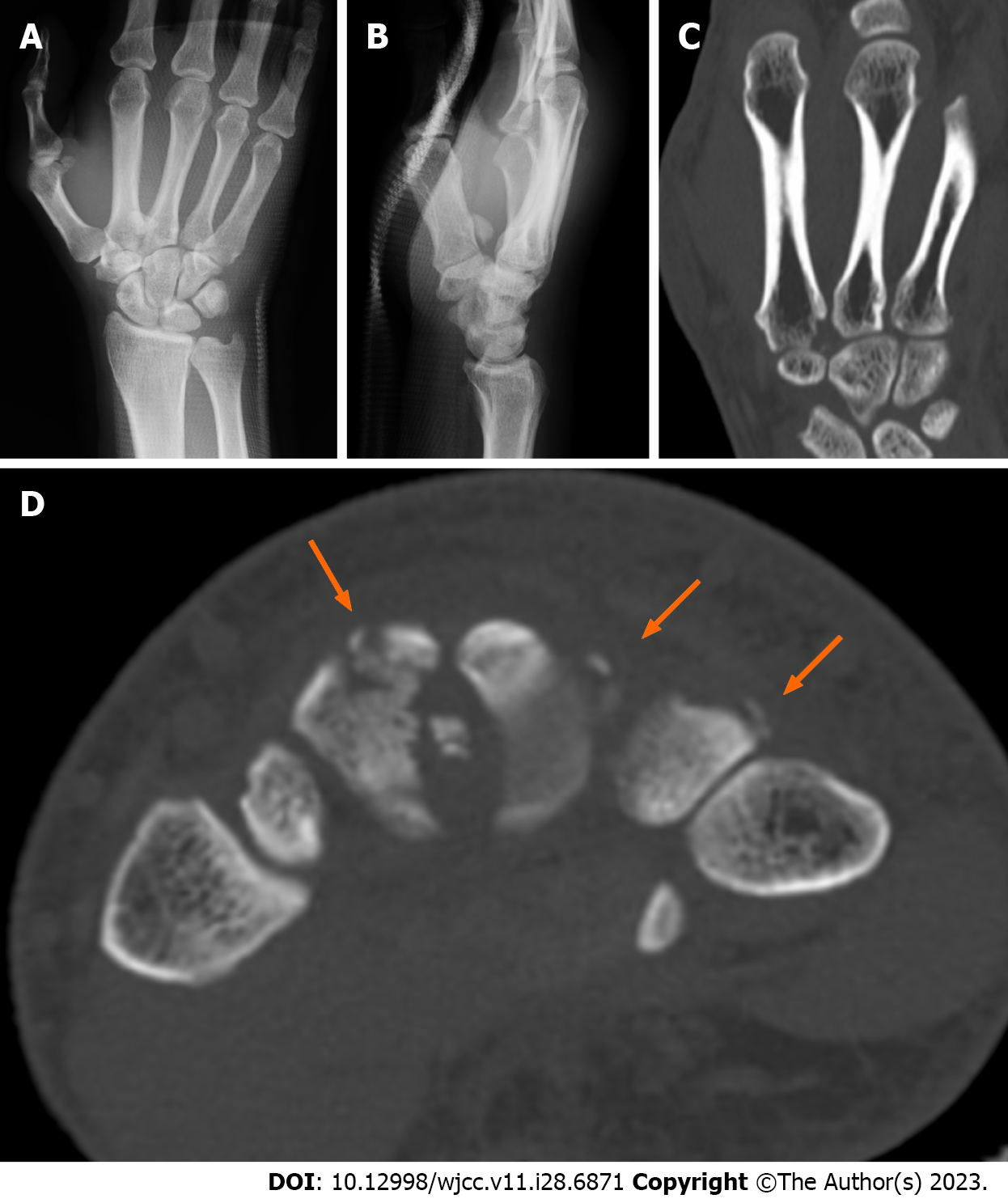
An anteroposterior radiograph showed a displaced fracture fragment between the bases of the second and third metacarpals (Figure 1A). On a lateral radiograph, the fragment was found to lie over the volar aspect of the CMC joint (Figure 1B). Computed tomography revealed that approximately one-third of the CMC joint surface of the second metacarpal was damaged (Figure 1C). In addition, we found small avulsion fragments on the dorsal aspects of the second to fourth metacarpal bases (Figure 1D).
We provisionally diagnosed an intra-articular fracture with significant instability of the CMC joints and performed open reduction and internal fixation.
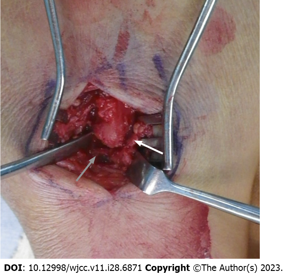
During surgery, we made a dorsal longitudinal incision over the CMC joint between the second and third metacarpals. The interosseous ligament between the second and third metacarpals was intact. The dorsal ligament of the third CMC joint was torn, the tendon of the extensor carpi radialis brevis was partially detached, and the third CMC joint showed some degree of instability (Figure 2). We considered that it had been dislocated to the volar side and then spontaneously reduced to that position.
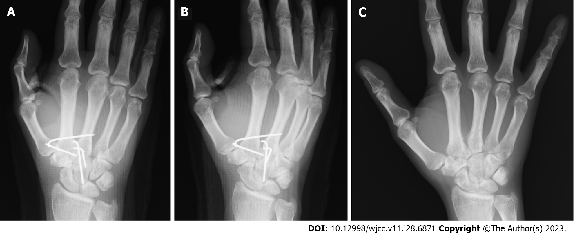
The fracture fragment could not be easily observed; thus, we divided the interosseous ligament between the second and third metacarpals partially, enlarged the metacarpal interspaces using the spreader, and then pulled out and reduced the fragment anatomically. The fragment was stabilized in an anatomical position using Kirschner wires (K-wires) (Figure 3A and B).
The K-wires were removed 6 wk postoperatively, and the patient returned to full activities of daily living. Union of the second metacarpal was confirmed radiographically at 12 wk postoperatively (Figure 3C). At 9 mo postoperative, the patient’s disabilities of the arm, shoulder, and hand scores were as follows: Disability/symptom: 1.19, Sports/music: 0.00, and Work: 0.00.
We reported a case with the displacement of an articular fracture fragment of the base of the second metacarpal from the ulnar side to the volar side, which was treated via the dorsal approach. Although volar dislocations of the CMC joint fractures have been reported[1-13], only six reports focused on the second and third metacarpals[7-10,12,13]. Among these, three reports[7,8,13] described complete dislocations of the CMC joint. The findings in the remaining three cases[9,10,12] were similar to those in our case with spontaneous reduction of the dislocated CMC joint.
Thomas et al[9] reported a case of isolated fracture dislocation of the base of the second metacarpal with the displacement of the base fragment into the palm. The remaining articular surface of the index metacarpal, as well as the metacarpal shaft, was displaced slightly palmarly, and dorsal ligamentous disruption was observed via a dorsal approach.
Han et al[12] reported a case of isolated volar fracture-dislocation of the second metacarpal caused by indirect injury. The capsule and attached ligaments around the second CMC joint were torn, and the ulnar side of the CMC joint ligament was almost entirely torn. The injury was treated with an open reduction using a volar approach.
Takami et al[10] reported a case of displaced fracture of the ulnar condyle at the base of the second metacarpal without dislocation of the second CMC joint. It seems probable that the fracture was caused by simultaneous volar subluxation of the base of the third metacarpal, which reduced spontaneously with the disruption of the interosseous ligament between the second and third metacarpals. During surgery, they used a volar incision paralleling the thenar crease.
In the first two cases, the second metacarpal was displaced in a volar direction and reduced spontaneously. The findings in the third case were similar to those in our case: the third metacarpal was displaced in a volar direction, and the interosseous ligament between the second and third metacarpals was torn. However, in our case, the interosseous ligament between the second and third metacarpals was intact, and the ligament of the third CMC joint was torn. Therefore, the articular fracture of the second metacarpal was likely to have occurred due to the dislocation of the third metacarpal around it with the interosseous ligament as a hinge. Thus, the dislocation mechanism in this case was different from that in previously reported cases. The dorsal approach is a good for observing ligament damage.
Of the three similar cases, the two most recent cases were treated using the palmar approach. We treated this lesion via the dorsal approach. The dorsal approach can be safely used with thin subcutaneous tissue.
The dorsal approach is a good option for cases involving displacement of an articular fracture fragment from the ulnar side to the volar side, not only because it allows direct observation of ligament damage and fixation of bone fragments, but also because the thin subcutaneous tissue makes the approach easier.
Provenance and peer review: Unsolicited article; Externally peer reviewed.
Peer-review model: Single blind
Specialty type: Orthopedics
Country/Territory of origin: Japan
Peer-review report’s scientific quality classification
Grade A (Excellent): 0
Grade B (Very good): 0
Grade C (Good): C, C, C
Grade D (Fair): 0
Grade E (Poor): 0
P-Reviewer: Li JM, China; Primadhi RA, Indonesia S-Editor: Liu JH L-Editor: A P-Editor: Liu JH
| 1. | Kleinman WB, Grantham SA. Multiple volar carpometacarpal joint dislocation. Case report of traumatic volar dislocation of the medial four carpometacarpal joint in a child and review of the literature. J Hand Surg Am. 1978;3:377-382. [RCA] [PubMed] [DOI] [Full Text] [Cited by in Crossref: 20] [Cited by in RCA: 15] [Article Influence: 0.3] [Reference Citation Analysis (0)] |
| 2. | Jameel J, Zahid M, Abbas M, Khan AQ. Volar dislocation of second, third, and fourth carpometacarpal joints: a rare and easily missed diagnosis. J Orthop Traumatol. 2013;14:67-70. [RCA] [PubMed] [DOI] [Full Text] [Full Text (PDF)] [Cited by in Crossref: 8] [Cited by in RCA: 7] [Article Influence: 0.5] [Reference Citation Analysis (0)] |
| 3. | Pundkare GT, Patil AM. Carpometacarpal Joint Fracture Dislocation of Second to Fifth Finger. Clin Orthop Surg. 2015;7:430-435. [RCA] [PubMed] [DOI] [Full Text] [Full Text (PDF)] [Cited by in Crossref: 16] [Cited by in RCA: 21] [Article Influence: 2.1] [Reference Citation Analysis (0)] |
| 4. | Schaefer N, Elliott D, Loveridge J. Volar dislocation of the index, middle, and ring carpometacarpal joints: a review. Plast Reconstr Surg Glob Open. 2015;3:e330. [RCA] [PubMed] [DOI] [Full Text] [Full Text (PDF)] [Reference Citation Analysis (0)] |
| 5. | Cates RA, Rhee PC, Kakar S. Multiple Volar Carpometacarpal Dislocations: Case Report/Review of the Literature. J Wrist Surg. 2016;5:236-240. [RCA] [PubMed] [DOI] [Full Text] [Cited by in Crossref: 4] [Cited by in RCA: 6] [Article Influence: 0.7] [Reference Citation Analysis (0)] |
| 6. | Ardente PDF, Biayna JC, Sarrias JS, Muñoz AN, Coll GF, Vergara P. Volar Dislocation of Second, Third and Fourth Carpometacarpal Joints in Association with a Bennet's Fracture of the Thumb Carpo-Metacarpal Dislocation: A Case Report. Open Orthop J. 2017;11:1035-1040. [RCA] [PubMed] [DOI] [Full Text] [Full Text (PDF)] [Cited by in Crossref: 1] [Cited by in RCA: 1] [Article Influence: 0.1] [Reference Citation Analysis (0)] |
| 7. | Harwin SF, Fox JM, Sedlin ED. Volar dislocation of the bases of the second and third metacarpals. A case report. J Bone Joint Surg Am. 1975;57:849-851. [PubMed] |
| 8. | Schutt Jr RC, Boswick Jr JA, Scott FA. Volar fracture-dislocation of the carpometacarpal joint of the index finger treated by delayed open reduction. J Trauma. 1981;21:986-987. [RCA] [DOI] [Full Text] [Cited by in Crossref: 14] [Cited by in RCA: 15] [Article Influence: 0.3] [Reference Citation Analysis (0)] |
| 9. | Thomas WO, Gottliebson WM, D'Amore TF, Harris CN, Parry SW. Isolated palmar displaced fracture of the base of the index metacarpal: a case report. J Hand Surg Am. 1994;19:455-456. [RCA] [PubMed] [DOI] [Full Text] [Cited by in Crossref: 13] [Cited by in RCA: 11] [Article Influence: 0.4] [Reference Citation Analysis (0)] |
| 10. | Takami H, Takahashi S, Ando M. Isolated volar displaced fracture of the ulnar condyle at the base of the index metacarpal: a case report. J Hand Surg Am. 1997;22:1064-1066. [RCA] [PubMed] [DOI] [Full Text] [Cited by in Crossref: 5] [Cited by in RCA: 6] [Article Influence: 0.2] [Reference Citation Analysis (0)] |
| 11. | Dillon JP, Laing AJ, Thakral R, Buckley JM, Mahalingam K. Volar dislocation of the index carpometacarpal joint in association with a Bennett's fracture of the thumb: a rare injury pattern. Emerg Med J. 2006;23:e23. [RCA] [PubMed] [DOI] [Full Text] [Cited by in Crossref: 3] [Cited by in RCA: 3] [Article Influence: 0.2] [Reference Citation Analysis (0)] |
| 12. | Han KJ, Lee J, Seo H. Isolated volar fracture-dislocation of the base of the second metacarpal bone by indirect injury. J Clin Orthop Trauma. 2015;6:42-45. [RCA] [PubMed] [DOI] [Full Text] [Cited by in RCA: 1] [Reference Citation Analysis (0)] |
| 13. | Silk G, Vetharajan N, Nagata H. Volar dislocation of the second and third carpometacarpal joints - the Lisfranc injury of the hand? Hand Surg Rehabil. 2018;37:320-323. [RCA] [PubMed] [DOI] [Full Text] [Cited by in Crossref: 2] [Cited by in RCA: 2] [Article Influence: 0.3] [Reference Citation Analysis (0)] |


