Copyright
©The Author(s) 2023.
World J Clin Cases. May 6, 2023; 11(13): 3076-3085
Published online May 6, 2023. doi: 10.12998/wjcc.v11.i13.3076
Published online May 6, 2023. doi: 10.12998/wjcc.v11.i13.3076
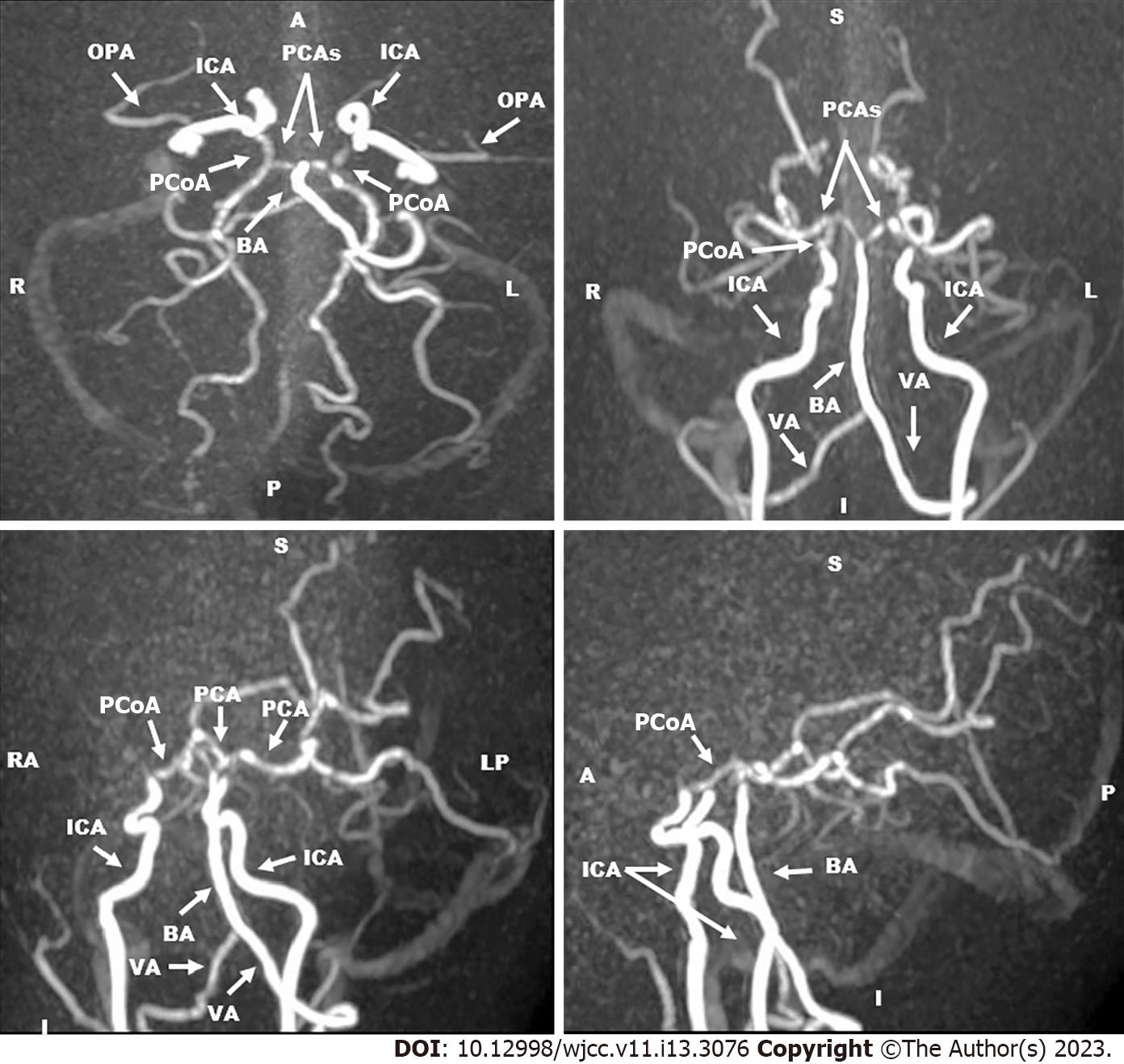
Figure 3 Magnetic resonance angiography of cerebral vessels three-dimension technique and maximum intensity projection images (Date: October 2019).
They revealed occlusion of the supraclinoid portion of internal carotid arteries. The right and left anterior cerebral arteries and middle cerebral arteries were not visualized. The basilar artery and the right and left posterior cerebral arteries had normal sizes and calibers. Collaterals were predominantly from the posterior circulation. OPA: The ophthalmic artery; ICA: The internal carotid artery; PCAs: The posterior cerebral arteries; PCoA: The posterior communicating artery; BA: The basilar artery; VA: The vertebral artery.
- Citation: Hamed SA, Yousef HA. Idiopathic steno-occlusive disease with bilateral internal carotid artery occlusion: A Case Report. World J Clin Cases 2023; 11(13): 3076-3085
- URL: https://www.wjgnet.com/2307-8960/full/v11/i13/3076.htm
- DOI: https://dx.doi.org/10.12998/wjcc.v11.i13.3076


