Copyright
©The Author(s) 2023.
World J Clin Cases. May 6, 2023; 11(13): 3070-3075
Published online May 6, 2023. doi: 10.12998/wjcc.v11.i13.3070
Published online May 6, 2023. doi: 10.12998/wjcc.v11.i13.3070
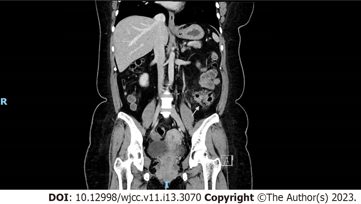
Figure 1 Computed tomography of abdomen pelvis coronal cut, showing signs of pericolonic fat stranding and extraluminal air pockets fluid density with peritoneal thickening at the sigmoid colon, likely representing a sealed perforation.
- Citation: Liew JJL, Lim WS, Koh FH. Unusual phenomenon-“polyp” arising from a diverticulum: A case report. World J Clin Cases 2023; 11(13): 3070-3075
- URL: https://www.wjgnet.com/2307-8960/full/v11/i13/3070.htm
- DOI: https://dx.doi.org/10.12998/wjcc.v11.i13.3070


