Published online May 6, 2023. doi: 10.12998/wjcc.v11.i13.3022
Peer-review started: November 7, 2022
First decision: January 20, 2023
Revised: February 3, 2023
Accepted: March 31, 2023
Article in press: March 31, 2023
Published online: May 6, 2023
Processing time: 168 Days and 21.5 Hours
Primary dedifferentiated chondrosarcoma (DDCS) of the lung is extremely rare and has a poor prognosis, especially in patients with a history of carcinomas and related treatment. Herein, we report a case of primary DDCS of the lung in a patient with a 4-year history of breast cancer and related treatment.
A 49-year-old woman was admitted to our hospital with complaints of headache, dizziness, slurred speech, and dyskinesia in May 2021. Computed tomography (CT) examinations showed multiple nodules in the brain, vertebral body, and both lungs with multiple enlarged lymph nodes in the right hilum and mediastinum, which were considered metastases of breast cancer. No obvious mass was discovered in the right hilum. After several months of related administration, the patient's headache disappeared, and her condition improved. However, new problems of asthma, dyspnea, cough, and restricted activity appeared in late November 2021. Although the CT scan indicated that the lesions in the brain, lung, and vertebral body had shrunk or disappeared, a soft tissue density lesion appeared in her right hilum and blocked the bronchial lumen. To relieve her dyspnea, part of the mass was resected, and a stent was placed via fiberoptic bronchoscopy. Following a complete pathological examination of the tumor, it was confirmed to be a primary DDCS of the lung. The patient then received two rounds of systemic chemotherapy with a regimen of cisplatin + ifosfamide + doxorubicin hydrochloride liposome, palliative radiotherapy for the tumor in her right lung, and four cycles of systemic chemotherapy and targeted therapy with a regimen of temozolomide combined with bevacizumab successively. She was in stable condition after the completion of the systemic chemotherapy and targeted therapy but underwent rapid progression after lung radiotherapy. The CT examinations showed multiple nodules in the brain and in both lungs, and the tumor in the right hilum was increased in size.
This case revealed a rare primary DDCS of the lung with a medical history of breast cancer, meaning a worse prognosis and making it more difficult to treat.
Core Tip: Dedifferentiated chondrosarcoma (DDCS) is a rare and high-grade malignant tumor. Here, we report a case of primary DDCS of the lung with a 4-year history of breast cancer and related treatment, which is extremely rare and easily misdiagnosed. It lacks a specific clinical manifestation and a precise imaging diagnosis. Thus, an additional pathological examination is beneficial. In addition, we found that radiotherapy accelerates the progression of DDCS. Altogether, this case created a more comprehensive understanding of this tumor and will provide a reference for future diagnosis, treatment, and prognosis estimations.
- Citation: Wen H, Gong FJ, Xi JM. Primary dedifferentiated chondrosarcoma of the lung with a 4-year history of breast cancer: A case report. World J Clin Cases 2023; 11(13): 3022-3028
- URL: https://www.wjgnet.com/2307-8960/full/v11/i13/3022.htm
- DOI: https://dx.doi.org/10.12998/wjcc.v11.i13.3022
Dedifferentiated chondrosarcoma (DDCS) is a tumor with a high degree of malignancy, poor curative effect, easy recurrence, and metastasis. Further, it is insensitive to chemoradiotherapy[1,2]. It usually occurs in people aged 40–60 years and is more common in males. The most common locations of DDCS are the femur and pelvis. It mainly manifests as localized pain, limited activity, and rapid enlargement of the mass. DDCS occurring outside the bone is extremely rare. Although primary DDCSs outside the bone have been observed in the lung[1-5], orbit[6], pleura[7], and throat[8], they were individual cases, and there are only four cases of DDCS occurring in the lung. Here, we present a case of primary DDCS of the lung with a 4-year history of breast cancer and related treatment.
A 49-year-old woman was admitted to our hospital to be evaluated due to headaches, dizziness, slurred speech, and dyskinesia on May 7, 2021.
The patient’s symptoms started approximately 2 wk ago.
The patient had a 4-year history of invasive ductal cancer of the left breast and underwent surgery, adjuvant chemoradiotherapy, and endocrine therapy. The specific circumstances are unclear. There is no additional remarkable past medical history about the patient.
No specific cancer history was recorded on her pedigree.
During admission, she was pale, weak, and walking unsteadily. The other physical examinations were normal.
Tumor-related biomarkers, including carcinoembryonic antigen, carbohydrate antigen (CA) 125, and CA199, were within the normal ranges. Other laboratory indicators were generally normal or slightly abnormal.
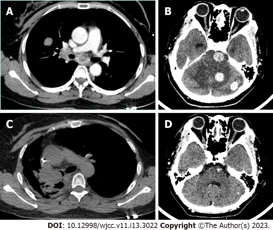
In May 2021, the computed tomography (CT) examinations showed multiple nodules in the brain, vertebral body, and both lungs, with multiple enlarged lymph nodes in the right hilum and mediastinum, which were considered metastases of breast cancer. No obvious mass was seen in the right hilum of the lung (Figure 1A and B). Subsequently, the patient was subjected to four cycles of systemic chemotherapy [Abraxane 260 mg/m2 intravenously (IV) on day 1 of a 21-d cycle] combined with targeted therapy (Bevacizumab 15 mg/kg IV on day 1 of a 21-d cycle), endocrine therapy (oral Exemestane tablet 25 mg once a day), and palliative radiotherapy for the brain metastasis successively. After administration, the patient's headache disappeared, and her condition improved. However, she was admitted to our hospital again due to the occurrence of asthma, dyspnea, cough, and restricted activity in late November 2021. The CT scan indicated that the lesions in the patient’s brain, lung, and vertebral body had shrunk or disappeared, but a soft tissue density lesion appeared at her right hilum of the lung and blocked the bronchial lumen (Figure 1C and D).
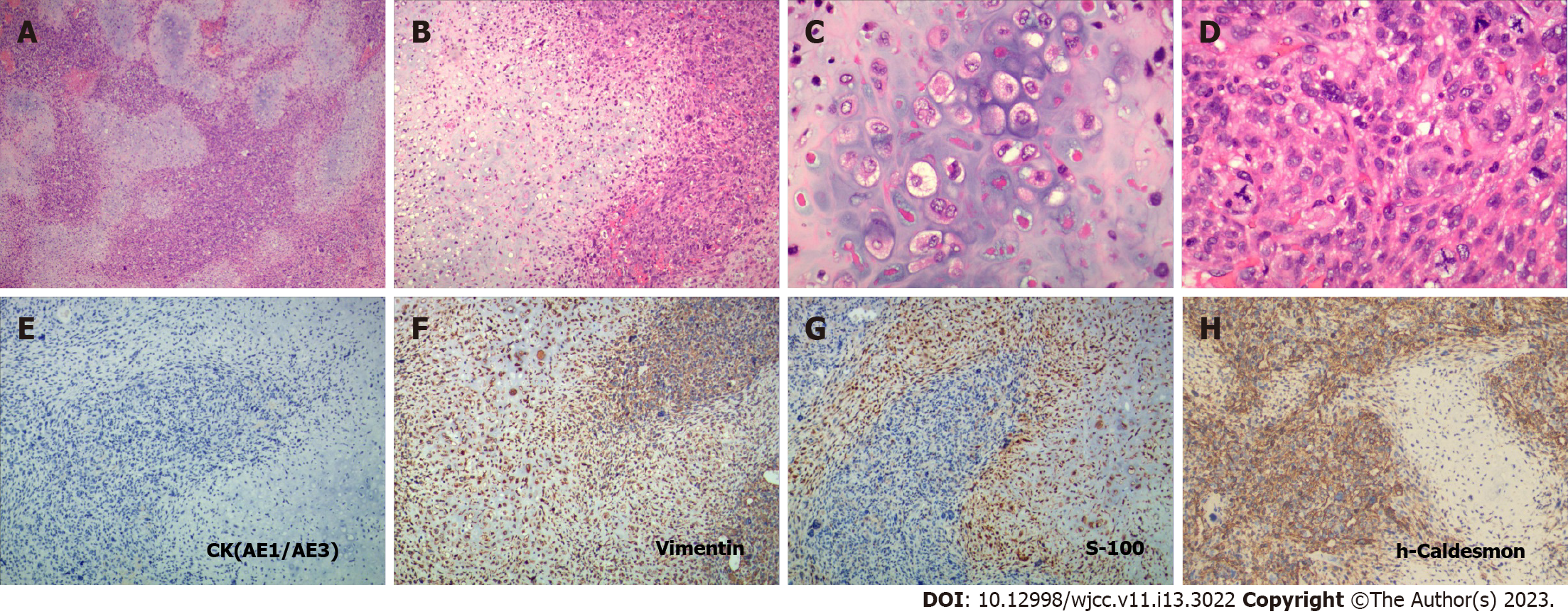
Since the patient presented symptoms of obvious dyspnea, a fiberoptic bronchoscopy was performed, part of the mass was resected, and a stent was placed. The collected specimen was sent for pathological examination. Histologically, the three pieces of gray and taupe tissue of the right lung (2.5 cm × 2 cm × 1.8 cm in size) showed two kinds of tumor components. One part was a well-differentiated chondrosarcoma—the tumor cells were round or oval, with mild atypia, a mitotic appearance, thin cytoplasm, and cartilaginous pits. The other part was a poorly differentiated sarcoma—the tumor cells were fusiform, large atypia, rich in tumor giant cells, and mitotic images. In addition, the two tumor components were well demarcated without transition and with hemorrhage and necrosis (Figure 2A–D). Immunohistochemistry staining showed that the tumor cells in both parts were positive for Vimentin, negative for Cytokeratin, and with a mutated p53 gene. S-100 tested positive in the chondrosarcoma, and h-Caldesmon, smooth muscle actin, cluster of differentiation antigen (CD) 99, and CD68 were partly positive in the dedifferentiated sarcoma (Figure 2E–H). The positive rate of Ki67 in the dedifferentiated sarcoma was approximately 30%.
Since careful clinical and radiologic examinations showed no evidence of further bone tumor, the tumor was confirmed to be a primary DDCS of the lung after a complete histologic preparation and examination.
The patient was diagnosed with DDCS of the lung in early December 2021. Subsequently, systemic chemotherapy was administrated for the patient with a regimen of cisplatin (75 mg/m2 IV on day 1 of a 21-d cycle) + ifosfamide (2 g IV on day 1 to 3 of a 21-d cycle) + doxorubicin hydrochloride liposome (40 mg/m2 IV on day 1 of a 21-d cycle ) in December 2021 and January 2022. Since the patient continued to suffer from asthma, palliative radiotherapy for the tumor in her right lung was performed in February 2022. She recently underwent chemotherapy and targeted therapy with a regimen of temozolomide (150 mg/m2 IV on days 1–5 of a 28-d cycle) combined with bevacizumab (7.5 mg/kg IV on day 1 of a 14-d cycle) from April to July 2022.
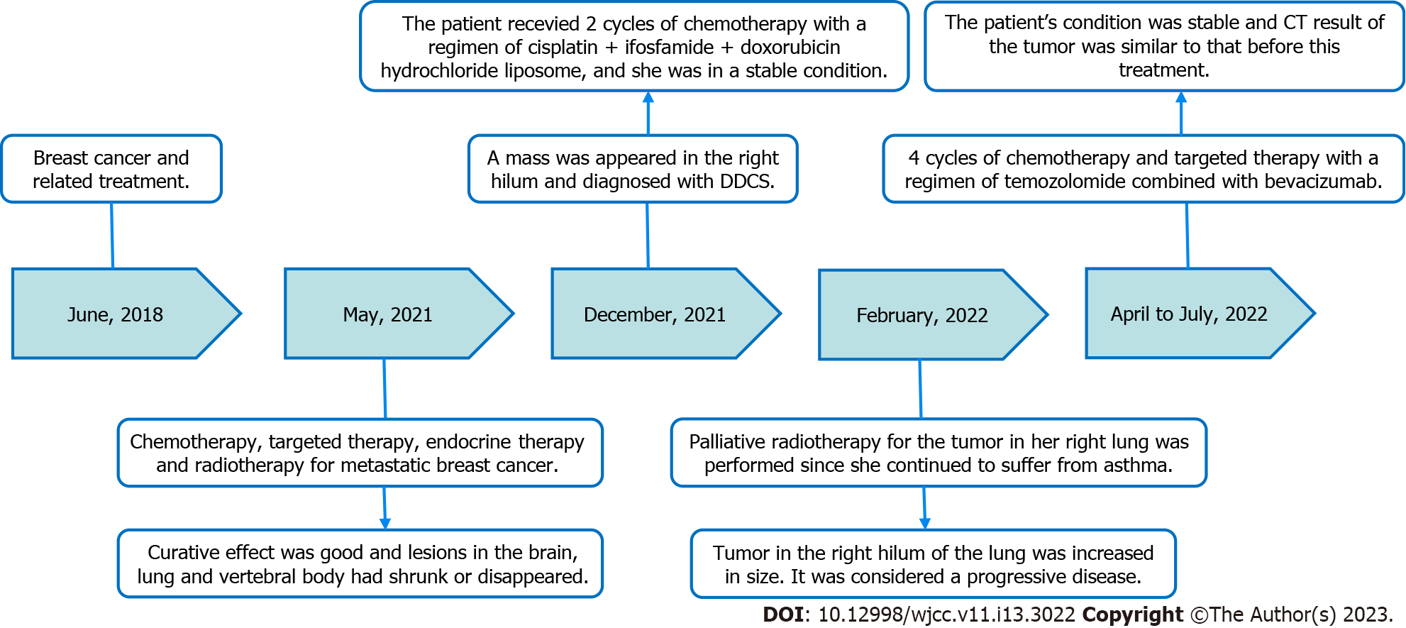
The patient was in a stable condition when two cycles of chemotherapy with a regimen of cisplatin + ifosfamide + doxorubicin hydrochloride liposome were completed. However, her condition significantly worsened after 30 sessions of radiation therapy for the tumor in her right lung. The CT examinations showed multiple nodules in the brain and both lungs, and the tumor in the right hilum of the lung was increased in size. It was considered a progressive disease based on the response evaluation criteria for solid tumors. Subsequently, she received four cycles of chemotherapy and targeted therapy with a regimen of temozolomide combined with bevacizumab. Her condition was stable, and the CT result of the tumor was similar to that before this treatment. No additional follow-up information was obtained from the patient at the time of manuscript writing. The timeline summarizing the main treatment and outcome of this case report is shown in Figure 3.
Regarding the origin of DDCS, there are two theories. Most studies believe that DDCS is derived from the differentiation of a stem cell with multi-directional differentiation potential, while others hold that it is differentiated from two different types of tumor cells independently, which is controversial[9-11]. Regarding molecular genetics, Yang et al[11] reported that isocitrate dehydrogenase (IDH) 1 and IDH2 were mutated in chondrosarcoma and had the same mutation in both components of DDCS, but there was no mutation in other mesenchymal tumors, which also supported that the tumor may originate from the same primary mesenchymal cells.
DDCS often presents a "biphasic sign" on CT, meaning it has the characteristics of soft tissue sarcoma and punctate and annular calcifications of chondrosarcoma in the same tumor. Histologically, the tumor is composed of the following two components: chondrosarcoma is usually grade I–II, and dedifferentiated sarcoma is high-grade spindle cell sarcoma with obvious atypia and frequent mitotic figures, which commonly include malignant fibrous histiocytoma, pleomorphic undifferentiated sarcoma, osteosarcoma, fibrosarcoma, rhabdomyosarcoma, etc. In terms of immunohistochemistry, chondrosarcoma components express the S-100 protein, and dedifferentiated components express corresponding sarcoma markers. In this case, the CT examination did not show punctate and annular calcifications, which are characteristic of chondrosarcoma, but instead, showed soft tissue density lesions. However, the pathological examination showed that the tumor is characterized by the following two distinct histopathologic components: A low-grade chondrosarcoma region sharply juxtaposed with a high-grade noncartilaginous sarcoma component, which correlated with the diagnosis of DDCS. Meanwhile, immunohistochemical expression of the tumor also supported this conclusion. Since the patient had a history of breast cancer and related treatment, if there were metaplastic cancer components in the breast tumor tissue, then the DDCS of the lung may have been transferred from the previous breast metaplastic carcinoma. Therefore, we reviewed her previous pathological sections of breast cancer but found no metaplastic cancer component and excluded the possibility of breast cancer metastasis. Furthermore, there was no evidence of further bone tumor according to the imaging examinations; therefore, we believe the tumor was a primary DDCS of the lung.
At present, surgical treatment remains the preferred choice for DDCS. Those who don’t have the opportunity for surgery can be treated with radiotherapy and chemotherapy. However, most scholars believe that DDCS is radiation resistant, and radiotherapy often has no obvious effect. Some scholars even found that DDCS after radiotherapy reduces the stability of tumor cells and deletes the PTEN gene, which promotes the proliferation potential of tumor cells[12]. The use of chemotherapy in the treatment of DDCS remains controversial, but most scholars believe that it has no obvious effect[13]. However, some scholars believe that when the dedifferentiated components are sensitive to chemotherapy and the patient's physical condition is good, additional chemotherapy can be considered[14,15]. Recent research found that a combination of surgery and chemotherapy showed a trend toward higher overall survival in non-metastatic patients with DDCS[16]. Therefore, whether to perform chemotherapy or radiotherapy should be determined by the physicians according to the specific condition of the patient. In this case, the patient was considered to have metastatic breast cancer. She had undergone relevant chemoradiotherapy, targeted therapy, and endocrine therapy. This caused the masses in the brain, vertebral body, and the other lung to shrink and disappear, but the hilar mass appeared and grew rapidly. After a diagnostic confirmation of DDCS, the patient received two rounds of systemic chemotherapy with a regimen of cisplatin + ifosfamide + doxorubicin hydrochloride liposome and four rounds of chemotherapy and targeted therapy with a regimen of temozolomide combined with bevacizumab successively. Consequently, her condition remains stable. The therapies of the patient in the present case were changed several times due to the diagnosis change and intolerance to chemotherapy; however, their effectiveness was unclear. Thus, whether the chemoradiotherapy suppressed the progression of DDCS is still unknown and must be investigated by further studies. These are the limitations of this rare case. The patient also underwent palliative radiotherapy for the tumor, which to the progression of the disease, meaning radiotherapy probably plays a negative role in the development of DDCS. Molecular targeted therapy for DDCS is still under study, and it has been reported that immunotherapy was effective for the tumor which is programmed death-ligand 1-positive[17].
The case of a primary DDCS of the lung with a 4-year history of breast cancer and related treatment is extremely rare. It lacks a specific clinical manifestation to distinguish it from other lung tumors, and the CT examination may not clearly show the “biphasic sign” characteristic. Moreover, when the tumor occurs in a patient who has a history of another malignant tumor, it is easily considered a recurrence or metastasis of the previous tumor by the oncologist, which results in misdiagnosis. Thus, clinical findings, image examination and pathological examination are indispensable to further confirm the DDCS and improve the recognition of this tumor.
In conclusion, DDCS is an extremely rare and high-grade malignant tumor. The tumor has a worse prognosis and more difficulties in treatment, especially in patients with a history of another carcinoma. Further, radiotherapy is likely to accelerate the progression of DDCS.
We thank the patient and her family for their support.
Provenance and peer review: Unsolicited article; Externally peer reviewed.
Peer-review model: Single blind
Specialty type: Medicine, research and experimental
Country/Territory of origin: China
Peer-review report’s scientific quality classification
Grade A (Excellent): 0
Grade B (Very good): 0
Grade C (Good): C, C
Grade D (Fair): 0
Grade E (Poor): 0
P-Reviewer: Ralic M, North Macedonia; Tangsuwanaruk T, Thailand S-Editor: Liu GL L-Editor: A P-Editor: Liu GL
| 1. | Li XF, Zhou HB, Zhao XL, Dai F, Li T, Wang L, Xu WM. [Primary dedifferentiated chondrosarcoma of lung: report of a case]. Zhonghua Binglixue Zazhi. 2011;40:127-128. [PubMed] |
| 2. | Gusho CA, Lee L, Zavras A, Seikel Z, Miller I, Colman MW, Gitelis S, Blank AT. Dedifferentiated Chondrosarcoma: A Case Series and Review of the Literature. Orthop Rev (Pavia). 2022;14:35448. [RCA] [PubMed] [DOI] [Full Text] [Cited by in Crossref: 5] [Cited by in RCA: 6] [Article Influence: 2.0] [Reference Citation Analysis (0)] |
| 3. | Boueiz A, Abougergi MS, Noujeim C, Bousamra A, Sfeir P, Zaatari G, Bou-Khalil P. Primary dedifferentiated chondrosarcoma of the lung. South Med J. 2009;102:861-863. [RCA] [PubMed] [DOI] [Full Text] [Cited by in Crossref: 6] [Cited by in RCA: 6] [Article Influence: 0.4] [Reference Citation Analysis (0)] |
| 4. | Steurer S, Huber M, Lintner F. Dedifferentiated chondrosarcoma of the lung: case report and review of the literature. Clin Lung Cancer. 2007;8:439-442. [RCA] [PubMed] [DOI] [Full Text] [Cited by in Crossref: 13] [Cited by in RCA: 14] [Article Influence: 0.8] [Reference Citation Analysis (1)] |
| 5. | Kawano D, Yoshino I, Shoji F, Ito K, Yano T, Maehara Y. Dedifferentiated chondrosarcoma of the lung: report of a case. Surg Today. 2011;41:251-254. [RCA] [PubMed] [DOI] [Full Text] [Cited by in Crossref: 4] [Cited by in RCA: 2] [Article Influence: 0.1] [Reference Citation Analysis (0)] |
| 6. | Qi XM, Zhang RR. Orbital soft tissue dedifferentiated chondrosarcoma: a case report. Zhonghua Yixue Zazhi. 2021;101:667-668. [DOI] [Full Text] |
| 7. | Liu Y, He CY, Liu J. Pleural dedifferentiate chondrosarcoma case. Fangshexue Shijian. 2021;4:279-280. [DOI] [Full Text] |
| 8. | Fidai SS, Ginat DT, Langerman AJ, Cipriani NA. Dedifferentiated Chondrosarcoma of the Larynx. Head Neck Pathol. 2016;10:345-348. [RCA] [PubMed] [DOI] [Full Text] [Cited by in Crossref: 7] [Cited by in RCA: 9] [Article Influence: 0.9] [Reference Citation Analysis (0)] |
| 9. | Bovée JV, Cleton-Jansen AM, Rosenberg C, Taminiau AH, Cornelisse CJ, Hogendoorn PC. Molecular genetic characterization of both components of a dedifferentiated chondrosarcoma, with implications for its histogenesis. J Pathol. 1999;189:454-462. [RCA] [PubMed] [DOI] [Full Text] [Cited by in RCA: 1] [Reference Citation Analysis (0)] |
| 10. | Meijer D, de Jong D, Pansuriya TC, van den Akker BE, Picci P, Szuhai K, Bovée JV. Genetic characterization of mesenchymal, clear cell, and dedifferentiated chondrosarcoma. Genes Chromosomes Cancer. 2012;51:899-909. [RCA] [PubMed] [DOI] [Full Text] [Cited by in Crossref: 72] [Cited by in RCA: 72] [Article Influence: 5.5] [Reference Citation Analysis (0)] |
| 11. | Yang T, Bai Y, Chen J, Sun K, Luo Y, Huang W, Zhang H. Clonality analysis and IDH1 and IDH2 mutation detection in both components of dedifferentiated chondrosarcoma, implicated its monoclonal origin. J Bone Oncol. 2020;22:100293. [RCA] [PubMed] [DOI] [Full Text] [Full Text (PDF)] [Cited by in Crossref: 7] [Cited by in RCA: 7] [Article Influence: 1.4] [Reference Citation Analysis (0)] |
| 12. | Gao L, Hong X, Guo X, Cao D, Gao X, DeLaney TF, Gong X, Chen R, Ni J, Yao Y, Wang R, Chen X, Tian P, Xing B. Targeted next-generation sequencing of dedifferentiated chondrosarcoma in the skull base reveals combined TP53 and PTEN mutations with increased proliferation index, an implication for pathogenesis. Oncotarget. 2016;7:43557-43569. [RCA] [PubMed] [DOI] [Full Text] [Full Text (PDF)] [Cited by in Crossref: 15] [Cited by in RCA: 18] [Article Influence: 2.0] [Reference Citation Analysis (0)] |
| 13. | Cranmer LD, Chau B, Mantilla JG, Loggers ET, Pollack SM, Kim TS, Kim EY, Kane GM, Thompson MJ, Harwood JL, Wagner MJ. Is Chemotherapy Associated with Improved Overall Survival in Patients with Dedifferentiated Chondrosarcoma? Clin Orthop Relat Res. 2022;480:748-758. [RCA] [PubMed] [DOI] [Full Text] [Cited by in Crossref: 12] [Cited by in RCA: 13] [Article Influence: 4.3] [Reference Citation Analysis (0)] |
| 14. | Dhinsa BS, DeLisa M, Pollock R, Flanagan AM, Whelan J, Gregory J. Dedifferentiated Chondrosarcoma Demonstrating Osteosarcomatous Differentiation. Oncol Res Treat. 2018;41:456-460. [RCA] [PubMed] [DOI] [Full Text] [Cited by in Crossref: 11] [Cited by in RCA: 11] [Article Influence: 1.6] [Reference Citation Analysis (0)] |
| 15. | Mavrogenis AF, Ruggieri P, Mercuri M, Papagelopoulos PJ. Dedifferentiated chondrosarcoma revisited. J Surg Orthop Adv. 2011;20:106-111. [PubMed] |
| 16. | Gonzalez MR, Bryce-Alberti M, Portmann-Baracco A, Inchaustegui ML, Castillo-Flores S, Pretell-Mazzini J. Appendicular dedifferentiated chondrosarcoma: A management and survival study from the SEER database. J Bone Oncol. 2022;37:100456. [RCA] [PubMed] [DOI] [Full Text] [Full Text (PDF)] [Cited by in RCA: 5] [Reference Citation Analysis (0)] |
| 17. | Singh A, Thorpe SW, Darrow M, Carr-Ascher JR. Case report: Treatment of metastatic dedifferentiated chondrosarcoma with pembrolizumab yields sustained complete response. Front Oncol. 2022;12:991724. [RCA] [PubMed] [DOI] [Full Text] [Cited by in Crossref: 3] [Cited by in RCA: 3] [Article Influence: 1.0] [Reference Citation Analysis (0)] |


