Copyright
©The Author(s) 2023.
World J Clin Cases. Apr 16, 2023; 11(11): 2567-2575
Published online Apr 16, 2023. doi: 10.12998/wjcc.v11.i11.2567
Published online Apr 16, 2023. doi: 10.12998/wjcc.v11.i11.2567
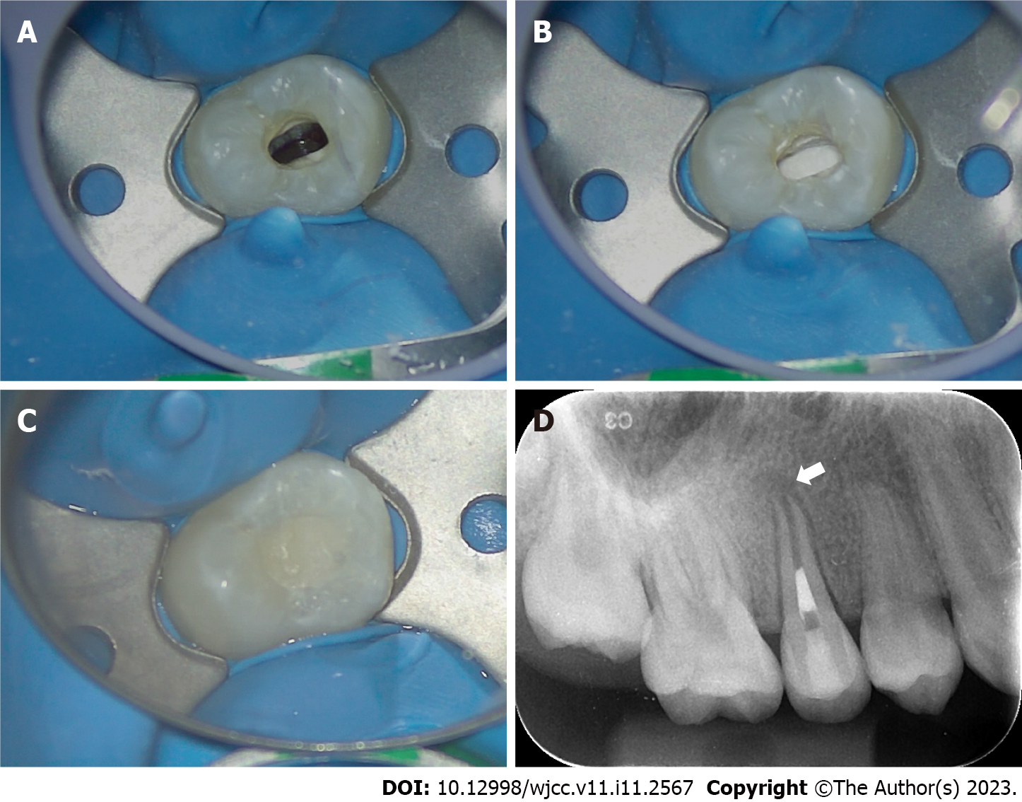
Figure 2 Treatment process of revascularization in the right maxillary second premolar.
A: Blood filled in the root canal space after penetration of the periapical tissue by a #20 K-file; B: The coronal third of the root canal was sealed with iRoot BP plus; C: The access cavity was temporarily restored with glass ionomer cement; D: Postoperative periapical radiograph of the right maxillary second premolar. Note the obvious hypodense area around the root apex and the wide-open apex (white arrow); the root canal is wide, and the dentinal wall is relatively thin.
- Citation: Chai R, Yang X, Zhang AS. Different endodontic treatments induced root development of two nonvital immature teeth in the same patient: A case report. World J Clin Cases 2023; 11(11): 2567-2575
- URL: https://www.wjgnet.com/2307-8960/full/v11/i11/2567.htm
- DOI: https://dx.doi.org/10.12998/wjcc.v11.i11.2567


