Published online Apr 6, 2023. doi: 10.12998/wjcc.v11.i10.2276
Peer-review started: December 26, 2022
First decision: January 20, 2023
Revised: January 29, 2023
Accepted: March 3, 2023
Article in press: March 3, 2023
Published online: April 6, 2023
Processing time: 94 Days and 2.7 Hours
Paragangliomas are rare neuroendocrine tumors. We hereby report a case of a localized paraganglioma found in the abdominal cavity, and review the relevant literature to improve the understanding of this disease.
A 29-year-old Chinese female patient was referred to our hospital due to an abdominal mass found on physical examination. Imaging revealed a mass in the left upper abdomen, suggestive of either a benign stromal tumor or an ectopic accessory spleen. Laparoscopic radical resection was subsequently performed, and histopathological analysis confirmed the diagnosis of a paraganglioma. The patient was followed up 3 months post-operation, and reported good recovery with no metastasis.
Radical resection can effectively treat intra-abdominal paragangliomas, with few side effects and low recurrence risk. In addition, early and accurate diagnosis and timely intervention are essential for the prognosis of this disease.
Core Tip: Intraperitoneal paraganglioma is clinically rare without characteristic imaging findings, and many clinicians may never encounter it. However, clinicians must remain vigilant to suspect, identify, locate, and remove the tumor, as the associated symptoms and hypertension can be cured by surgical resection. If the tumor is not diagnosed and removed, there is a risk of death and heart disease. Therefore, due to the small number of cases, the lack of understanding of its clinical features and imaging signs, especially easy to miss diagnosis.
- Citation: Guo W, Li WW, Chen MJ, Hu LY, Wang XG. Primary intra-abdominal paraganglioma: A case report. World J Clin Cases 2023; 11(10): 2276-2281
- URL: https://www.wjgnet.com/2307-8960/full/v11/i10/2276.htm
- DOI: https://dx.doi.org/10.12998/wjcc.v11.i10.2276
Paragangliomas are rare neuroendocrine tumors that can occur at any age. It represents an important cause of secretory hypertension as it is often characterized by the excessive production of catecholamines. This study aimed to report a case of an intra-abdominal paraganglioma, and improve the understanding of this disease by elucidating the clinical features of the patient, while reviewing the relevant literature.
A 29-year-old woman was referred to the Department of General Surgery of the Second Hospital of Jiaxing on July 20, 2022 following findings of an abdominal mass 5 d ago.
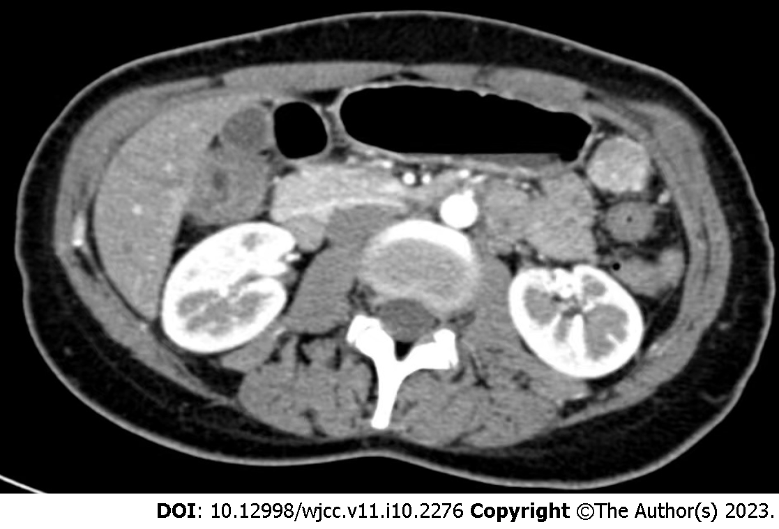
Initial abdominal computed tomography (CT) revealed soft tissue nodules with central calcification in the left upper abdomen, which warranted further imaging with contrast-enhanced CT.
The patient had no surgical or tumor history. Besides a 2-year history of Hashimoto’s thyroiditis and hyperthyroidism, no other underlying diseases such as hypertension and diabetes were reported.
The patient denied any family history of cancer.
Physical examination was grossly unremarkable. No gastrointestinal symptoms such as abdominal pain, distension, nausea and vomiting, jaundice, abnormal bowel movements, or abnormal stool forms were noted. Urinary symptoms such as gross hematuria, frequency, urgency, and dysuria were not noted as well. The patient was not pyrexic, and denied any chest tightness or shortness of breath.
Laboratory results were as follows: Thyroid stimulating hormone, 0.003 µIU/mL; anti-thyroglobulin, 282.92 IU/mL, anti-thyroid peroxidase antibody, 149.91 IU/mL (reference range, 0–34 IU/mL); and CA19-9, 2.0 U/mL (reference range 0–37 U/m). The remaining laboratory test results were otherwise unremarkable.
Pathological examination of the lesion suggested a paraganglioma (Figure 1). Immunohistochemical staining was subsequently performed, which demonstrated the following: Syn (-), CgA (-), CD56 (-), Ki-67 (+ 5%), CD10 (-), S-100 (+), NES (+), AE1/AE3 (-), SOX10 (-), and P53 (wild type). A diagnosis of an intra-abdominal paraganglioma was thus confirmed.
Contrast-enhanced CT confirmed a mass in the left upper abdomen. At this stage, a benign stromal tumor of mesenchymal origin or an ectopic accessory spleen was considered (Figure 2).
The patient was diagnosed with a primary intra-abdominal paraganglioma.
Laparoscopic radical resection of the abdominal mass was indicated. During the operation, the mass was found adhered to the surrounding omentum in the left upper abdominal region, in close proximity to the spleen, stomach, small intestine, and colon. The lesion was approximately 22 mm × 26 mm in size. Postoperative chemotherapy was not indicated.
The patient recovered well without any discomfort. At 3 mo follow-up, no complications such as recurrence or metastasis were observed.
Paragangliomas are rare neuroendocrine tumors originating from either the adrenal medulla, or the extra-adrenal sympathetic and parasympathetic ganglia. Symptoms involve the classic triad of headache, palpitation, and sweating. Adrenal and extra-adrenal sympathetic paragangliomas are often characterized by the excessive production of catecholamines, which can not only result in hypertension, but also associate with the risk of acute cardiovascular complications. Diagnosis is often based on plasma or urinary metanephrine measurements and nuclear imaging. Moreover, normal catecholamine levels have been reported to virtually exclude the presence of a sympathetic paraganglioma[1]. Intra-abdominal paragangliomas are particularly rare, with incidence of approximately 1 in 500000.
Parasympathetic paragangliomas are usually located in the head and neck, while sympathetic paragangliomas are more commonly located in the abdomen, followed by the chest and pelvis[2]. And the tumer that may present with cranial neuropathies when located along the skull base[3]. Head and neck paragangliomas are usually painless and slow growing, and are thus often an incidental clinical finding. They mainly manifest as carotid body tumors and vagal paragangliomas. As parasympathetic paragangliomas are non-secretory, symptoms are usually secondary to mass effects. These may include neck pain and dysphagia, conductive hearing loss and pulsatile tinnitus in cervical tympanic paragangliomas, as well as lower cranial nerve defects in advanced tumors[4]. Paragangliomas located outside of the head and neck may also be non-secretory, and are often accompanied by mild symptoms[5].
Due to the catecholamine-secreting nature of abdominal paragangliomas, the common clinical symptoms may include malignant hypertension, palpitation, headache, dizziness, anxiety, metabolic disorder syndrome, orthopnea, oliguria, anuria, and hepatic encephalopathy, among others. In rare cases, patients may experience paraganglioma crises, an endocrine emergency resulting in life-threatening hemodynamic instability and end-organ damage[6]. This can often be misdiagnosed as septic shock, heart failure, thyroid storm, and malignant hyperthermia[7,8]. Given its mortality rate of approximately 15%, early recognition of the signs and symptoms of paragangliomas is thereby critical[7,9]. It was found that in patients with paraganglioma, the metaiodobenzylguanidine (MIBG) uptake intensity ratio was significantly higher in malignant lesions than in benign lesions. Therefore, iodine-131 MIBG uptake was able to distinguish between benign and malignant diseases, which was not helped by magnetic resonance imaging[10]. Furthermore, one study used radio-labeled MIBG and somatostatin analogues for scintillation imaging for correct localization. The results showed that MIBG was more accurate in imaging pheochromocytoma than somatostatin analogues. But somatostatin analogues are more accurate than MIBGs in detecting neuroendocrine tumors[11]. While symptoms are often paroxysmal, the clinical manifestations of paragangliomas can vary based on catecholamine subtypes, and may range from asymptomatic to life-threatening[12]. As exemplified in our case, the lack of obvious clinical abnormalities such as abdominal pain, abdominal distension, hypertension, and dizziness could have led to misdiagnosis or missed diagnosis.
Paragangliomas are mostly benign in nature, with surgical resection being the main treatment of choice. In contrast, malignant paragangliomas often warrant a multidisciplinary approach, involving endocrinology, oncology, surgery, nuclear medicine, radiotherapy, interventional radiology, and histology. So far, there are no standardized treatment regimens for metastatic diseases. Current treatment measures mainly involve beta-blockers and catecholamine synthesis inhibitors (A-methyl-p-tyrosine) to prevent tumor progression and minimize catecholamine-induced symptoms[13].
The diagnosis of paraganglioma in our case was mainly based on pathology, and was confirmed upon findings of Syn (-) and S-100 (+) on immunohistochemical staining. While morphologically similar to malignant perivascular epithelioid cell and stromal tumors, S-100 played a role as a differentiating factor. As a neurogenic index, the positive expression of S-100 observed in our patient was in keeping with the origin of paragangliomas from chromaffin cells of the neural crest. In contrast, perivascular epithelioid cell tumors are mostly malignant in nature, and are characterized by positive HMB45, SMA, and Desmin, the latter 2 of which are myogenic indices. Stromal tumors are also commonly malignant, and are distinguished by the expression of CD117. In our case, the low expression of Ki-67 indicated a benign tumor.
In conclusion, intra-abdominal paragangliomas are clinically rare with no characteristic imaging findings, and are, as such, easily missed. However, surgical resection can associate with good clinical prognosis.
We thank the patient for his contribution to this case report. We also thank the Department of General Surgery of the Second Affiliated Hospital of Jiaxing University for their support with the treatment of this case.
Provenance and peer review: Unsolicited article; Externally peer reviewed.
Peer-review model: Single blind
Specialty type: Surgery
Country/Territory of origin: China
Peer-review report’s scientific quality classification
Grade A (Excellent): 0
Grade B (Very good): B, B
Grade C (Good): 0
Grade D (Fair): 0
Grade E (Poor): 0
P-Reviewer: Lucke-Wold B, United States; Maurea S, Italy S-Editor: Liu JH L-Editor: A P-Editor: Liu JH
| 1. | Cornu E, Belmihoub I, Burnichon N, Grataloup C, Zinzindohoué F, Baron S, Billaud E, Azizi M, Gimenez-Roqueplo AP, Amar L. [Phaeochromocytoma and paraganglioma]. Rev Med Interne. 2019;40:733-741. [RCA] [PubMed] [DOI] [Full Text] [Cited by in Crossref: 5] [Cited by in RCA: 5] [Article Influence: 0.8] [Reference Citation Analysis (0)] |
| 2. | Lenders JW, Duh QY, Eisenhofer G, Gimenez-Roqueplo AP, Grebe SK, Murad MH, Naruse M, Pacak K, Young WF Jr; Endocrine Society. Pheochromocytoma and paraganglioma: an endocrine society clinical practice guideline. J Clin Endocrinol Metab. 2014;99:1915-1942. [RCA] [PubMed] [DOI] [Full Text] [Cited by in Crossref: 1592] [Cited by in RCA: 1745] [Article Influence: 158.6] [Reference Citation Analysis (0)] |
| 3. | Moor R, Goutnik M, Lucke-Wold B, Laurent D, Chen S, Friedman W, Rahman M, Allen N, Rivera-Zengotita M, Koch M. Clival Paraganglioma, Case Report and Literature Review. OBM Neurobiol. 2022;6. [RCA] [PubMed] [DOI] [Full Text] [Full Text (PDF)] [Cited by in Crossref: 3] [Reference Citation Analysis (0)] |
| 4. | Smith JD, Harvey RN, Darr OA, Prince ME, Bradford CR, Wolf GT, Else T, Basura GJ. Head and neck paragangliomas: A two-decade institutional experience and algorithm for management. Laryngoscope Investig Otolaryngol. 2017;2:380-389. [RCA] [PubMed] [DOI] [Full Text] [Full Text (PDF)] [Cited by in Crossref: 38] [Cited by in RCA: 67] [Article Influence: 8.4] [Reference Citation Analysis (0)] |
| 5. | Fishbein L. Pheochromocytoma/Paraganglioma: Is This a Genetic Disorder? Curr Cardiol Rep. 2019;21:104. [RCA] [PubMed] [DOI] [Full Text] [Cited by in Crossref: 9] [Cited by in RCA: 9] [Article Influence: 1.5] [Reference Citation Analysis (0)] |
| 6. | Meijs AC, Snel M, Corssmit EPM. Pheochromocytoma/paraganglioma crisis: case series from a tertiary referral center for pheochromocytomas and paragangliomas. Hormones (Athens). 2021;20:395-403. [RCA] [PubMed] [DOI] [Full Text] [Full Text (PDF)] [Cited by in Crossref: 8] [Cited by in RCA: 19] [Article Influence: 4.8] [Reference Citation Analysis (0)] |
| 7. | Whitelaw BC, Prague JK, Mustafa OG, Schulte KM, Hopkins PA, Gilbert JA, McGregor AM, Aylwin SJ. Phaeochromocytoma [corrected] crisis. Clin Endocrinol (Oxf). 2014;80:13-22. [RCA] [PubMed] [DOI] [Full Text] [Cited by in Crossref: 72] [Cited by in RCA: 82] [Article Influence: 7.5] [Reference Citation Analysis (0)] |
| 8. | Juszczak K, Drewa T. Adrenergic crisis due to pheochromocytoma - practical aspects. A short review. Cent European J Urol. 2014;67:153-155. [RCA] [DOI] [Full Text] [Full Text (PDF)] [Cited by in Crossref: 4] [Cited by in RCA: 9] [Article Influence: 0.8] [Reference Citation Analysis (0)] |
| 9. | Martucci VL, Pacak K. Pheochromocytoma and paraganglioma: diagnosis, genetics, management, and treatment. Curr Probl Cancer. 2014;38:7-41. [RCA] [PubMed] [DOI] [Full Text] [Cited by in Crossref: 130] [Cited by in RCA: 119] [Article Influence: 10.8] [Reference Citation Analysis (0)] |
| 10. | Maurea S, Cuocolo A, Imbriaco M, Pellegrino T, Fusari M, Cuocolo R, Liuzzi R, Salvatore M. Imaging characterization of benign and malignant pheochromocytoma or paraganglioma: comparison between MIBG uptake and MR signal intensity ratio. Ann Nucl Med. 2012;26:670-675. [RCA] [PubMed] [DOI] [Full Text] [Cited by in Crossref: 7] [Cited by in RCA: 13] [Article Influence: 1.0] [Reference Citation Analysis (0)] |
| 11. | Lastoria S, Maurea S, Vergara E, Acampa W, Varrella P, Klain M, Muto P, Bernardy JD, Salvatore M. Comparison of labeled MIBG and somatostatin analogs in imaging neuroendocrine tumors. Q J Nucl Med. 1995;39:145-149. [PubMed] |
| 12. | Petrák O, Rosa J, Holaj R, Štrauch B, Krátká Z, Kvasnička J, Klímová J, Waldauf P, Hamplová B, Markvartová A, Novák K, Michalský D, Widimský J, Zelinka T. Blood Pressure Profile, Catecholamine Phenotype, and Target Organ Damage in Pheochromocytoma/Paraganglioma. J Clin Endocrinol Metab. 2019;104:5170-5180. [RCA] [PubMed] [DOI] [Full Text] [Cited by in Crossref: 15] [Cited by in RCA: 34] [Article Influence: 5.7] [Reference Citation Analysis (0)] |
| 13. | Corssmit EPM, Snel M, Kapiteijn E. Malignant pheochromocytoma and paraganglioma: management options. Curr Opin Oncol. 2020;32:20-26. [RCA] [PubMed] [DOI] [Full Text] [Cited by in Crossref: 12] [Cited by in RCA: 24] [Article Influence: 4.8] [Reference Citation Analysis (0)] |


