Copyright
©The Author(s) 2022.
World J Clin Cases. Oct 16, 2022; 10(29): 10755-10762
Published online Oct 16, 2022. doi: 10.12998/wjcc.v10.i29.10755
Published online Oct 16, 2022. doi: 10.12998/wjcc.v10.i29.10755
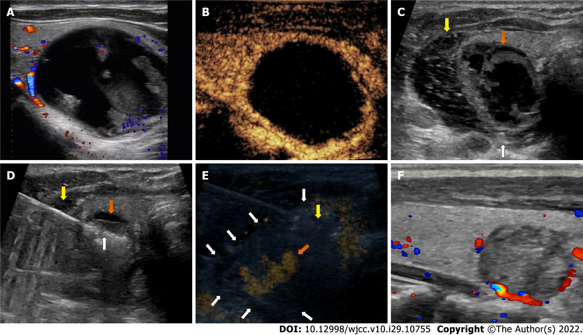
Figure 2 Case 2: A 45-year-old male with a cystic benign thyroid nodule in the left lobe.
A: Color Doppler ultrasonography showed the nodule with little vascularity and a minimal peripheral blood supply; B: Preoperative contrast-enhanced ultrasonography showed the nodule with only peripheral blood supply and almost no enhancement inside; C: After the first aspiration of the intranodular fluid, the nodule showed minimal shrinkage (orange arrow). Then, the needle for hydrodissection was placed in the location between the dorsal capsule of lateral lobe and the recurrent laryngeal nerve (white arrow), and the nodule was separated from the surrounding structures (yellow arrow); D: The needle for hydrodissection remained in place, and the radiofrequency electrode was inserted into the nodule (white arrow) for ablation; E: During the ablation, contrast-enhanced ultrasonography showed a perithyroidal hematoma (white arrow) around the nodule and an active hemorrhage near the location where the needle for hydrodissection was placed (orange arrow) after the needle was withdrawn. Compression was applied to achieve hemostasis. Then, radiofrequency ablation was performed to stop the bleeding; F: At the 1-mo follow-up, the perithyroidal hematoma had disappeared on ultrasonography, and the nodule showed shrinkage with a volume reduction ratio of 86.4%.
- Citation: Zheng BW, Wu T, Yao ZC, Ma YP, Ren J. Perithyroidal hemorrhage caused by hydrodissection during radiofrequency ablation for benign thyroid nodules: Two case reports. World J Clin Cases 2022; 10(29): 10755-10762
- URL: https://www.wjgnet.com/2307-8960/full/v10/i29/10755.htm
- DOI: https://dx.doi.org/10.12998/wjcc.v10.i29.10755


