Published online Oct 16, 2022. doi: 10.12998/wjcc.v10.i29.10529
Peer-review started: May 27, 2022
First decision: July 13, 2022
Revised: July 21, 2022
Accepted: August 24, 2022
Article in press: August 24, 2022
Published online: October 16, 2022
Processing time: 124 Days and 18.1 Hours
Shoulder injuries caused by trauma are common, including clavicle fractures. Even so, bipolar segmental fracture of the clavicle is extremely rare and seldom reported. Therefore, there is still a controversial issue about how to treat these bipolar segmental clavicle fractures.
A 56-year-old security guard arrived at our emergency room after falling on his electric bicycle. There was no loss of consciousness or other pain. He had no numbness in his fingers. X-rays and 3D computed tomography revealed that the patient's right shoulder had a bipolar segmental clavicle fracture. The surgical procedure included both open reduction and internal fixation. At the 1-year follow-up, he had a full range of motion and minimal discomfort in the injured shoulder.
We provide a rare case of bipolar clavicle facture in the right clavicle. We hold the opinion that such patients would get better clinical and radiological outcomes by early and correct operation.
Core Tip: A rare case of bipolar clavicle fracture due to trauma is reported. Early operation provides a possibility for such patients to recover well later.
- Citation: Liang L, Chen XL, Chen Y, Zhang NN. Surgical treatment of bipolar segmental clavicle fracture: A case report. World J Clin Cases 2022; 10(29): 10529-10534
- URL: https://www.wjgnet.com/2307-8960/full/v10/i29/10529.htm
- DOI: https://dx.doi.org/10.12998/wjcc.v10.i29.10529
Clavicle fractures are a frequent occurrence in clinical practice. Considering the anatomy of the clavicle, more than 70% of clavicle fractures occur in the midshaft region[1]. However, due to the low occupancy of the clavicle, simultaneous fractures of the proximal and distal ends of the clavicle, also known as the bipolar clavicle fracture, are not frequently mentioned in the medical literature. It is frequently caused by high-energy trauma, such as automobile accidents and falls from great heights[2-4]. Despite multiple managements ranging from non-operative to surgical available, it remains controversial as to which is the optimal treatment. Historically, a figure-8 sling was most frequently used for traditional conservative treatment[5-7]. However, as people's knowledge and beliefs evolve, more and more evidence demonstrates that surgical management results in a more reliable, earlier, and complication-free return to full function. In this article, we report a patient with a right bipolar segmental clavicle fracture treated by bipolar fixation.
A 56-year-old security guard presented to our hospital with right shoulder pain after a 1-d history of fall from an electric bicycle.
The patient fell onto his right shoulder 1 d ago and felt pain and swelling in his right shoulder without coma, dyspnea, or numbness in his upper extremities. He did not immediately go to the hospital. Subsequently, the patient complained of increased edema, pain in the right shoulder, and limited range of motion at the right sternoclavicular joint. He visited our hospital's emergency room and was sent for imaging.
Previously, the patient was in good health. He denied having a history of diabetes, hypertension, coronary heart disease, other significant surgical injuries, and food and drug allergies.
The patient had no relevant personal or family history.
The patient’s right shoulder and sternoclavicular joint were swollen and exhibited local congestion. It was impossible for him to raise the right upper limb above the shoulder. There was no limb numbness. The injured limb's muscle strength and radial artery pulse were normal.
Blood analysis revealed the following: White blood cell count, 5.5 × 10⁹/L; red blood cell count, 3.11 × 10¹²/L; haemoglobin, 113 g/L; and platelet count, 116 × 109/L. Biochemical analysis revealed the following: Total protein, 56.9 g/L; albumin, 34.8 g/L; creatinine, 100.4umol/L; urea nitrogen, 3.12 mol/L; prothrombin time, 12.0 s; prothrombin activity percentage, 100%; activated partial prothrombin time, 41.3 s; and D-dimer, 2.41 mg/L. The findings of routine urine and stool examinations were normal, as were the findings of electrocardiography.
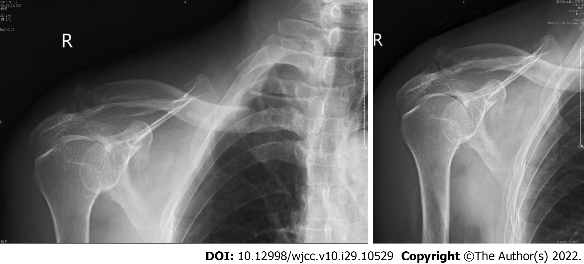
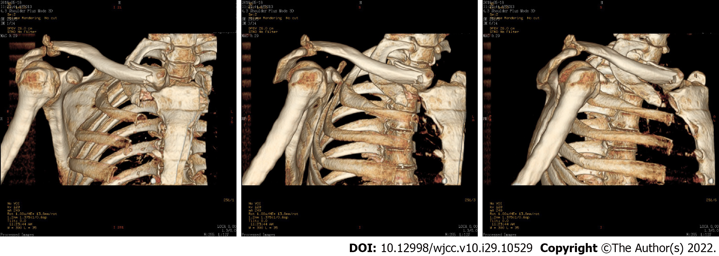
On standard radiograph of the patient, both the proximal and distal ends of the clavicle fractures were detached (Figure 1). In addition, 3D computed tomography (CT) revealed the degree of the displa
Bipolar segmental clavicle fracture.
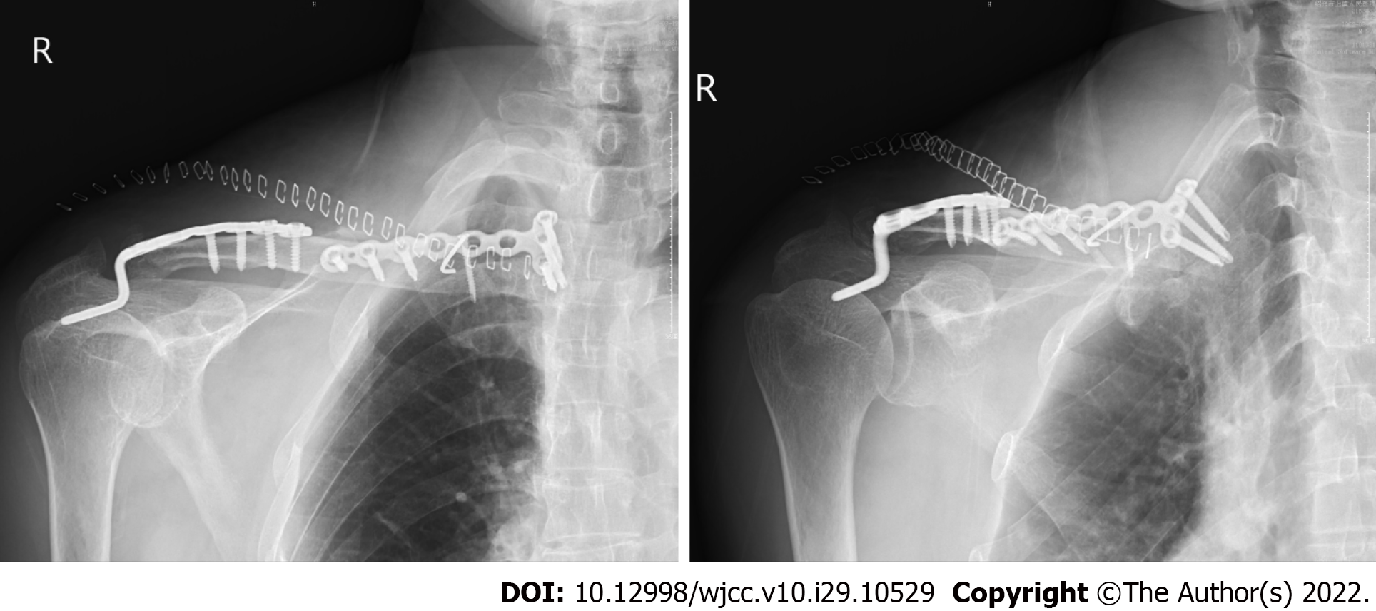
We decided to treat the patient by open reduction and internal fixation after admission. The patient consented to the surgery 4 d after the injury, which was performed with his permission. The patient underwent surgery under general anesthesia with a 30-degree angle of support. After a skin incision was made over the proximal and distal ends of the clavicle, the fractures were observable upon direct examination. At the proximal end of the free bone fragment, one cortical screw and a Kirschner wire were used to fix it, followed by a fragment-contoured T plate. The distal aspect of the clavicle was elevated. The rupture of the acromioclavicular ligament caused the subluxation of the acromioclavicular joint. To stabilize the fracture and dislocation, an acromioclavicular hook plate was placed.
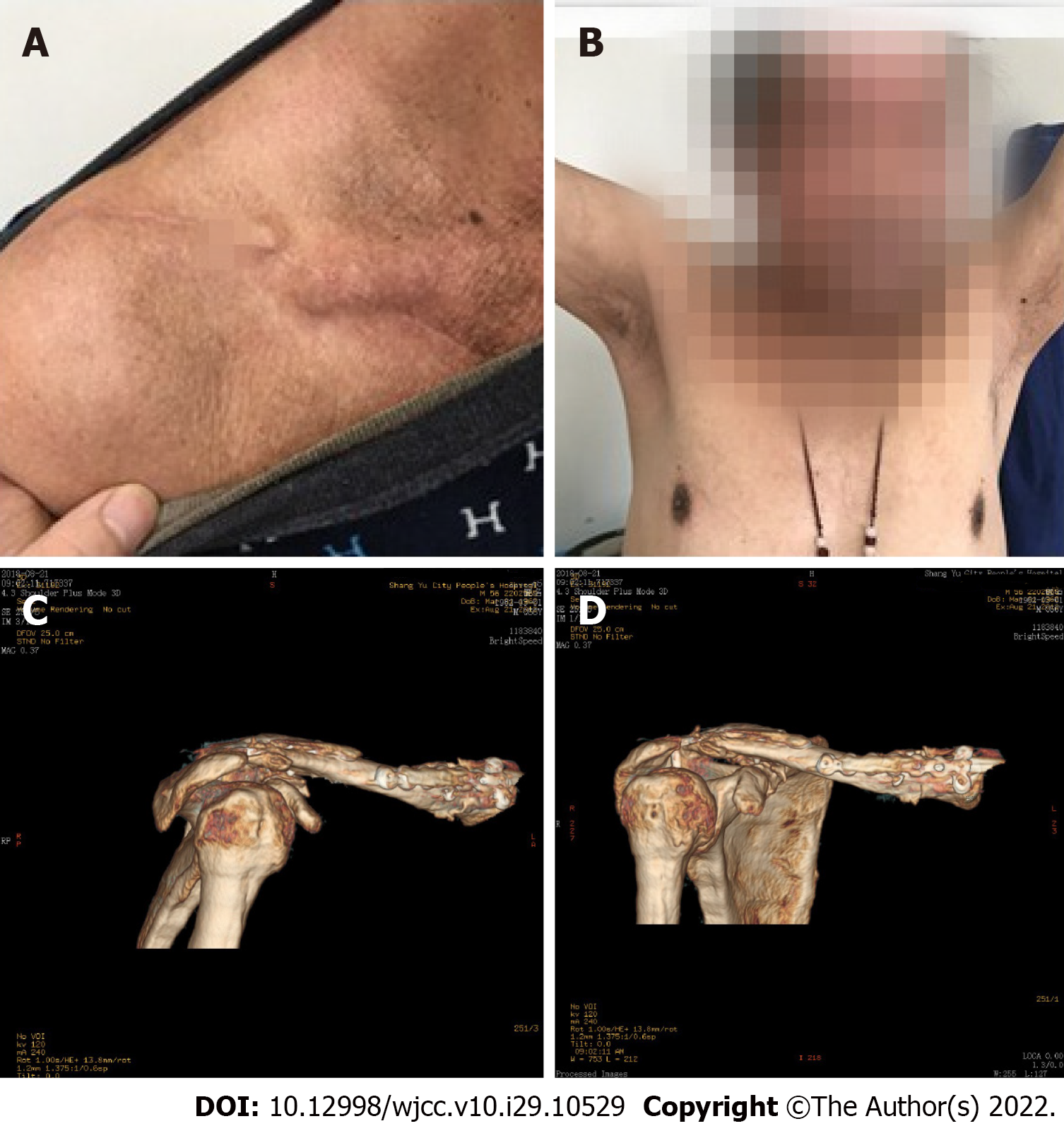
On postoperative radiographic evaluations, excellent fracture line and dislocation reduction were observed (Figure 3). The patient performed horizontal right shoulder range of motion exercises under supervision. Six weeks after surgery, the active motion of the right shoulder joint was gradually permitted as tolerated. After 10 wk of recovery, the patient returned to work. At the 4-mo follow-up, the patient could flex his right shoulder forward up to 150 degrees. X-rays and 3D CT revealed that bipolar segmental clavicle fractures had healed completely (Figure 4). At the 1-year follow-up, the implant was removed due to implant-related discomfort.
Clavicle fractures represent 2% to 5% of all fractures and 5% to 10% of fractures in young adults[8]. However, bipolar clavicle fractures are uncommonly reported in the medical literature. Due to their rarity, there is no consensus regarding treating bipolar clavicle fractures. It suggests using age and activity demands to determine the treatment approach. Elderly patients should be treated with caution. Young and active individuals are often operated on. However, there is a counterargument. Some articles report that non-surgical treatment results in gross clavicle deformity and poor function. Even if a low-energy trauma causes shoulder instability, the condition may persist in the patients. Therefore, surgical intervention was chosen for our patient.
Additionally, the selection of the internal fixation material is debatable. A locking plate is frequently used to treat fractures of the clavicle. In their report, Takahisa Ogawa et al decided to internally fixate only the distal end of the bipolar segmental clavicle fracture, while the proximal end was treated conservatively[9]. After surgery, a month-long arm sling was used to prevent displacement of the proximal clavicle fracture. After 3 years, the patient experienced neither pain nor hypofunction. In their case of a left segmental bipolar clavicle fracture, Varelas et al opted for conservative treatment due to the patient's advanced age and low functional demand. CT revealed a displaced medial fracture with clavicle bone shortening. Therefore, they chose two anatomical compression plates with a lateral extension[10].
In our case, one 3.5 mm cortical screw and one Kirschner wire were utilized to immobilize the proximal fractured bone. Another 9-hole reconstruction locking plate was used to prevent rotational displacement from securing the proximal clavicle fracture. The fracture of the distal end was treated with a seven-hole locking hook plate. Wu et al concluded that hook plate fixation was statistically associated with better shoulder function and stability[11,12]. On the first postoperative day, the patient was instructed to activate the shoulder joint and gradually raise the right upper limb above the shoulder. Three months later, the clavicle fracture line had healed and disappeared.
We provide a rare case of bipolar clavicle facture in the right clavicle. We hold the opinion that such patients would get better clinical and radiological outcomes by early and correct operation.
Provenance and peer review: Unsolicited article; Externally peer reviewed.
Peer-review model: Single blind
Specialty type: Medicine, research and experimental
Country/Territory of origin: China
Peer-review report’s scientific quality classification
Grade A (Excellent): 0
Grade B (Very good): 0
Grade C (Good): C, C
Grade D (Fair): 0
Grade E (Poor): 0
P-Reviewer: Khalifa AA, Egypt;
| 1. | Nordqvist A, Petersson C. The incidence of fractures of the clavicle. Clin Orthop Relat Res. 1994;300:127-132. [RCA] [DOI] [Full Text] [Cited by in Crossref: 199] [Cited by in RCA: 204] [Article Influence: 6.6] [Reference Citation Analysis (0)] |
| 2. | Eni-Olotu DO, Hobbs NJ. Floating clavicle--simultaneous dislocation of both ends of the clavicle. Injury. 1997;28:319-320. [RCA] [PubMed] [DOI] [Full Text] [Cited by in Crossref: 25] [Cited by in RCA: 29] [Article Influence: 1.0] [Reference Citation Analysis (0)] |
| 3. | Heywood R, Clasper J. An unusual case of segmental clavicle fracture. J R Army Med Corps. 2005;151:93-94. [RCA] [PubMed] [DOI] [Full Text] [Cited by in Crossref: 24] [Cited by in RCA: 24] [Article Influence: 1.2] [Reference Citation Analysis (0)] |
| 4. | Madhuri V, Gangadharan S, Gibikote S. Bipolar physeal injuries of the clavicle in a child. Indian J Orthop. 2012;46:593-595. [RCA] [PubMed] [DOI] [Full Text] [Full Text (PDF)] [Cited by in Crossref: 5] [Cited by in RCA: 5] [Article Influence: 0.4] [Reference Citation Analysis (0)] |
| 5. | Post M. Current concepts in the treatment of fractures of the clavicle. Clin Orthop Relat Res. 1989;245:89-101. [DOI] [Full Text] |
| 6. | Robinson CM, Court-Brown CM, McQueen MM, Wakefield AE. Estimating the risk of nonunion following nonoperative treatment of a clavicular fracture. J Bone Joint Surg Am. 2004;86:1359-1365. [RCA] [PubMed] [DOI] [Full Text] [Cited by in Crossref: 477] [Cited by in RCA: 426] [Article Influence: 20.3] [Reference Citation Analysis (0)] |
| 7. | Grassi FA, Tajana MS, D'Angelo F. Management of midclavicular fractures: comparison between nonoperative treatment and open intramedullary fixation in 80 patients. J Trauma. 2001;50:1096-1100. [RCA] [PubMed] [DOI] [Full Text] [Cited by in Crossref: 110] [Cited by in RCA: 105] [Article Influence: 4.4] [Reference Citation Analysis (0)] |
| 8. | Postacchini F, Gumina S, De Santis P, Albo F. Epidemiology of clavicle fractures. J Shoulder Elbow Surg. 2002;11:452-456. [RCA] [DOI] [Full Text] [Cited by in Crossref: 588] [Cited by in RCA: 577] [Article Influence: 25.1] [Reference Citation Analysis (0)] |
| 9. | Ogawa T, Sasaki T, Masayuki-Kawashima MK, Okawa A, Mahito-Kawashima MK. Internal Fixation of Only the Distal End in a Bipolar Segmental Clavicle Fracture: A Case Report. Malays Orthop J. 2017;11:47-49. [RCA] [PubMed] [DOI] [Full Text] [Full Text (PDF)] [Cited by in Crossref: 3] [Cited by in RCA: 7] [Article Influence: 0.9] [Reference Citation Analysis (0)] |
| 10. | Varelas N, Joosse P, Zermatten P. Operative Treatment of an Atypical Segmental Bipolar Fracture of the Clavicle. Arch Trauma Res. 2015;4:e29923. [RCA] [PubMed] [DOI] [Full Text] [Full Text (PDF)] [Cited by in Crossref: 10] [Cited by in RCA: 12] [Article Influence: 1.2] [Reference Citation Analysis (0)] |
| 11. | Wu K, Chang CH, Yang RS. Comparing hook plates and Kirschner tension band wiring for unstable lateral clavicle fractures. Orthopedics. 2011;34:e718-e723. [RCA] [PubMed] [DOI] [Full Text] [Cited by in Crossref: 18] [Cited by in RCA: 20] [Article Influence: 1.4] [Reference Citation Analysis (0)] |
| 12. | Kiefer H, Claes L, Burri C, Holzwarth J. The stabilizing effect of various implants on the torn acromioclavicular joint. A biomechanical study. Arch Orthop Trauma Surg (1978). 1986;106:42-46. [RCA] [PubMed] [DOI] [Full Text] [Cited by in Crossref: 45] [Cited by in RCA: 45] [Article Influence: 1.2] [Reference Citation Analysis (0)] |


