Copyright
©The Author(s) 2022.
World J Clin Cases. Sep 26, 2022; 10(27): 9760-9767
Published online Sep 26, 2022. doi: 10.12998/wjcc.v10.i27.9760
Published online Sep 26, 2022. doi: 10.12998/wjcc.v10.i27.9760
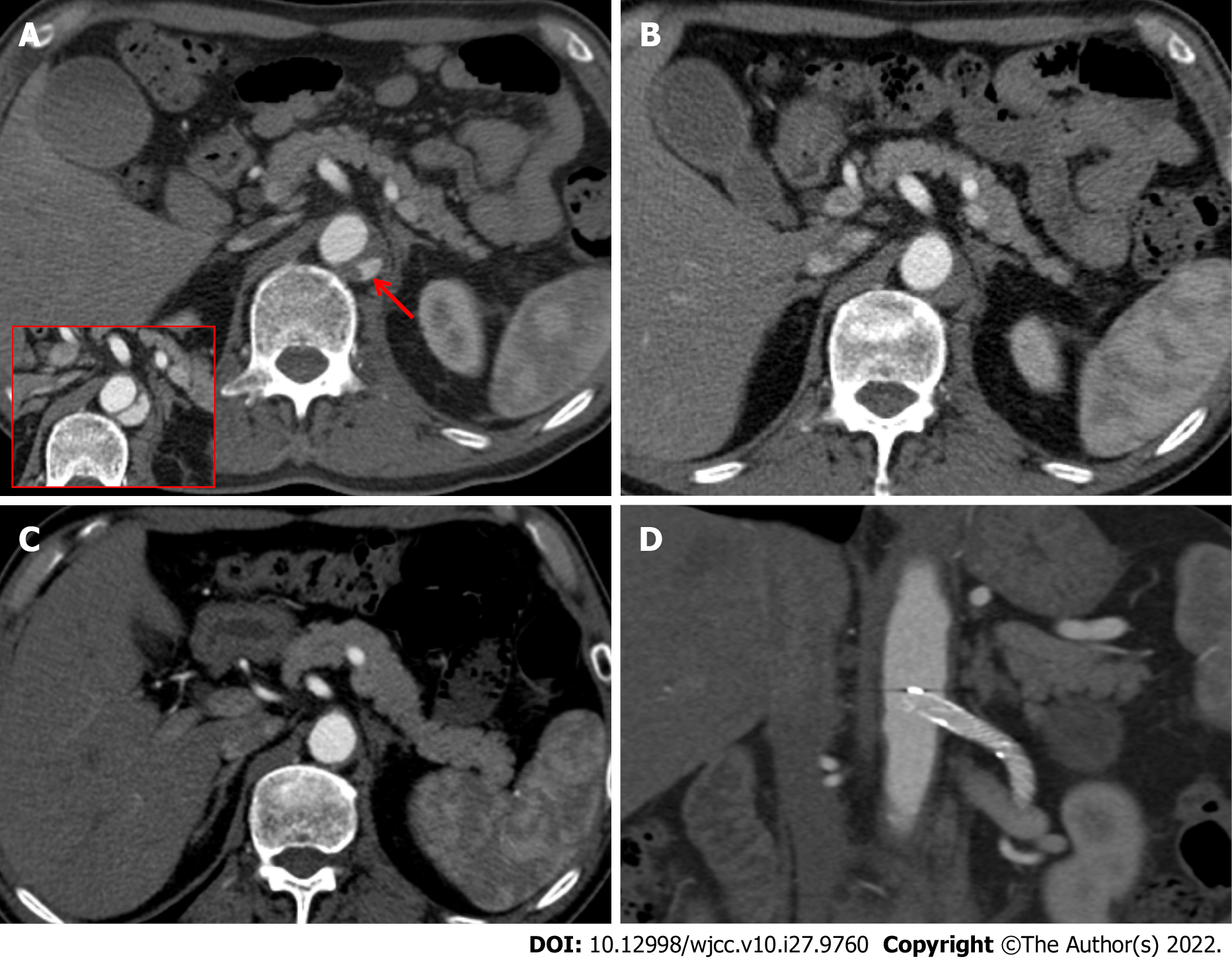
Figure 3 Consecutive computed tomography scans performed 26 d, 45 d, and 7 mo after injury.
A: The computed tomography (CT) scan taken 26 d after injury revealed a decrease in the size and extent of the intramural blood collection (red arrow) compared with the CT scan taken seven days prior (figure in red-border box); B: The CT scan taken 45 d after injury showed that the intramural blood collection markedly decreased in size; C: In the 7-mo CT scan, the intramural hematoma (IMH) had completely disappeared; D: There was no evidence of stent-related complications such as perforation, obstruction, and stent migration.
- Citation: Kim Y, Lee JY, Lee JS, Ye JB, Kim SH, Sul YH, Yoon SY, Choi JH, Choi H. Endovascular treatment of traumatic renal artery pseudoaneurysm with a Stanford type A intramural haematoma: A case report. World J Clin Cases 2022; 10(27): 9760-9767
- URL: https://www.wjgnet.com/2307-8960/full/v10/i27/9760.htm
- DOI: https://dx.doi.org/10.12998/wjcc.v10.i27.9760


