Published online Sep 26, 2022. doi: 10.12998/wjcc.v10.i27.9783
Peer-review started: March 4, 2022
First decision: May 30, 2022
Revised: June 16, 2022
Accepted: August 14, 2022
Article in press: August 14, 2022
Published online: September 26, 2022
Processing time: 196 Days and 2.2 Hours
With aging, four major facial retaining ligaments become elongated, leading to facial sagging and wrinkling. Even though synthetic fillers are popular, however, it cannot address the problems of soft tissue descent alone, and injection of these fillers requires knowledge of the injection technique including the selection of injection sites, the amount of filler, and the dosage used per injection site.
This report aimed to assess the safety and efficacy of a nonsurgical retightening technique to lift and tighten the true ligaments of the face, to improve age-related skin sagging and wrinkling. We objectively quantified the aesthetic lifting effect of a nonsurgical facial retightening procedure that strategically injected high G’ fillers into the base of the true retaining ligaments of the face in two female patients. Facial images were recorded with a three-dimensional facial imaging system for comparison of the clinical outcome. The primary efficacy outcome was the change in facial anthropometric measurements obtained prior to and after injection. The patients were followed for 6 mo after the procedure. Skin retightening was observed, with an evident lift in the orbital, zygomatic, and mandibular regions, and the lifting effect was still observable at the 6-mo follow-up. Few mild adverse events, such as mild-to-moderate pain, tenderness, and itching, occurred during the 1st week after the procedure. No adverse events were reported 1 mo post-procedure.
The results of this study demonstrated that our nonsurgical retightening procedure with strategically placed high G’ fillers achieved quantifiable aesthetic improvements in the orbital, zygomatic, and mandibular regions of two patients. Future research with a larger sample could provide a more in-depth evaluation and validation of the aesthetic improvements observed in this study.
Core Tip: We objectively quantified the aesthetic lifting effect using facial anthropometric measurements. Our results showed direct retightening of the true retaining ligaments by strategically planned high G’ fillers to the base of the ligaments achieved immediate lifting effect.
- Citation: Huang P, Li CW, Yan YQ. Efficacy evaluation of True Lift®, a nonsurgical facial ligament retightening injection technique: Two case reports. World J Clin Cases 2022; 10(27): 9783-9789
- URL: https://www.wjgnet.com/2307-8960/full/v10/i27/9783.htm
- DOI: https://dx.doi.org/10.12998/wjcc.v10.i27.9783
Four major true facial retaining ligaments suspend the superficial muscular aponeurotic system (SMAS) of the face[1]. With aging, elongation of these true ligaments results in facial skin sagging and wrinkling. Recent advances in synthetic fillers, including non-animal stabilized hyaluronic acid, have gained popularity in noninvasive facial lifting and volume restoration procedures[2]. However, volume restoration using fillers alone does not address the problems of soft tissue descent. In addition, administering non-animal stabilized hyaluronic acid as a filler requires experience with this technique, and a satisfactory aesthetic outcome requires precise selection of the injection site as well as knowledge of the injection technique, the amount of filler, and the dosage used per injection site[3].
We have previously published an article describing a novel nonsurgical facial retightening procedure using the True Lift® technique with strategically planned 5-point injections of fillers at the base of facial true retaining ligaments. This resulted in direct tightening of ligaments and indirect lifting of the SMAS layer[4]. In this study, we objectively quantified the aesthetic lifting effect of this facial retightening technique 3 mo and 6 mo after the procedure, using a three-dimensional (3D) photogrammetric facial imaging system (Morpheus 3D® scanner, Morpheus Co., Ltd, Gyeonggi-do, Republic of South Korea)[5].
Two female participants visited our clinic for esthetic improvement of skin firmness.
No concurrent esthetic procedure was given during the study.
They both had prior histories of receiving facial dermatologic procedures for esthetic purposes.
They were 28 years and 29 years of age at the time of the procedure and were healthy with no medical conditions. The esthetic procedures were performed in January 2018.
Three-dimensional facial images were taken with a commercial 3D scanner (Morpheus 3D Scanner®, Morpheus Co., Ltd., Gyeonggi-do, Republic of Korea)[5]. An LED white light was used as the light source in the imaging unit, and the entire scanning procedure took approximately 0.8 sec. The patients were seated with the head in a natural position and with the lips slightly closed. For each subject, 3 images were taken from 3 different horizontal angles (from the front, right, and left sides at a 45-degree angle). These images were then merged into a single 3D facial image.
Both subjects were treated in the same manner. Five injections were made to each side of the face. These injection points are illustrated in Supplementary Figure 1. A total of 1 mL of high G’ filler (Restylane® Lyft Lidocaine, Galderma Laboratories, L.P., Fort Worth, TX, United States) was used on each side of the individual’s face, and the exact amount allocated to each injection site was standardized. High G’ filler was injected with a sharp needle to ensure accurate placement to the base of 5 ligaments (Supple
Briefly, the injection technique for the orbital retaining ligament (point 1) and the zygomatic retaining ligaments (points 2 and 3) involves elevating the skin and soft tissues at these points to expose the base of the ligaments. The filler is then injected perpendicularly to the periosteum using a sharp needle to ensure accurate placement at the base of the ligaments. Injection of the buccal-maxillary retaining ligament (point 4) does not require any additional elevation of the skin and soft tissues in this region. The filler is placed at the base of the buccal-maxillary retaining ligament with a needle at a 45-degree angle to the canine fossa. Injection of the mandibular retaining ligament (point 5) is similar to injection at point 4. The bolus injection is placed at the base of this ligament, just medial to the Marionette lines near the base of the mandibular border.
Before and after comparison of the 3D facial images taken with the commercial 3D scanner were performed using superimposition evaluation. Measurements taken between each facial landmark were then made on 3D facial images using the Morpheus 3D “line-length” tool, which enables measurements of the direct distance between two points. Photogrammetric analysis was performed by a single trained operator.
The primary outcome was the change in facial anthropometric measurements, based on the images taken with the 3D facial imaging system. After a baseline measurement (T0), the measurements were made at 3 time points after the injections: immediately after the procedure (T1); at 3 mo (T2); and at 6 mo (T3). Then, the facial anthropometric measurements representing improvements in the orbital, zygomatic, and mandibular regions were determined. The secondary outcome was any self-reported post-injection adverse event (AE), which were recorded after each return visit (V) for 14 consecutive days: V1 at 14 d; V2 at 1 mo; V3 at 3 mo; and V4 at 6 mo post-treatment.
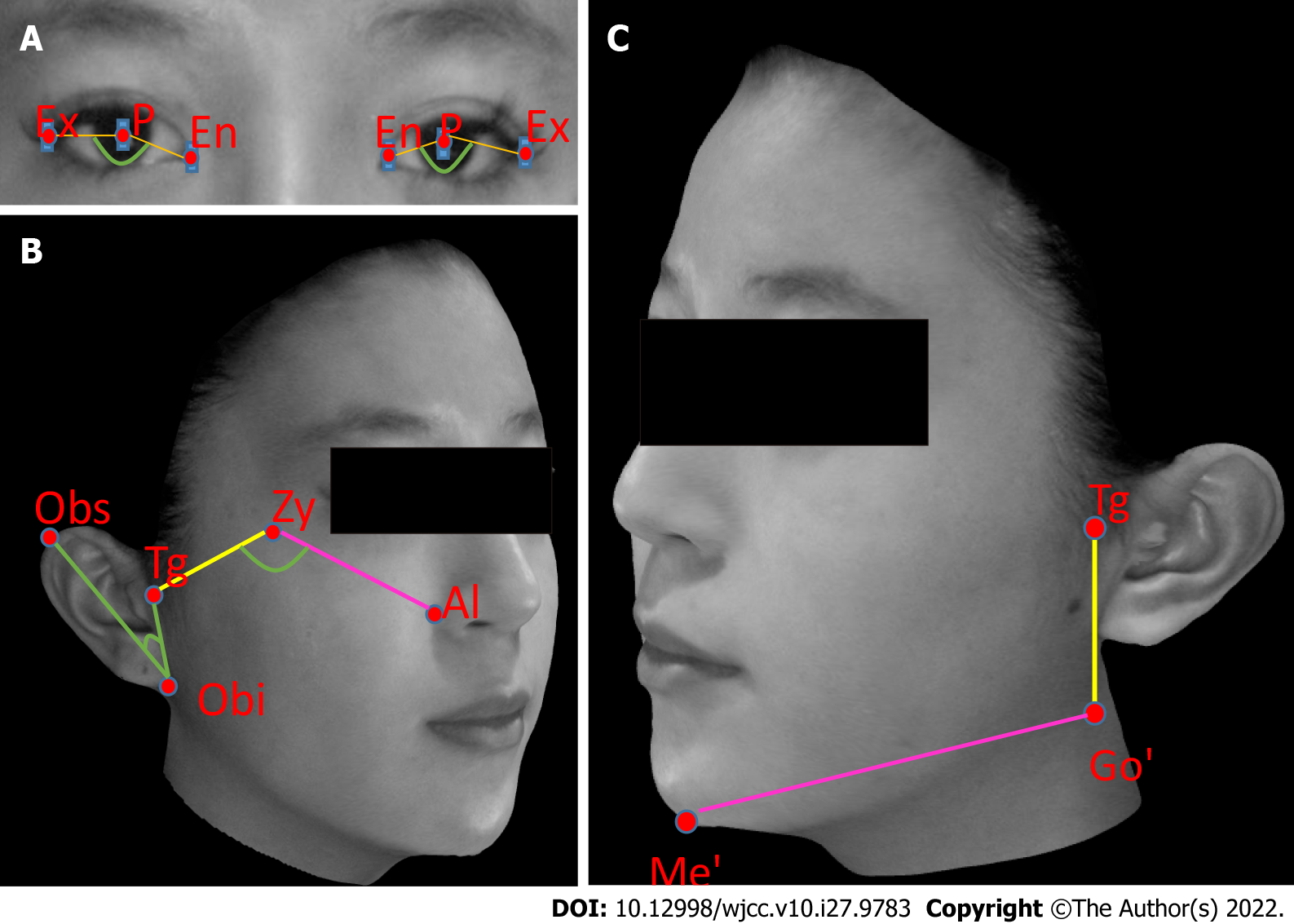
Retightening in the orbital region was defined by the angle formed between landmarks endocanthion, pupil, and exocanthion. Retightening in the zygomatic region (mid-face) was defined by the angle formed by tragion (Tg)-zygion-ala. Retightening in the mandibular region was defined by measuring the distance between soft tissue gonion (Go’) and soft tissue menton. The reference for the mid-face was defined by the angle formed between landmarks otobasion superius-otobasion inferius–Tg. The reference for the lower face was defined by the distance between landmark Tg to soft tissue Go’. These landmarks are illustrated in Figure 1.
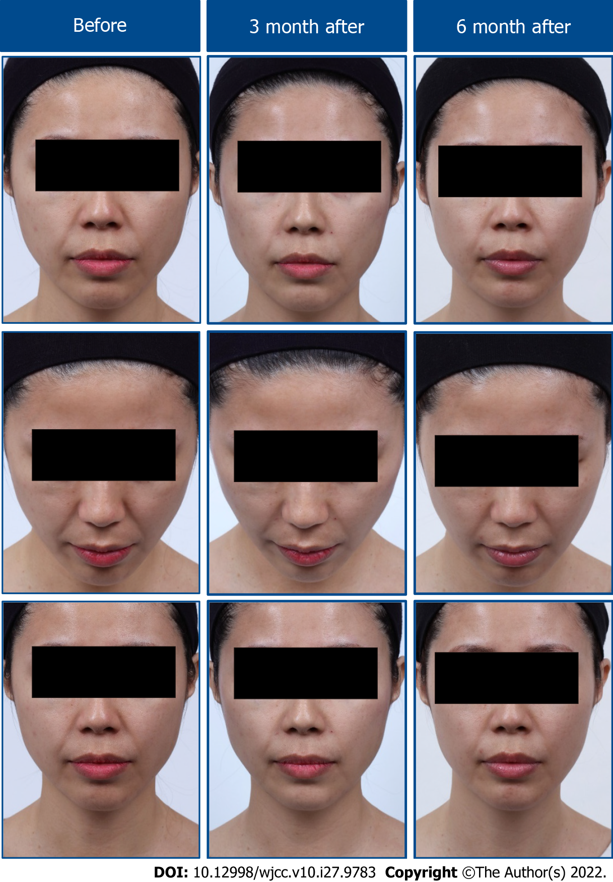
In both patients, the angle formed by exocanthion-pupil-endocanthion increased immediately after the procedure (T1) compared to the angle at baseline (T0). This represented retightening of the orbital retaining ligaments, and the increase in the angle was still evident at the final follow-up visit 6 mo post-injection (Table 1). The angle formed by Tg-zygion-ala decreased immediately after the procedure, and lasted until the final observation point in both cases, demonstrating the lifting effects in the zygomatic region (Table 1). The distance between Go’ and soft tissue menton was substantially shortened, demonstrating retightening in the mandibular retaining ligaments, and these improvements lasted for at least 6 mo (Table 1). In contrast, the reference angle (otobasion superius-otobasion inferius-Tg) and distance (Tg-Go’) used in the mid- and lower face, respectively, were unchanged by the treatment procedure. Representative photographs of changes in the patients’ facial contours before and after the procedure are shown in Figure 2. Post-injection AE, including mild-to-moderate bruising, pain, tenderness, and itching, were reported in both subjects during the 1st week after the procedure. No other AEs of any grades were noted 1 mo later or in any later follow-up visits (Supplementary Table 2).
| T0 (baseline) | T1 | T2 | T3 | Change in angle | ||||
| (post tx) | (3 mo) | (6 mo) | T0 to T1 | T0 to T2 | T0 to T3 | |||
| Orbital ligament | ||||||||
| Ex-P-En angle in degrees | ||||||||
| Right eye | Case 1 | 140.5 | 149.4 | 148.8 | 147.9 | 8.9 | 8.3 | 7.4 |
| Case 2 | 140.0 | 145.3 | 144.7 | 146.7 | 5.3 | 4.7 | 6.7 | |
| Left eye | Case 1 | 143.3 | 150.2 | 150.1 | 149.1 | 6.9 | 6.8 | 5.8 |
| Case 2 | 143.3 | 147.4 | 146.0 | 148.0 | 4.1 | 2.7 | 4.7 | |
| Zygomatic ligament | ||||||||
| Tg-Zy-Al angle in degrees | ||||||||
| Right cheek | Case 1 | 122.1 | 109.9 | 113.4 | 115.6 | -12.2 | -8.7 | -6.5 |
| Case 2 | 122.1 | 111.1 | 111.7 | 112.4 | -11.0 | -10.4 | -9.7 | |
| Left cheek | Case 1 | 116.3 | 110.1 | 111.8 | 112.5 | -6.2 | -4.5 | -3.8 |
| Case 2 | 128.9 | 120.6 | 121.1 | 123.6 | -8.3 | -7.8 | -5.3 | |
| Obs-Obi-Tg angle in degrees | ||||||||
| Right ear | Case 1 | 39.6 | 39.6 | 39.6 | 39.6 | 0 | 0 | 0 |
| Case 2 | 45.6 | 45.6 | 45.6 | 45.6 | 0 | 0 | 0 | |
| Left ear | Case 1 | 47.7 | 47.7 | 47.7 | 47.7 | 0 | 0 | 0 |
| Case 2 | 47.7 | 47.7 | 47.7 | 47.7 | 0 | 0 | 0 | |
| Mandibular retaining ligament | ||||||||
| Go’-Me’ curved distance in mm | ||||||||
| Right cheek | Case 1 | 110.0 | 103.5 | 104.7 | 106.1 | -6.5 | -5.3 | -3.9 |
| Case 2 | 110.0 | 103.5 | 104.2 | 104.9 | -6.5 | -5.8 | -5.1 | |
| Left cheek | Case 1 | 108.3 | 101.2 | 103.2 | 104.8 | -7.1 | -5.1 | -3.5 |
| Case 2 | 105.9 | 99.8 | 100.8 | 101.7 | -6.1 | -5.1 | -4.2 | |
| Tg-Go’ curved distance in mm | ||||||||
| Right ear | Case 1 | 49.8 | 49.8 | 49.8 | 49.8 | 0 | 0 | 0 |
| Case 2 | 49.8 | 49.8 | 49.8 | 49.8 | 0 | 0 | 0 | |
| Left ear | Case 1 | 50.8 | 50.8 | 50.8 | 50.8 | 0 | 0 | 0 |
| Case 2 | 61.9 | 61.9 | 61.9 | 6.19 | 0 | 0 | 0 | |
Results from this study illustrated visible and lasting aesthetic improvements in the orbital, zygomatic, and mandibular regions, which are major focuses when addressing SMAS sagging from facial aging[6]. As in previous reports[7], there was no AE suggesting hypersensitivity in our patients, and no AEs were reported 1 wk post-injection.
Facial aging is a multifactorial process, involving displacement of fat compartments, elongation of the facial ligaments, attenuation of the SMAS layer, loss of bony support, and thinning of subcutaneous and dermal tissues[4]. In the early stage of facial aging, elongation of the facial ligaments results in descent of the overlying tissues and hence sagging of the skin.
Our novel nonsurgical facial retightening procedure is aimed at addressing the structural descent of the soft tissues due to facial ligament laxity. This is accomplished by injecting high G’ filler at the base of the true retaining ligaments. Because this technique is based on retightening of the true retaining ligaments, it is suitable for improving facial laxity during the early aging process but is not suitable for mature patients at the later stages of the facial aging process. Instead, later in the aging process rejuvenation requires more than facial retightening, such as facial volume restoration or correction of bony support.
The success of this facial retightening procedure is dependent upon the precision of injections of high G’ fillers into the base of the true retaining ligaments. Therefore, a high level of skill, experience, and an excellent knowledge of facial anatomy are required to achieve optimum placement of the filler.
Limitations of this study included the small sample size and the age of the participants. The two patients in this study were relatively young and likely to be in very early stages of facial aging, which may partly explain the observed improvements. The aesthetic improvements observed in our study should be validated with a larger sample size. In addition, the effectiveness of the procedure for patients with different stages of facial aging should also be investigated in the future.
This report demonstrated that application of a nonsurgical retightening procedure with use of a high G’ filler achieved quantifiable aesthetic improvements in the orbital, zygomatic, and mandibular regions in two female patients. Future research with a larger sample size could provide a more in-depth evaluation and could better substantiate the validity of the aesthetic improvements observed in this study.
Provenance and peer review: Unsolicited article; Externally peer reviewed.
Peer-review model: Single blind
Specialty type: Dermatology
Country/Territory of origin: Taiwan
Peer-review report’s scientific quality classification
Grade A (Excellent): A
Grade B (Very good): B
Grade C (Good): C
Grade D (Fair): 0
Grade E (Poor): E
P-Reviewer: Anzola F LK, Colombia; Liu L, China; Wang P, China; Zhang YN, China S-Editor: Ma YJ L-Editor: Filipodia P-Editor: Ma YJ
| 1. | Kang MS, Kang HG, Nam YS, Kim IB. Detailed anatomy of the retaining ligaments of the mandible for facial rejuvenation. J Craniomaxillofac Surg. 2016;44:1126-1130. [RCA] [PubMed] [DOI] [Full Text] [Cited by in Crossref: 8] [Cited by in RCA: 8] [Article Influence: 0.9] [Reference Citation Analysis (0)] |
| 2. | Weiss RA, Moradi A, Bank D, Few J, Joseph J, Dover J, Lin X, Nogueira A, Mashburn J. Effectiveness and Safety of Large Gel Particle Hyaluronic Acid With Lidocaine for Correction of Midface Volume Deficit or Contour Deficiency. Dermatol Surg. 2016;42:699-709. [RCA] [PubMed] [DOI] [Full Text] [Cited by in Crossref: 16] [Cited by in RCA: 21] [Article Influence: 2.3] [Reference Citation Analysis (0)] |
| 3. | Wu W, Carlisle I, Huang P, Ribé N, Russo R, Schaar C, Verpaele A, Strand A. Novel administration technique for large-particle stabilized hyaluronic acid-based gel of nonanimal origin in facial tissue augmentation. Aesthetic Plast Surg. 2010;34:88-95. [RCA] [PubMed] [DOI] [Full Text] [Cited by in Crossref: 10] [Cited by in RCA: 8] [Article Influence: 0.5] [Reference Citation Analysis (0)] |
| 4. | Huang P. The True Lift Technique™: facial ligament retightening, an anatomical approach. PMFA Journal. 2018;5. |
| 5. | Kim SH, Jung WY, Seo YJ, Kim KA, Park KH, Park YG. Accuracy and precision of integumental linear dimensions in a three-dimensional facial imaging system. Korean J Orthod. 2015;45:105-112. [RCA] [PubMed] [DOI] [Full Text] [Full Text (PDF)] [Cited by in Crossref: 24] [Cited by in RCA: 29] [Article Influence: 2.9] [Reference Citation Analysis (0)] |
| 6. | de Maio M, Wu WTL, Goodman GJ, Monheit G; Alliance for the Future of Aesthetics Consensus Committee. Facial Assessment and Injection Guide for Botulinum Toxin and Injectable Hyaluronic Acid Fillers: Focus on the Lower Face. Plast Reconstr Surg. 2017;140:393e-404e. [RCA] [PubMed] [DOI] [Full Text] [Cited by in Crossref: 47] [Cited by in RCA: 64] [Article Influence: 8.0] [Reference Citation Analysis (0)] |
| 7. | Hamilton RG, Strobos J, Adkinson NF Jr. Immunogenicity studies of cosmetically administered nonanimal-stabilized hyaluronic acid particles. Dermatol Surg. 2007;33 Suppl 2:S176-S185. [RCA] [PubMed] [DOI] [Full Text] [Cited by in Crossref: 13] [Cited by in RCA: 16] [Article Influence: 0.9] [Reference Citation Analysis (0)] |


