Published online Sep 26, 2022. doi: 10.12998/wjcc.v10.i27.9641
Peer-review started: May 6, 2022
First decision: May 30, 2022
Revised: June 11, 2022
Accepted: August 15, 2022
Article in press: August 15, 2022
Published online: September 26, 2022
Processing time: 133 Days and 1 Hours
The tibial stop of anterior cruciate ligament (ACL) is fan-shaped and attached to the medial groove in front of the intercondylar spine, which is located between the anterior horn of the medial and lateral meniscus. The incidence of this fracture is low previously reported, which is common in children and adolescents. With the increase of sports injury and traffic injury and the deepening of under-standing, it is found that the incidence of the disease is high at present.
To explore the difference between open reduction and internal fixation with small incision and high-intensity non-absorbable suture under arthroscopy in the treatment of tibial avulsion fracture of ACL.
Seventy-six patients with tibial avulsion fracture of anterior cruciate ligament diagnosed and treated in Guanyun County People's Hospital from April 2018 to June 2020 were retrospectively analyzed. According to the surgical methods, they were divided into group A (40 cases) and group B (36 cases). Patients in group A were treated with arthroscopic high-strength non-absorbable suture, and patients in group B were treated with small incision open reduction and internal fixation. The operation time, fracture healing time, knee joint activity and functional score before and after operation, and surgical complications of the two groups were compared.
The operation time of group A was higher than that of group B, and the difference was statistically significant (P < 0.05); the fracture healing time of group A was compared with that of group B, and the difference was not statistically significant (P > 0.05); The knee joint function activity was compared between two groups before operation, 3 mo and 6 mo after operation, and the difference was not statistically significant (P > 0.05); the knee joint function activity of group A and group B at 3 mo and 6 mo after operation was significantly higher than that before operation (P < 0.05); the limp, support, lock, instability, swelling, upstairs, squatting, pain and Lysholm score were compared between the two groups before and 6 mo after operation, and the difference was not statistically significant (P > 0.05); the scores of limp, support, lock, instability, swelling, upstairs, squatting, pain and Lysholm in group A and group B at 6 mo after operation were significantly higher than those before operation (P > 0.05); the surgical complication rate of group A was 2.63%, which was lower than 18.42% of group B, and the difference was statistically significant (P > 0.05).
Both small incision open reduction and internal fixation and arthroscopic high-strength non-absorbable sutures can achieve good results in the treatment of anterior cruciate ligament tibial avulsion fractures. The operation time of arthroscopic high-strength non-absorbable sutures is slightly longer, but the complication rate is lower.
Core Tip: Anterior cruciate ligament tibial avulsion fracture is a common type of anterior cruciate ligament injury, which has a serious impact on the quality of life and physical and mental health of patients. In this study, we compared the effect of small incision open reduction and internal fixation and arthroscopic high-strength non-absorbable suture in the treatment of tibial avulsion fracture of anterior cruciate ligament, in order to provide a basis for clinical practice.
- Citation: Niu HM, Wang QC, Sun RZ. Therapeutic effect of two methods on avulsion fracture of tibial insertion of anterior cruciate ligament. World J Clin Cases 2022; 10(27): 9641-9649
- URL: https://www.wjgnet.com/2307-8960/full/v10/i27/9641.htm
- DOI: https://dx.doi.org/10.12998/wjcc.v10.i27.9641
Anterior cruciate ligament tibial avulsion fracture is a common type of anterior cruciate ligament injury, which has a serious impact on the quality of life and physical and mental health of patients. Due to the difficulty of closed reduction in patients with tibial avulsion fracture, anterior cruciate ligament injury is often accompanied by medial meniscus rupture, and early surgical treatment is necessary[1]. The traditional treatment method is to take open reduction and internal fixation for repair, which can achieve the anatomical reconstruction effect in the reconstruction of cruciate ligament and restore the stability of patients’ joints. However, it is impossible to observe the posterior horn of meniscus during the operation. Therefore, X-ray observation needs to be repeated during the operation, and some patients may damage the epiphysis during the treatment. With the popularization of arthroscopy, the intraoperative trauma to patients is smaller, but the traditional Kirschner wire fixation method has poor fixation effect, while the effect of absorbable suture on the epiphysis of patients is slight, and the fixation effect is better. However, there are different opinions on how to choose the two surgical methods in clinic[2]. Therefore, this study compared the effect of small incision open reduction and internal fixation and arthroscopic high-strength non-absorbable suture in the treatment of tibial avulsion fracture of anterior cruciate ligament, in order to provide a basis for clinical practice.
A total of 76 patients with tibial avulsion fracture of anterior cruciate ligament diagnosed and treated in Yunxian People’s Hospital from April 2018 to June 2020 were selected for retrospective analysis. According to the surgical methods, they were divided into group A (n = 40) and group B (n = 36).
Inclusion criteria: (1) Patients aged 19-65 years; (2) Patients with tibial avulsion fracture of anterior cruciate ligament caused by trauma were confirmed by X-ray, computed tomography and magnetic resonance imaging after admission; (3) Patients had obvious lower limb pain on the affected side during hospitalization and limited activity. The interval between injury and operation was within 2 wk; (4) The classification standard was type II or type III (Meyers-McKeever standard)[3]; (5) All operations were performed by the same group of orthopedic medical staff in our hospital; and (6) The research programme receives informed consent from patients and their families. Exclusion criteria: (1) Severe osteoporosis; (2) Patients with long-term hormone therapy; (3) Cancer patients; (4) Bone tuberculosis; (5) A history of drug use or addiction; and (6) Lower limb nerve and muscle atrophy disease.
Group A: Patients were treated with high-intensity non-absorbable suture under arthroscopy. Patients were placed in supine position after general anesthesia satisfaction, and the articular cartilage, meniscus and anterior cruciate ligament were explored. After placement of arthroscopic instruments, the posterior medial and posterolateral approaches were established to expose the stokes fracture block of anterior cruciate ligament. After inserting probes into the lateral approach, the fracture block was pried and reset. The blood clot was cleared by planer. A 2 cm incision was performed on the lateral side of tibial nodules. The insertion locator was used to drill the bone tunnel from the lateral side of tibial nodules along the anterior cruciate ligament, and another bone tunnel was drilled under the outside of the fracture surface. The tendon line was introduced through the tunnel to pass through the joint. The knotter was used to knot at the root of the anterior cruciate ligament, and the tunnel under the fracture block was passed to the outside of the joint. The fracture block was reset by arthroscopy, and the bone bridge between the outer mouth of the tunnel was knotted at the risk of tendon tightening.
Group B: Open reduction and internal fixation with small incision was performed, and the prone position was taken when the patient was satisfied with anesthesia. A 3-4 cm incision was made in the transverse striation of the median popliteal fossa posterior to the knee. The subcutaneous tissue and fascia were separated to expose the gap between the medial head and the lateral head of the gastrocnemius muscle. The lateral head of the gastrocnemius muscle and the vascular nerve of the popliteal fossa were pulled by the hook. The medial head of the gastrocnemius muscle was pulled inward by the hook. The posterior articular capsule was exposed, and the fracture block was reset after exposure. The Kirschner wire was used to vertical fracture line for temporary fixation. X-ray confirmed that the fracture reduction was satisfactory. The hollow nail was screwed along the Kirschner wire to perform compression fixation, and the incision was sutured to end the operation.
The operation time, fracture healing time, knee joint activity and functional score before and after operation, and surgical complications of the two groups were compared.
Knee joint function evaluation using Lysholm score[4], Lysholm score scale, claudication total score of 5 points, support total score of 5 points, lock total score of 15 points, instability total score of 25 points, swelling total score of 10 points, upstairs total score of 10 points, squat total score of 5 points, pain total score of 25 points, were scored according to the scale grading standard.
In this study, the measurement indexes such as operation time and fracture healing time were tested by normal distribution, which were in line with approximate normal distribution or normal distribution, and expressed as mean ± SD. The t test was used for comparison between the two groups. Non-counting data were represented by percentage, and χ2 test was used for comparison. Professional SPSS 21.0 software for data processing, test level α = 0.05.
Age, height, weight, time from injury to operation, gender, affected side distribution, Meyers-McKeever classification and tibial instability were compared between group A and group B, and the difference was not statistically significant (P > 0.05) (Table 1).
| Basic information | Group A (n = 40) | Group B (n = 38) | t/χ2 value | P value |
| Age (yr) | 36.4 ± 8.5 | 38.4 ± 9.0 | -1.009 | 0.316 |
| Height (cm) | 167.8 ± 4.6 | 167.2 ± 5.0 | 0.552 | 0.583 |
| Weight (kg) | 65.1 ± 6.8 | 67.4 ± 7.2 | -1.451 | 0.151 |
| Time from injury to surgery (d) | 7.4 ± 2.1 | 6.9 ± 1.7 | 1.152 | 0.253 |
| Sex | 0.601 | 0.438 | ||
| Male | 24 (60.00) | 26 (68.42) | ||
| Female | 16 (40.00) | 12 (31.58) | ||
| Affected side distribution | 1.990 | 0.158 | ||
| Left side | 20 (50.00) | 25 (65.79) | ||
| Right | 20 (50.00) | 13 (34.21) | ||
| Meyers-McKeever type | 0.020 | 0.887 | ||
| Type II | 29 (72.50) | 27 (71.05) | ||
| Type III | 11 (27.50) | 11 (28.95) | ||
| Tibia instability | 0.690 | 0.406 | ||
| Stage II | 26 (65.00) | 28 (73.68) | ||
| Stage III | 14 (35.00) | 10 (26.32) |
The operation time of group A was higher than that of group B, and the difference was statistically significant (P < 0.05). The fracture healing time of group A was compared with that of group B, and the difference was not statistically significant (P > 0.05) (Table 2).
| Groups | n | Operation time (min) | Fracture healing time (wk) |
| Group A | 40 | 108.5 ± 18.4 | 12.5 ± 1.4 |
| Group B | 38 | 59.2 ± 11.7 | 12.8 ± 1.5 |
| t value | 14.037 | -0.914 | |
| P value | 0.000 | 0.364 |
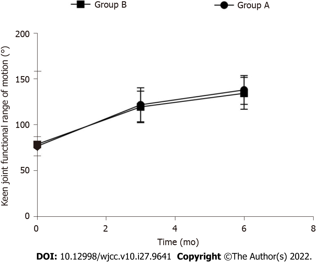
The knee joint functional activity was compared between the two groups before operation, 3 mo and 6 mo after operation, and the difference was not statistically significant (P > 0.05). The knee joint function activity of group A and group B at 3 mo and 6 mo after operation was significantly higher than that before operation (P < 0.05) (Table 3, Figure 1).
The total scores of lameness, support, strangulation, instability, swelling, upstairs, squat, pain and Lysholm were compared between the two groups before and 6 mo after operation, and the difference was not statistically significant (P > 0.05). The scores of claudication, support, lock, instability, swelling, upstairs, squatting, pain and Lysholm in group A and group B at 6 mo after operation were significantly higher than those before operation (P < 0.05) (Table 4).
| Project | Preoperative | t value | P value | 6 months after surgery | t value | P value | ||
| Group A (n = 40) | Group B (n = 38) | Group A (n = 40) | Group B (n = 38) | |||||
| Limp | 1.88 ± 0.30 | 1.94 ± 0.45 | -0.696 | 0.488 | 3.92 ± 0.74a | 4.11 ± 0.80a | -1.090 | 0.279 |
| Support | 1.90 ± 0.41 | 1.98 ± 0.48 | -0.793 | 0.430 | 4.13 ± 0.68a | 4.26 ± 0.88a | -0.732 | 0.466 |
| Winch | 5.84 ± 1.20 | 6.12 ± 1.32 | -0.981 | 0.330 | 11.30 ± 2.57a | 10.93 ± 2.83a | 0.605 | 0.547 |
| Unstable | 12.67 ± 2.48 | 11.88 ± 2.27 | 1.465 | 0.147 | 20.38 ± 3.70a | 21.03 ± 3.58a | -0.788 | 0.433 |
| Swelling | 4.41 ± 0.86 | 4.67 ± 0.90 | -1.305 | 0.196 | 8.81 ± 0.89a | 8.48 ± 1.03a | 1.516 | 0.134 |
| Go upstairs | 3.36 ± 0.78 | 3.62 ± 0.85 | -1.409 | 0.163 | 7.96 ± 1.14a | 8.21 ± 1.26a | -0.920 | 0.361 |
| Squat | 1.67 ± 0.41 | 1.80 ± 0.48 | -1.288 | 0.202 | 3.88 ± 1.03a | 4.02 ± 0.89a | -0.641 | 0.524 |
| Pain | 13.52 ± 2.96 | 12.81 ± 2.56 | 1.130 | 0.262 | 20.86 ± 3.02a | 21.32 ± 3.31a | -0.642 | 0.523 |
| Total score | 45.25 ± 7.33 | 44.82 ± 6.81 | 0.268 | 0.789 | 81.24 ± 9.25a | 82.36 ± 8.90a | -0.544 | 0.588 |
The complication rate of group A was 2.63% lower than that of group B 18.42%, and the difference was statistically significant (P < 0.05) (Table 5).
| Groups | n | Incision infection | Loose internal fixation | Venous thrombosis of lower extremity | Complication rate |
| Group A | 40 | 0 | 0 | 1 | 1 (2.63) |
| Group B | 38 | 2 | 2 | 3 | 7 (18.42) |
| χ2 value | 5.367 | ||||
| P value | 0.021 |
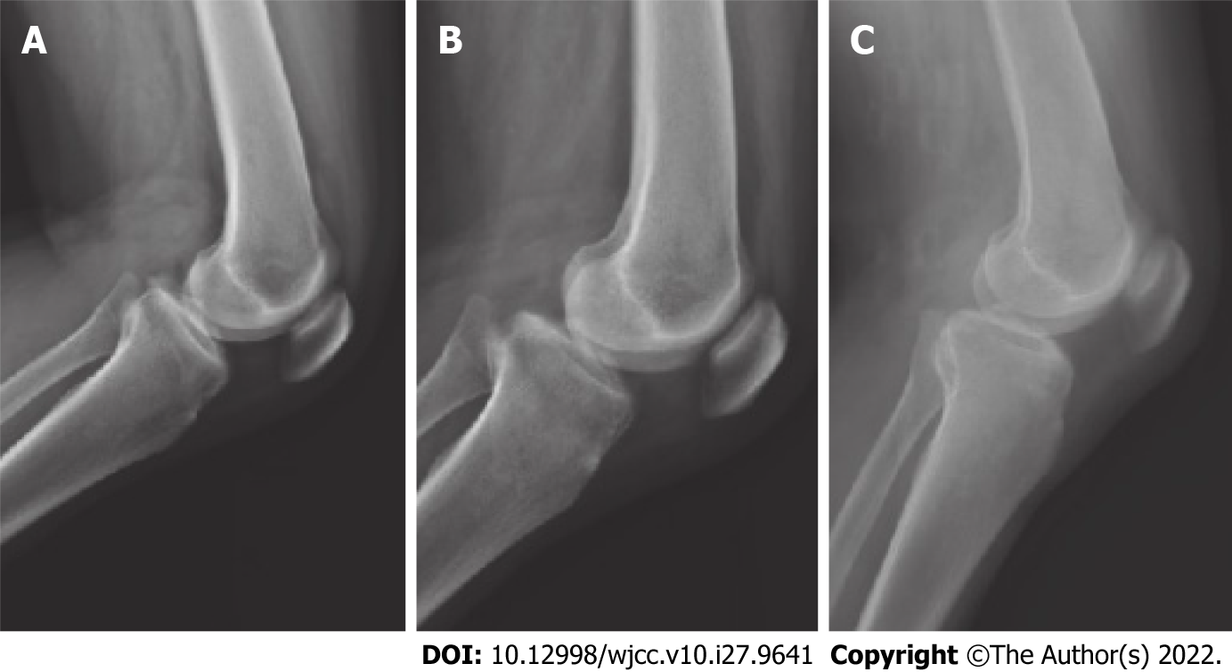
Two typical cases of tibial avulsion fracture of the left anterior cruciate ligament of the knee were reviewed (Figure 2 and Figure 3).
Avulsion fracture of tibial insertion of anterior cruciate ligament is generally caused by trauma, which causes avulsion injury of tibial attachment of anterior cruciate ligament. Children and adolescents are the most common in clinical practice. The incidence has increased in recent years, which has a serious impact on the quality of life and physical and mental health of patients[5]. The incidence of avulsion fracture of tibial insertion in the anterior cruciate ligament is lower than that of the belt body. Generally, violence causes excessive extension of the knee joint or excessive internal rotation and abduction of the tibia. At the same time, the strong contraction of the quadriceps femoris exceeds the maximum tension of the anterior cruciate ligament, which leads to the rupture of the anterior cruciate ligament pa
At present, with the development of minimally invasive technology, arthroscopic treatment has become a commonly used treatment method in clinical practice. In this study, the operation time of group A was higher than that of group B, indicating that arthroscopic treatment of tibial avulsion fracture of anterior cruciate ligament with high-intensity non-absorbable suture was more complicated, so the operation time was prolonged. However, there was no difference in the fracture healing time between the two groups, indicating that the two surgical methods had similar fracture healing effects. Arthroscopic surgery without incision, clear visual field under the microscope, can avoid nerve and vascular injury and have smaller trauma for patients, but the operation is relatively complex, so the operation time is prolonged. In the process of operation, it can deal with intra-articular meniscus, other ligaments and other structural damage[8,9]. In this study, the absorbable suture operation was a soft fixation material. The suture had a certain elasticity and high mechanical strength, so it was better in line with the principle of biological fixation. The fracture fixation was strong, and the patients recovered quickly after operation, which could be recovered early[10].
This study also found that two groups of patients after 3 mo, 6 mo of knee joint function activity than before surgery were significantly increased, knee function score than before surgery were markedly increased, suggesting that the two surgical methods in improving knee joint activity and recovery of knee function in patients with similar effect[11-13]. Previous studies have confirmed that the two surgical methods have no effect on the medium-term efficacy of patients. The knee joint function recovery after suture fixation and screw fixation is ideal, and the difference between the both is not statistically significant, which is consistent with the results of this study[14,15]. In this study, the surgical complication rate of group A was 2.63%, which was lower than that of group B (18.42%), suggesting that arthroscopic high-strength non-absorbable suture in the treatment of tibial avulsion fracture of anterior cruciate ligament can reduce the incidence of surgical complications. The suture technique can adjust the direction of the suture by controlling the direction of the bone tunnel and the outlet position of the bone tunnel joint above the epiphyseal line, which helps to enhance the accuracy and stability of the reduction, and ensures the firm fixation of the bone block. And it can adjust the tension of the ligament twice to reduce the occurrence of surgical complications[16,17]. Some scholars reported that in the fixation of anterior cruciate ligament fractures, the second band-line anchor was first fixed on both sides of the central slightly deviated back of the tibial bone bed by suture technique, and the outer row of one anchor was fixed to the anterior and lateral sides of the bone bed. The position of the outer row of anchor was adjusted according to the reduction status of the fracture block. The fracture block was fixed by suture intersection ‘fan type’ compression to avoid the front and back ends of the fracture block[18-20].
In this study, the effects of two surgical methods on tibial avulsion fracture of anterior cruciate ligament were compared, and it was confirmed that arthroscopic non-absorbable suture surgery can significantly reduce the occurrence of surgical complications. High-strength sutures have certain elasticity, and the mechanical strength is large. The bone has micro-motion, which is in line with the current treatment principle of biological fixation. The fracture fixation is strong, indicating that the clinical operation can be reasonably selected according to the actual situation of patients. However, the number of cases included in this study is small, and there may be some bias in the selection of cases, forming a certain bias on the surgical results. Moreover, this study failed to take long-term follow-up for patients. Although both surgical methods have achieved satisfactory early clinical efficacy, large sample size and long-term follow-up are needed to provide more reliable conclusions.
To sum up, both small incision open reduction and internal fixation and arthroscopic high-intensity non-absorbable suture can achieve good results in the treatment of tibial avulsion fracture of anterior cruciate ligament. The operation time of arthroscopic high-intensity non-absorbable suture is slightly longer, but the complication rate is lower.
With the increase of sports injury and traffic injury and the deepening of understanding, it is found that the incidence of the disease is high at present.
In this study, the therapeutic effect of two methods on avulsion fracture of tibial insertion of anterior cruciate ligament was investigated.
This study aimed to compare the difference between these two operations for the treatment of tibial avulsion fractures of the anterior cruciate ligament.
Patients were treated with high-intensity non-absorbable suture under arthroscopy.
Both small incision open reduction and internal fixation and arthroscopic high-strength non-absorbable sutures can achieve good results in the treatment of anterior cruciate ligament tibial avulsion fractures.
The operation time of arthroscopic high-intensity non-absorbable suture is slightly longer, but the complication rate is lower.
There are different opinions on how to choose between the two surgical methods in clinical practice. More studies are required in the future.
Provenance and peer review: Unsolicited article; Externally peer reviewed.
Peer-review model: Single blind
Specialty type: Orthopedics
Country/Territory of origin: China
Peer-review report’s scientific quality classification
Grade A (Excellent): 0
Grade B (Very good): B
Grade C (Good): C
Grade D (Fair): 0
Grade E (Poor): 0
P-Reviewer: Burden AM, Switzerland; Palacios S, Spain S-Editor: Wang JL L-Editor: A P-Editor: Wang JL
| 1. | Niu WJ, Huang LA, Zhou X, Yang YF, Liang HR, Song WJ, Liu Y, Duan WP. [Clinical effects of arthroscopy-assisted anterior cruciate ligament tibial eminence avulsion fracture compared with traditional open surgery:a Meta-analysis]. Zhongguo Gu Shang. 2022;35:292-299. [RCA] [PubMed] [DOI] [Full Text] [Cited by in RCA: 1] [Reference Citation Analysis (2)] |
| 2. | Lu HD, Zeng C, Dong YX, Cai DZ, Wen XY. [Treatment of tibial avulsion fracture of the posterior cruciate ligament with open reduction and steel-wire internal fixation]. Zhongguo Gu Shang. 2011;24:195-198. [PubMed] |
| 3. | Everhart JS, Klitzman RG. Editorial Commentary: Suspensory Fixation of Displaced Tibial Posterior Cruciate Ligament Avulsions: A Novel Application of a Familiar Technique. Arthroscopy. 2021;37:1881-1882. [RCA] [PubMed] [DOI] [Full Text] [Cited by in Crossref: 1] [Cited by in RCA: 1] [Article Influence: 0.3] [Reference Citation Analysis (0)] |
| 4. | Mao X, Hong Q, You R, Lu Y, Zhao F. Research on Influencing Factors of Clinical Efficacy of Meniscus Resection Based on Logistic Regression Analysis. Scanning. 2022;2022:4606139. [RCA] [PubMed] [DOI] [Full Text] [Cited by in RCA: 1] [Reference Citation Analysis (0)] |
| 5. | Gobbi A, Herman K, Grabowski R, Szwedowski D. Primary Anterior Cruciate Ligament Repair With Hyaluronic Scaffold and Autogenous Bone Marrow Aspirate Augmentation in Adolescents With Open Physes. Arthrosc Tech. 2019;8:e1561-e1568. [RCA] [PubMed] [DOI] [Full Text] [Full Text (PDF)] [Cited by in Crossref: 8] [Cited by in RCA: 8] [Article Influence: 1.3] [Reference Citation Analysis (0)] |
| 6. | Acebrón-Fabregat Á, Pino-Almero L, López-Lozano R, Mínguez-Rey M. [Treatment and evolution of chronic avulsion of the anterior tibial spine in the pediatric age]. Acta Ortop Mex. 2019;33:96-101. [PubMed] |
| 7. | Mutchamee S, Ganokroj P. Arthroscopic Transosseous Suture-bridge Fixation for Anterior Cruciate Ligament Tibial Avulsion Fractures. Arthrosc Tech. 2020;9:e1607-e1611. [RCA] [PubMed] [DOI] [Full Text] [Full Text (PDF)] [Cited by in Crossref: 7] [Cited by in RCA: 7] [Article Influence: 1.4] [Reference Citation Analysis (0)] |
| 8. | Leie M, Heath E, Shumborski S, Salmon L, Roe J, Pinczewski L. Midterm Outcomes of Arthroscopic Reduction and Internal Fixation of Anterior Cruciate Ligament Tibial Eminence Avulsion Fractures With K-Wire Fixation. Arthroscopy. 2019;35:1533-1544. [RCA] [PubMed] [DOI] [Full Text] [Cited by in Crossref: 10] [Cited by in RCA: 10] [Article Influence: 1.7] [Reference Citation Analysis (0)] |
| 9. | Bisping L, Lenz R, Lutter C, Schenck RC, Tischer T. Hyperflexion Knee Injury with Anterior Cruciate Ligament Rupture and Avulsion Fractures of Both Posterior Meniscal Attachments: A Case Report. JBJS Case Connect. 2020;10:e1900541. [RCA] [PubMed] [DOI] [Full Text] [Cited by in Crossref: 1] [Cited by in RCA: 1] [Article Influence: 0.2] [Reference Citation Analysis (0)] |
| 10. | Albtoush OM, Ghafel A, Al-Mnayyis A, Farah RI, Othman A, Springer F. Segond Fracture Associated with Avulsion of both Anterior and Posterior Cruciate Ligaments. Rofo. 2020;192:576-578. [RCA] [PubMed] [DOI] [Full Text] [Cited by in Crossref: 3] [Cited by in RCA: 3] [Article Influence: 0.6] [Reference Citation Analysis (0)] |
| 11. | Gilmer BB. Editorial Commentary: Anterior Cruciate Ligament Tibial Eminence Avulsion Fractures: Are They Trying to Tell Us Something? Arthroscopy. 2019;35:1545-1546. [RCA] [PubMed] [DOI] [Full Text] [Cited by in Crossref: 3] [Cited by in RCA: 3] [Article Influence: 0.5] [Reference Citation Analysis (0)] |
| 12. | Mortazavi SMJ, Hasani Satehi S, Vosoughi F, Rezaei Dogahe R, Besharaty S. Arthroscopic Fixation of Anterior Cruciate Ligament Avulsion Fracture Using FiberWire Suture With Suture Disc. Arthrosc Tech. 2021;10:e1709-e1715. [RCA] [PubMed] [DOI] [Full Text] [Full Text (PDF)] [Cited by in Crossref: 2] [Cited by in RCA: 4] [Article Influence: 1.0] [Reference Citation Analysis (0)] |
| 13. | Bode L, Kühle J, Brenner AS, Freigang V, Eberbach H, Niemeyer P, Südkamp NP, Schmal H, Bode G. Patellofemoral cartilage defects are acceptable in patients undergoing high tibial osteotomy for medial osteoarthritis of the knee. BMC Musculoskelet Disord. 2022;23:489. [RCA] [PubMed] [DOI] [Full Text] [Full Text (PDF)] [Cited by in Crossref: 2] [Cited by in RCA: 8] [Article Influence: 2.7] [Reference Citation Analysis (0)] |
| 14. | Xuan Q, Ruan Y, Cao C, Yin Z, Du J, Lv C, Fang F, Gu W. Effect of Ultrasonic Penetration with Volatile Oil of Olibanum and Chuanxiong Rhizoma on Acute Knee Synovitis Induced by Sports Training: An Open-Label Randomized Controlled Study. Pain Res Manag. 2022;2022:6806565. [RCA] [PubMed] [DOI] [Full Text] [Full Text (PDF)] [Cited by in Crossref: 1] [Cited by in RCA: 2] [Article Influence: 0.7] [Reference Citation Analysis (0)] |
| 15. | Arkader A, Schur M, Refakis C, Capraro A, Woon R, Choi P. Unicortical Fixation is Sufficient for Surgical Treatment of Tibial Tubercle Avulsion Fractures in Children. J Pediatr Orthop. 2019;39:e18-e22. [RCA] [PubMed] [DOI] [Full Text] [Cited by in Crossref: 16] [Cited by in RCA: 24] [Article Influence: 4.0] [Reference Citation Analysis (0)] |
| 16. | Rammelt S, Bartoníček J, Schepers T, Kroker L. Fixation of anterolateral distal tibial fractures: the anterior malleolus. Oper Orthop Traumatol. 2021;33:125-138. [RCA] [PubMed] [DOI] [Full Text] [Cited by in Crossref: 8] [Cited by in RCA: 18] [Article Influence: 4.5] [Reference Citation Analysis (0)] |
| 17. | Zhao Z, Deng Y, Chen Y, Bai XW. [Arthroscopic treatment of tibial avulsion fracture of posterior cruciate ligament with a knot-free anchor and Endobuton titanium plate]. Zhongguo Gu Shang. 2021;34:1136-1140. [RCA] [PubMed] [DOI] [Full Text] [Cited by in RCA: 1] [Reference Citation Analysis (0)] |
| 18. | Flatman M, Harb Z. Bilateral anterior superior iliac spine apophysis avulsion fractures in a skeletally mature patient: case report and literature review. Ann R Coll Surg Engl. 2021;103:e74-e75. [RCA] [PubMed] [DOI] [Full Text] [Cited by in Crossref: 1] [Cited by in RCA: 1] [Article Influence: 0.3] [Reference Citation Analysis (0)] |
| 19. | Guven MF, Karaismailoglu B, Kara E, Ahmet SH, Guler C, Tok O, Ozsahin MK, Aydıngöz Ö. Does posterior cruciate ligament sacrifice influence dynamic balance after total knee arthroplasty? J Orthop Surg (Hong Kong). 2021;29:23094990211061610. [RCA] [PubMed] [DOI] [Full Text] [Cited by in Crossref: 1] [Cited by in RCA: 1] [Article Influence: 0.3] [Reference Citation Analysis (0)] |
| 20. | Petersen W. Editorial Commentary: Medial and Lateral Meniscus Root Injuries Are Distinct, and Indications for Repair May Differ: Get Down to the Root of the Problem! Arthroscopy. 2021;37:2217-2219. [RCA] [PubMed] [DOI] [Full Text] [Cited by in Crossref: 1] [Cited by in RCA: 1] [Article Influence: 0.3] [Reference Citation Analysis (0)] |


