Copyright
©The Author(s) 2022.
World J Clin Cases. May 26, 2022; 10(15): 4998-5004
Published online May 26, 2022. doi: 10.12998/wjcc.v10.i15.4998
Published online May 26, 2022. doi: 10.12998/wjcc.v10.i15.4998
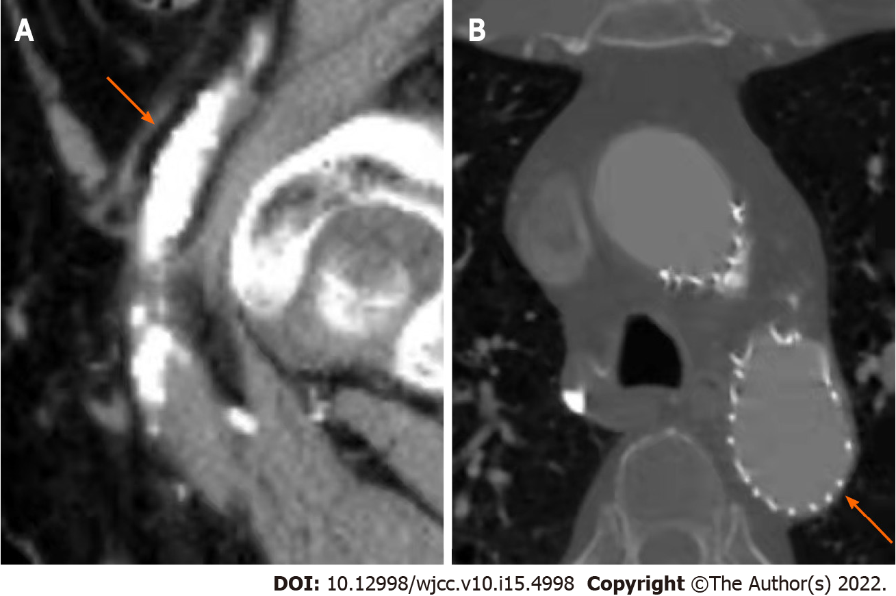
Figure 3 Postoperative computed tomography angiography after four years.
A: On the sagittal view, an intact vascular lumen without a double lumen structure can be seen (arrow); B: On the transverse view, the covered stent was well formed, and no obvious endo-leak was found (arrow).
- Citation: Fang XX, Wu XH, Chen XF. Blunt aortic injury–traumatic aortic isthmus pseudoaneurysm with right iliac artery dissection aneurysm: A case report. World J Clin Cases 2022; 10(15): 4998-5004
- URL: https://www.wjgnet.com/2307-8960/full/v10/i15/4998.htm
- DOI: https://dx.doi.org/10.12998/wjcc.v10.i15.4998


