Copyright
©The Author(s) 2022.
World J Clin Cases. May 16, 2022; 10(14): 4580-4585
Published online May 16, 2022. doi: 10.12998/wjcc.v10.i14.4580
Published online May 16, 2022. doi: 10.12998/wjcc.v10.i14.4580
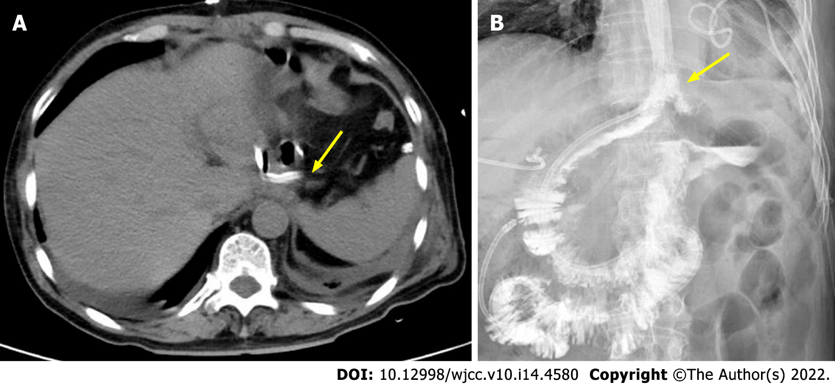
Figure 2 Imaging manifestations after negative pressure drainage via computed tomography.
A: Computed tomography showed the drainage tube performed in the thoracic cavity near the leakage of the anastomosis (arrow); B: No anastomotic leakage on fluoroscopic examination (arrow).
- Citation: Jiang ZY, Tao GQ, Zhu YF. Computer tomography-guided negative pressure drainage treatment of intrathoracic esophagojejunal anastomotic leakage: A case report . World J Clin Cases 2022; 10(14): 4580-4585
- URL: https://www.wjgnet.com/2307-8960/full/v10/i14/4580.htm
- DOI: https://dx.doi.org/10.12998/wjcc.v10.i14.4580


