Copyright
©The Author(s) 2022.
World J Clin Cases. May 16, 2022; 10(14): 4519-4527
Published online May 16, 2022. doi: 10.12998/wjcc.v10.i14.4519
Published online May 16, 2022. doi: 10.12998/wjcc.v10.i14.4519
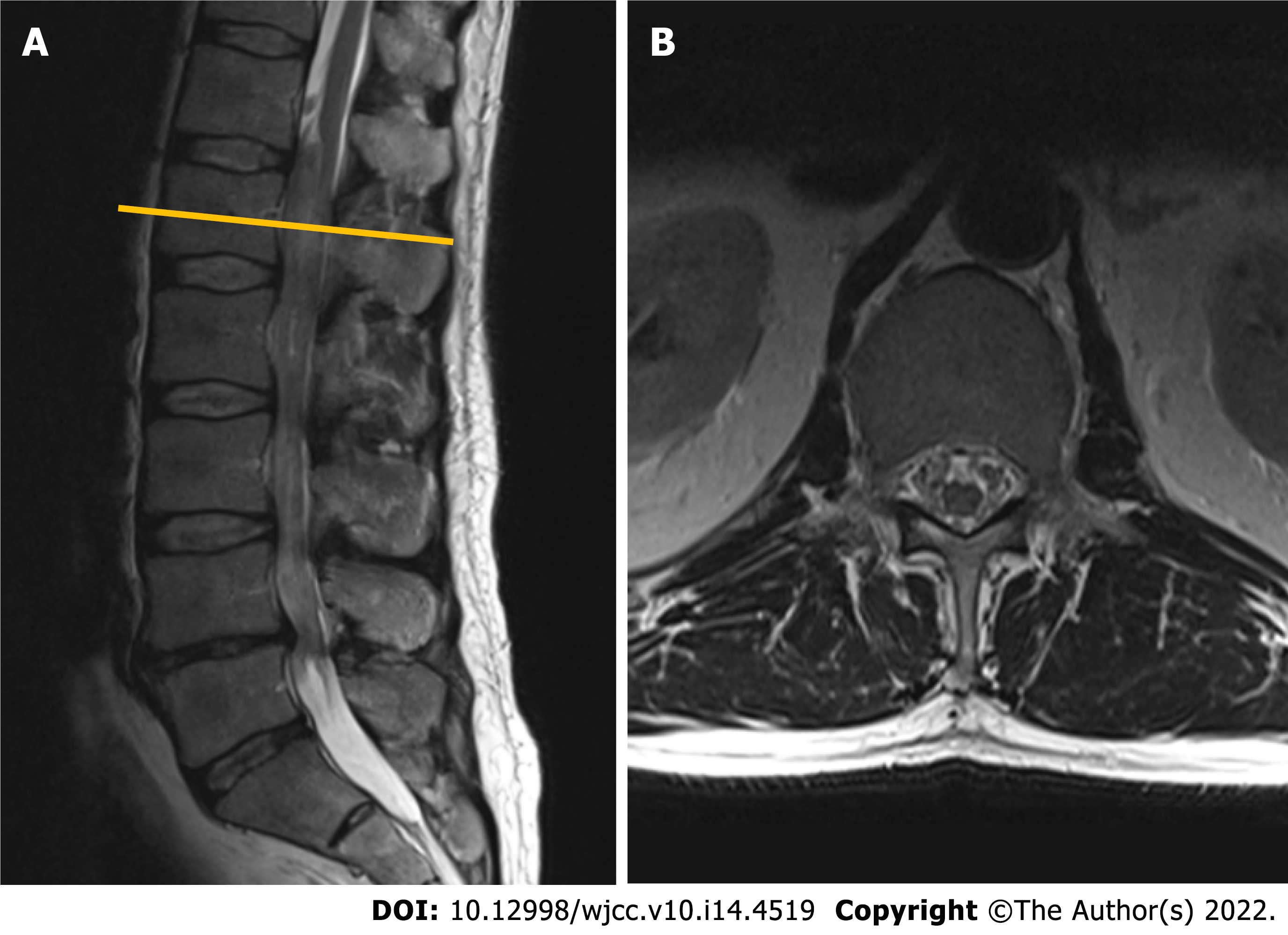
Figure 1 Contrast-enhanced magnetic resonance imaging (T2-weighted imaging sequence) performed in 2010 revealed a non-enhancing intradural tumor extending throughout the Th12-L4 vertebral levels.
A: Sagittal plane: a non-enhancing intradural tumor extending throughout the Th12-L4 vertebral levels; B: Axial plane. Tumor at the level of L1 vertebral body. Note few enlarged lumbar nerve roots engulfing conus medullaris.
- Citation: Chomanskis Z, Juskys R, Cepkus S, Dulko J, Hendrixson V, Ruksenas O, Rocka S. Plexiform neurofibroma of the cauda equina with follow-up of 10 years: A case report. World J Clin Cases 2022; 10(14): 4519-4527
- URL: https://www.wjgnet.com/2307-8960/full/v10/i14/4519.htm
- DOI: https://dx.doi.org/10.12998/wjcc.v10.i14.4519


