Published online May 16, 2022. doi: 10.12998/wjcc.v10.i14.4632
Peer-review started: November 6, 2021
First decision: December 27, 2021
Revised: December 28, 2021
Accepted: March 6, 2022
Article in press: March 6, 2022
Published online: May 16, 2022
Processing time: 187 Days and 18.1 Hours
The incidence rate of severely curved root canals in mandibular molars is low, and the root canal treatment of mandibular molars with this aberrant canal anatomy may be technically challenging.
A 26-year-old Chinese female patient presented with intermittent and occlusal pain in the left mandibular second molar. The patient had undergone filling restoration for caries before endodontic consultation. With the aid of cone beam computed tomography (CBCT), a large periapical radiolucency was observed, and curved root canals in a mandibular second molar were confirmed, depicting a severe and curved distolingual root. Nonsurgical treatments, including novel individualized preparation skills and techniques and the use of bioceramic materials as an apical barrier, were performed, and complete healing of the periapical lesion and a satisfactory effect were achieved.
A case of severely curved root canals in a mandibular second molar was successfully treated and are reported herein. The complex anatomy of the tooth and the postoperative effect were also evaluated via the three-dimensional reconstruction of CBCT images, which accurately identified the aberrant canal morphology. New devices and biomaterial applications combined with novel synthesis techniques can increase the success rate of intractable endodontic treatment.
Core Tip: The treatment of patients with severely curved root canals is problematic. Herein, with the guidance of cone beam computed tomography, individualized preparation skills and techniques and the use of bioceramic materials as an apical barrier may aid in the treatment of such severely curved teeth.
- Citation: Xu LJ, Zhang JY, Huang ZH, Wang XZ. Successful individualized endodontic treatment of severely curved root canals in a mandibular second molar: A case report. World J Clin Cases 2022; 10(14): 4632-4639
- URL: https://www.wjgnet.com/2307-8960/full/v10/i14/4632.htm
- DOI: https://dx.doi.org/10.12998/wjcc.v10.i14.4632
To date, root canal therapy (RCT) is a preferred treatment for pulpitis and periapical disease, and its success rate is closely associated with the anatomical morphology of the root canal system[1]. Being familiar with internal canal morphology is crucial for endodontists. The anatomical variations existing in the root canal system, such as curvature, may result in severe complications, such as ledge formation, apical transportation, and perforation during root canal preparation, which increases the failure rates of treatment[2]. To reduce the occurrence of these complications, a comprehensive understanding of root canal curvature models, including the degree of curvature and radius, is important. Mandibular permanent molars are the most vulnerable to dental disease, but the anatomical structure of the root canal is usually complex and substantially varied, which is considerably challenging for clinicians. According to reports, the anatomical configuration of molar roots and canals varies by nation. For example, the proportions of Spanish, Iranian, and Indian people with permanent second mandibular molars that have two roots are 83%, 81.6%, and 79.35%, respectively[3-5]. Most mandibular second molars have a small degree of bifurcation or have conical roots that are fused on the buccal surface and separated on the lingual surface. This fused root is coined in a C-shaped root, which is an important feature of mandibular second molars. Kim et al[6] reported that the proportion of patients with a double root canal system in their mandibular second molars totaled 58% in Korea, while the proportion with the C-shaped type accounted for 40%, as analysed according to cone beam computed tomography (CBCT) data.
CBCT has been introduced as a high-resolution imaging modality in oral and maxillofacial radiology[7]. Analysing and displaying the curved root canal system in the sagittal, coronal, and axial planes allow for three-dimensional reconstruction of CBCT scans, providing high-resolution images of the root canal system to gain a better understanding of the direction of curvature. Thus, visualization of the canal anatomy can enable precise canal preparation and provide clinical guidance for the diagnosis and treatment of complex and curved canals. This clinical report describes three severely curved canals in the left mandibular second molar that successfully healed with individualized RCTs under dental microscopic and CBCT guidance. Herein, we propose preparation techniques with ultrasound systems and dental lasers, and provide evidence that filling with bioceramic materials as an apical barrier may aid in the treatment of severely curved teeth.
A 26-year-old Chinese female patient was referred for evaluation of the left mandibular second molar with the chief complaints of intermittent pain and occlusal pain in this tooth.
The patient was referred for evaluation of the left mandibular second molar with the chief complaints of intermittent pain and occlusal pain in this tooth.
The patient denied having a remarkable medical history or drug allergies, and she reported caries for which her dentist filled as restoration.
There was no personal or family history.
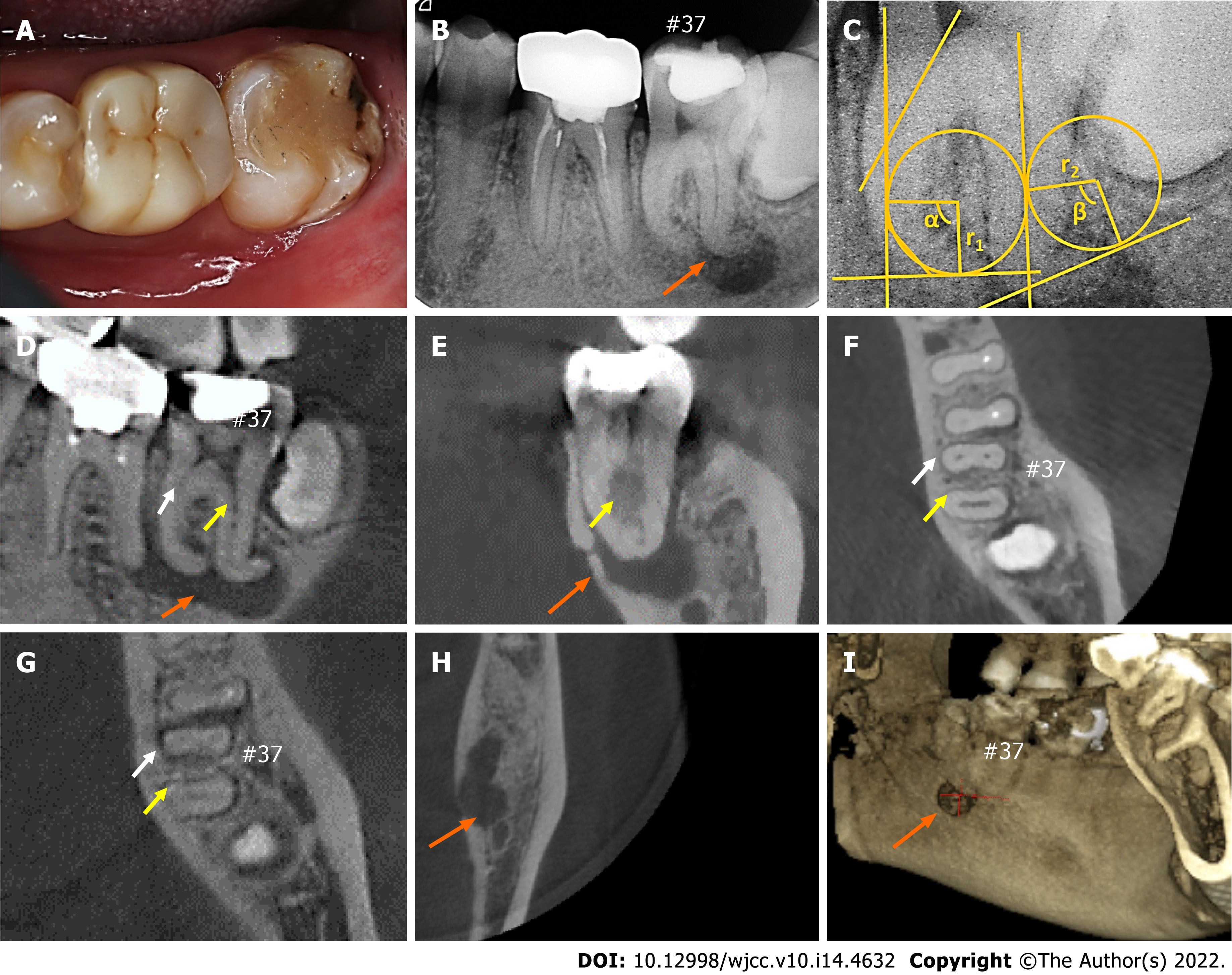
Upon extraoral examination, no significant signs were noted. The intraoral examination revealed that the left mandibular second molar (#37) had been restored with white material (Figure 1A) and showed no signs of swelling, no response to the pulp test, and no pathological mobility. Periodontal probing around the tooth showed a pocket within physiological limits without an intraoral sinus. However, there was severe pain from percussion and palpation. The first mandibular molar had a crown and no response to the cold test or percussion and was asymptomatic.
No laboratory examinations were performed.
Radiographic examination showed that tooth #37 had a large periapical radiolucency encompassing both the mesial and distal regions with a size of 11 mm × 6 mm × 6 mm (Figure 1B).
Chronic apical periodontitis.
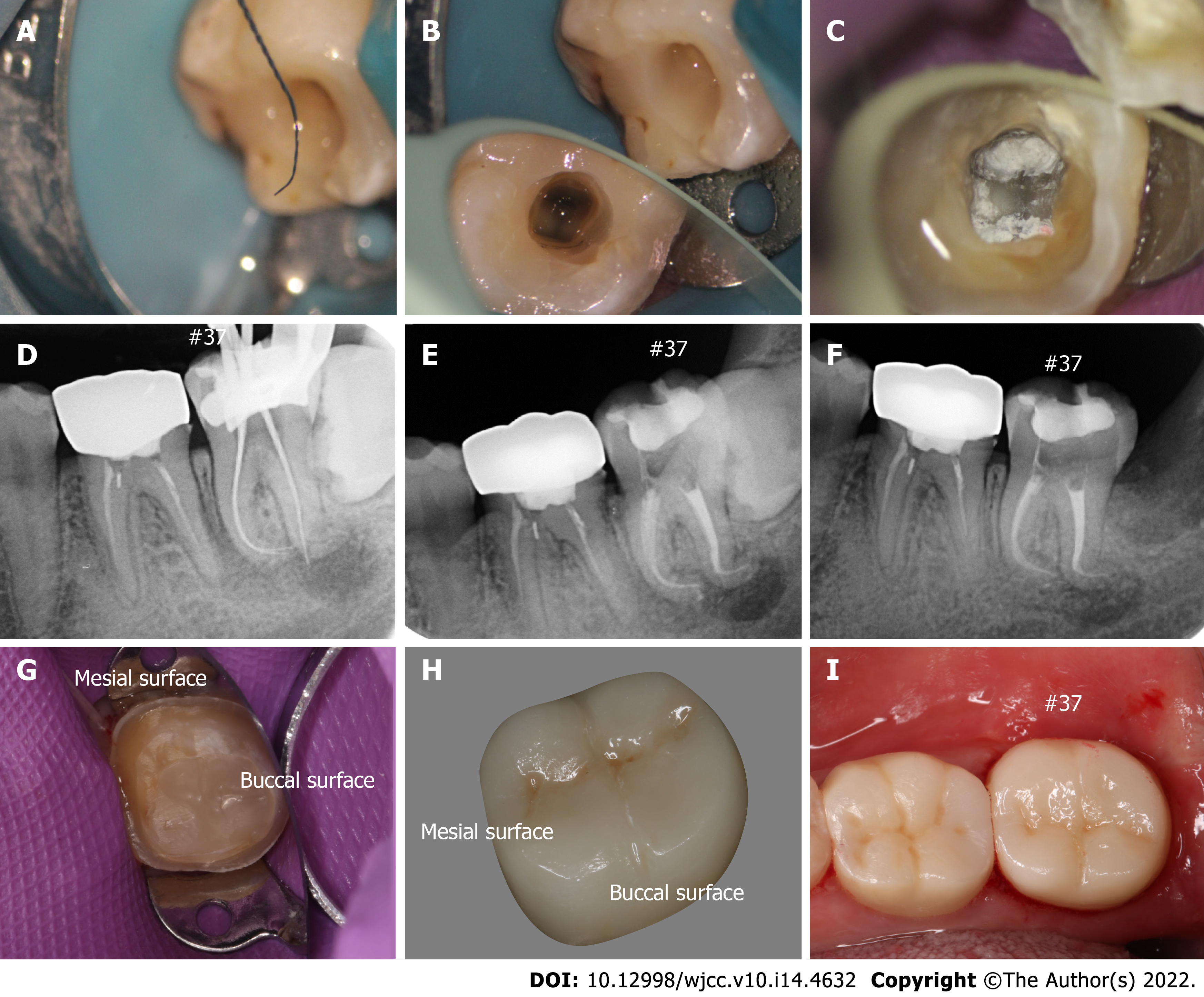
After discussing possible treatment options, the patient agreed to treatment for tooth #37 and signed an informed consent form. The tooth was isolated with a rubber dam, and the old fillings were removed before completely exposing the top pulp chamber. Endodontic access was completed using a diamond bur with a water spray. The entire procedure was performed under a dental microscope (ZUMAX, Suzhou, China) and with the guidance of CBCT. Three canals, namely, the mesiobuccal, mesiolingual, and distal canals, were identified under magnification, and a Ni-Ti file rotary system (Orodeka, PLEX, Italy) was used for root canal preparation. The preparation and process of cleaning and shaping the canals were divided into two parts: (1) During the initial stage of RCT, the orifices of the root canals were trimmed using ET18D (ACTEON, SATELEC, France), and coronal access was obtained using #15/08 (Orodeka, PLEX, Italy); and (2) for mesial root canals, after exploring and dredging the position of the canals with #08 and #10 K-files (Densply, United States), the initial working length (WL) was determined with #10 K-files at the end of the apex under magnification, which was confirmed by periapical radiographs (Figure 2A-D). Then, canals were shaped and enlarged using #15/03, #20/04, and #25/04, while for a distal root canal, the upper canal was used for the crown-down technique with #15/03, #20/04, and #25/06 according to the resistance. After that, #6 K-files were used to establish a straight path to the apex with EDTA gel (MD-ClelCream, Meta Biomed, United States), and the WL was determined according to the penetration of the #06 K-files (referring to the point on the crown edge to the apical foramen minus 1 mm)[8]. The step-back technique, using the 0.5-mm recession method with #08, #10, and #15 K-files, was used for apex preparation to maintain the original morphology and shape of the root canal. Finally, the canal was finished with #12/03 and #15/03. A total of 20 mL of 5.25% NaOCl combined with 17% EDTA solution was used to irrigate every root canal during preparation. An ultrasound system (P5 Newtron XS, SATELEC, France) was introduced to activate the irrigant, and a photon-initiated photoacoustic streaming (PIPS) technique (Er:YAG, SSP, 2 Hz, 20 mJ, 0.15 W, LightWalkerAT, Fotona, Germany) was used to further remove the deep smear layer and eliminate any remaining bacteria in the dentin canal tubes. Finally, paper points were used to dry the canals for inspection, calcium hydroxide paste was used as filler, and then the coronal was temporarily sealed with temporary filling material (Ceivitron, Taibei, China). All operations were carried out successfully under a dental operating microscope.
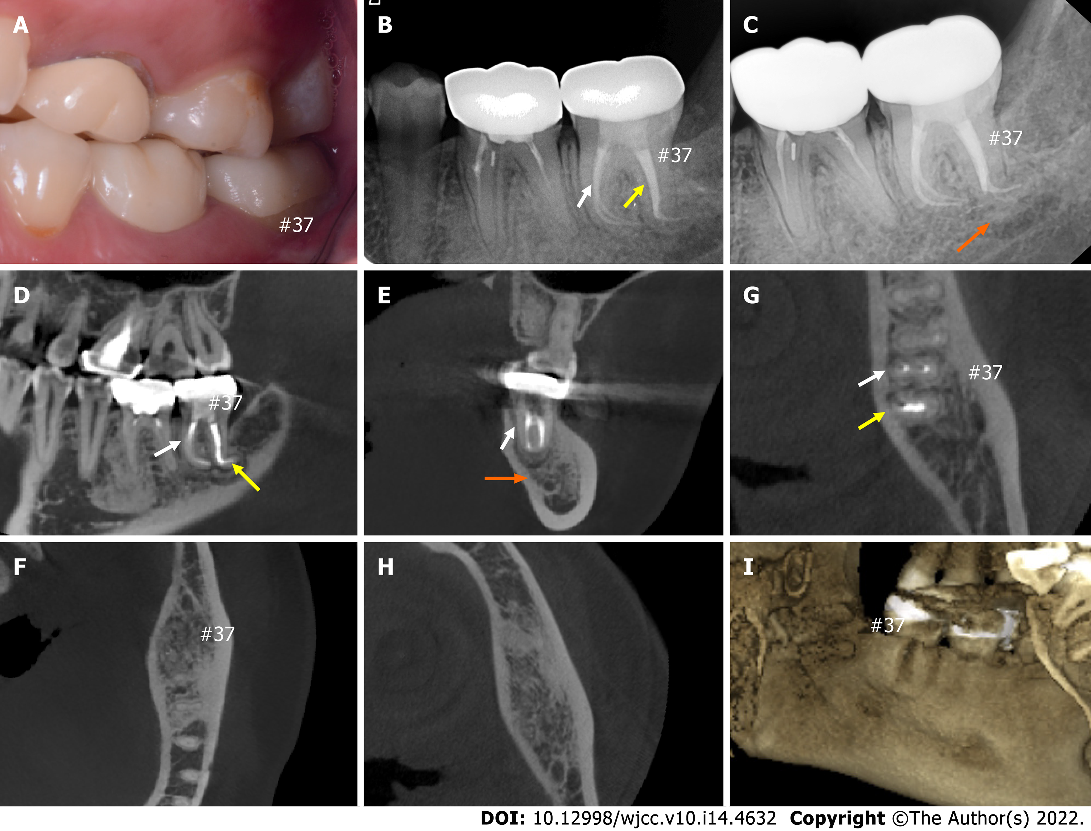
The tooth was re-examined 2 wk later, and the canals were copiously irrigated with 17% EDTA solution to remove calcium hydroxide paste. After cleaning with the PIPS technique and distilled water, the canals were dried with paper points. The main gutta-percha cones were selected (#25/04), and the mesial canals were filled with large taper gutta-perchas and root canal sealer iRoot SP (Innovative Bioceramix, Vancouver, BC, Canada). However, gutta-perchas could not reach the WL point in the distal canal due to the sharp curved apex. Therefore, the vertical condensation technique was used for the apical sites, in which iRoot BP Plus (Innovative Bioceramix, Vancouver, BC, Canada) was placed as a barrier to exert a better apical sealing effect after filling with iRoot SP (Figure 2E). Postoperative radiographs were taken to confirm that three canals were filled compactly, especially in the curved corners. After 3 mo of observation (Figure 3B), the patient had no spontaneous pain or other obvious abnormalities, and the tooth was restored with composite resin (Filtek Z350 XT, 3M ESPE). The patient was then referred for restorative treatment. The edge of the ceramic crown and occlusal was checked to ensure a proper fit (Figure 2G-I).
At the 3-mo and 1-year follow-ups, the treated mandibular molar showed complete healing of the periapical lesion and a satisfactory effect was achieved.
Endodontic treatment failure in mandibular molars is mostly due to the complexity and diversity of root canal configurations. In this case, three mandibular molar canals, namely, the mesiobuccal, mesiolingual, and distal canals, were separate and independent from each other. Interestingly, the CBCT images revealed that these canals were severely curved, showing highly rare degrees of curvature, illustrating the challenges that must be faced when dealing with the anatomical variations in canals. As studies have reported, most mandibular second molars have two roots or a fused root, with 55% having three canals[9]. Precisely understanding the positions, directions, and angles of these curvature canals is important for treatment. In this study, visible three-dimensional canal models based on CBCT datasets were found to facilitate the shaping and cleaning efficiency of root canal systems. The root canals in tooth #37 had two roots: The mesial root had two separate canals, the distal root had an oblate canal (Figure 1F-G), and a large periapical radiolucency that perforated the lingual cortical plate was observed in the apical region of #37 (Figure 1H-I). More importantly, all canals in both the mesial and distal roots had a sharp curvature mainly in the distal direction. Referring to the method of canal curvature, namely, the Pruett method[10], the degree of root canal curvature was measured using periapical radiographs, which showed that the curvature was mainly in the distal direction. The degree of curvature in the mesial and distal root was determined to be 91.5 (α) and 105 (β) degrees, with radii of curvature of 3.2 mm (r1) and 3 mm (r2), respectively (Figure 1C), indicating that the canals were severely curved, which made treatment difficult. Friedland et al[11] reported the use of three-dimensional reconstructions of CBCT images to efficiently and accurately observe and analyze anatomically curved canals. Hence, the precise assessment of root canal curvature is essential for guiding endodontic operations.
In this case, all the root canals were severely curved, especially the apical tip of the distal root canals (Figure 1), which was intractable to preparation and fillings. However, the small taper and flexibility of Ni-Ti files allow the original apical shape and position to be maintained[12]. In addition, files that are pre-bent into the root canal may retain more pericervical dentine and reduce dentin stress, instrument separation, and other complications[13]. The crown-down technique, which can be used to access canals, recommends a wide pathway to facilitate irrigation (Figure 2A-B). High concentrations of sodium hypochlorite with ultrasonic activation as a mechanochemical preparation can further eliminate infections of the lateral canals and curved apex. The use of lasers in dentistry fields confers many advantages, such as removing carious enamel and dentine and facilitating endodontic treatment and prosthetic procedures, including crown lengthening and sulcus uncovering[14]. Erbium laser-assisted working techniques in endodontic therapy can accelerate the healing processes via endodontic space decontamination and the removal of pathological tissues[15] and carious dental tissues, as well as through debridement and disinfection of periodontal tissue[16]. Photon-induced photoacoustic streaming (PIPS) is a new technique that requires the use of an Er:YAG laser to activate the water molecules in irrigants to remove dentin debris and smear layers due to the positive radial effect[17,18]. For these curved canals, PIPS can be used to clean the apical region as well as the narrow area of irregular canals (traffic and the gorge area) that the files cannot reach, which is a minimally invasive method to disinfect the tooth[19]. Great importance should be attached to the ability to fill the apex of curvature since conventional canal fillings cannot seal the irregular apex. iRoot BP Plus can be used for repairs such as pulpotomy, pulp floor perforation repair, and root perforation repair[20]. Interestingly, we filled the curved apex with iRoot BP Plus (Figure 2C-F) due to its good sealing ability and its capacity to absorb water from the dentinal tubules and to prevent oral fluid contamination[21]. The apical barrier using bioceramic materials in the apical regions showed good biocompatibility, was chemically bonded to the dentin, and reduced the number of microcracks generated by pressurized filling[20]. Finally, crown restoration was performed to protect the remaining tooth tissue (Figure 2G-I) and the natural occlusion was checked (Figure 3A). At the 3-mo (Figure 3B) and 1-year (Figure 3C-I) follow-ups, the treated mandibular molar showed complete healing of the periapical lesion and a satisfactory effect was achieved.
In conclusion, a thorough understanding of tooth and root canal morphology by CBCT during preoperative assessment is highly important in complicated cases. Exploring the root canals under magnification, making preparations with individual sequential techniques combined with new instruments such as ultrasonic activation and PIPS, and using fillings with bioceramics as an apical barrier are essential prerequisites to increase the success rate of this difficult endodontic treatment. Although the endodontic treatment of teeth with large periapical bone destruction and aberrant curved canals is difficult and intractable, nonsurgical root canal therapy was performed with novel devices and introduced skills in this case, resulting in a good prognosis (the periapical radiolucency disappeared without any symptoms). This report may also provide meaningful guidance and serve as a reference for other similar cases.
Provenance and peer review: Unsolicited article; Externally peer reviewed.
Peer-review model: Single blind
Specialty type: Dentistry, oral surgery and medicine
Country/Territory of origin: China
Peer-review report’s scientific quality classification
Grade A (Excellent): 0
Grade B (Very good): B, B
Grade C (Good): 0
Grade D (Fair): 0
Grade E (Poor): 0
P-Reviewer: Aoun G, Lebanon; Zafari N, Iran S-Editor: Xing YX L-Editor: Wang TQ P-Editor: Xing YX
| 1. | Zhang M, Xie J, Wang YH, Feng Y. Mandibular first premolar with five root canals: a case report. BMC Oral Health. 2020;20:253. [RCA] [PubMed] [DOI] [Full Text] [Full Text (PDF)] [Cited by in Crossref: 4] [Cited by in RCA: 4] [Article Influence: 0.8] [Reference Citation Analysis (0)] |
| 2. | Lin LM, Rosenberg PA, Lin J. Do procedural errors cause endodontic treatment failure? J Am Dent Assoc. 2005;136:187-93; quiz 231. [RCA] [PubMed] [DOI] [Full Text] [Cited by in Crossref: 88] [Cited by in RCA: 100] [Article Influence: 5.0] [Reference Citation Analysis (0)] |
| 3. | Pérez-Heredia M, Ferrer-Luque CM, Bravo M, Castelo-Baz P, Ruíz-Piñón M, Baca P. Cone-beam Computed Tomographic Study of Root Anatomy and Canal Configuration of Molars in a Spanish Population. J Endod. 2017;43:1511-1516. [RCA] [PubMed] [DOI] [Full Text] [Cited by in Crossref: 37] [Cited by in RCA: 48] [Article Influence: 6.0] [Reference Citation Analysis (0)] |
| 4. | Madani ZS, Mehraban N, Moudi E, Bijani A. Root and Canal Morphology of Mandibular Molars in a Selected Iranian Population Using Cone-Beam Computed Tomography. Iran Endod J. 2017;12:143-148. [RCA] [PubMed] [DOI] [Full Text] [Full Text (PDF)] [Cited by in RCA: 20] [Reference Citation Analysis (0)] |
| 5. | Pawar AM, Pawar M, Kfir A, Singh S, Salve P, Thakur B, Neelakantan P. Root canal morphology and variations in mandibular second molar teeth of an Indian population: an in vivo cone-beam computed tomography analysis. Clin Oral Investig. 2017;21:2801-2809. [RCA] [PubMed] [DOI] [Full Text] [Cited by in Crossref: 25] [Cited by in RCA: 32] [Article Influence: 4.0] [Reference Citation Analysis (0)] |
| 6. | Kim SY, Kim BS, Kim Y. Mandibular second molar root canal morphology and variants in a Korean subpopulation. Int Endod J. 2016;49:136-144. [RCA] [PubMed] [DOI] [Full Text] [Cited by in Crossref: 31] [Cited by in RCA: 37] [Article Influence: 3.7] [Reference Citation Analysis (0)] |
| 7. | Lambrecht JT, Berndt DC, Schumacher R, Zehnder M. Generation of three-dimensional prototype models based on cone beam computed tomography. Int J Comput Assist Radiol Surg. 2009;4:175-180. [RCA] [PubMed] [DOI] [Full Text] [Cited by in Crossref: 27] [Cited by in RCA: 23] [Article Influence: 1.4] [Reference Citation Analysis (0)] |
| 8. | Katz A, Tamse A. A combined radiographic and computerized scanning method to evaluate remaining dentine thickness in mandibular incisors after various intracanal procedures. Int Endod J. 2003;36:682-686. [RCA] [PubMed] [DOI] [Full Text] [Cited by in Crossref: 11] [Cited by in RCA: 11] [Article Influence: 0.5] [Reference Citation Analysis (0)] |
| 9. | Al-Qudah AA, Awawdeh LA. Root and canal morphology of mandibular first and second molar teeth in a Jordanian population. Int Endod J. 2009;42:775-784. [RCA] [PubMed] [DOI] [Full Text] [Cited by in Crossref: 65] [Cited by in RCA: 78] [Article Influence: 4.9] [Reference Citation Analysis (0)] |
| 10. | Pruett JP, Clement DJ, Carnes DL Jr. Cyclic fatigue testing of nickel-titanium endodontic instruments. J Endod. 1997;23:77-85. [RCA] [PubMed] [DOI] [Full Text] [Cited by in Crossref: 645] [Cited by in RCA: 624] [Article Influence: 22.3] [Reference Citation Analysis (0)] |
| 11. | Friedland B, Donoff B, Dodson TB. The use of 3-dimensional reconstructions to evaluate the anatomic relationship of the mandibular canal and impacted mandibular third molars. J Oral Maxillofac Surg. 2008;66:1678-1685. [RCA] [PubMed] [DOI] [Full Text] [Cited by in Crossref: 47] [Cited by in RCA: 44] [Article Influence: 2.6] [Reference Citation Analysis (0)] |
| 12. | Berutti E, Paolino DS, Chiandussi G, Alovisi M, Cantatore G, Castellucci A, Pasqualini D. Root canal anatomy preservation of WaveOne reciprocating files with or without glide path. J Endod. 2012;38:101-104. [RCA] [PubMed] [DOI] [Full Text] [Cited by in Crossref: 19] [Cited by in RCA: 47] [Article Influence: 3.4] [Reference Citation Analysis (0)] |
| 13. | Pacheco-Yanes J, Gazzaneo I, Pérez AR, Armada L, Neves MAS. Transportation assessment in artificial curved canals after instrumentation with Reciproc, Reciproc Blue, and XP-endo Shaper Systems. J Investig Clin Dent. 2019;10:e12417. [RCA] [PubMed] [DOI] [Full Text] [Cited by in Crossref: 5] [Cited by in RCA: 12] [Article Influence: 2.0] [Reference Citation Analysis (0)] |
| 14. | SteinerOliveira, Carolina, Ramalho, Müller K, BelloSilva, Stella M, Aranha, Corrêa AC, Eduardo, Paula CD. The use of lasers in restorative dentistry: truths and myths. Braz Dent Sci. 2012;3:1-15. |
| 15. | Doriana Agop-Forna MSCT. Dental lasers in restorative dentistry: A review. J RJOR. 2021;13:7-17. |
| 16. | Ozcelik O, Cenk Haytac M, Seydaoglu G. Enamel matrix derivative and low-level laser therapy in the treatment of intra-bony defects: a randomized placebo-controlled clinical trial. J Clin Periodontol. 2008;35:147-156. [RCA] [PubMed] [DOI] [Full Text] [Cited by in Crossref: 35] [Cited by in RCA: 42] [Article Influence: 2.3] [Reference Citation Analysis (0)] |
| 17. | DiVito E, Peters OA, Olivi G. Effectiveness of the erbium:YAG laser and new design radial and stripped tips in removing the smear layer after root canal instrumentation. Lasers Med Sci. 2012;27:273-280. [RCA] [PubMed] [DOI] [Full Text] [Cited by in Crossref: 137] [Cited by in RCA: 138] [Article Influence: 9.2] [Reference Citation Analysis (0)] |
| 18. | Mandras N, Pasqualini D, Roana J, Tullio V, Banche G, Gianello E, Bonino F, Cuffini AM, Berutti E, Alovisi M. Influence of Photon-Induced Photoacoustic Streaming (PIPS) on Root Canal Disinfection and Post-Operative Pain: A Randomized Clinical Trial. J Clin Med. 2020;9. [RCA] [PubMed] [DOI] [Full Text] [Full Text (PDF)] [Cited by in Crossref: 5] [Cited by in RCA: 10] [Article Influence: 2.0] [Reference Citation Analysis (0)] |
| 19. | Swimberghe RCD, Buyse R, Meire MA, De Moor RJG. Efficacy of different irrigation technique in simulated curved root canals. Lasers Med Sci. 2021;36:1317-1322. [RCA] [PubMed] [DOI] [Full Text] [Cited by in Crossref: 5] [Cited by in RCA: 12] [Article Influence: 3.0] [Reference Citation Analysis (0)] |
| 20. | Yang Y, Xia B, Xu Z, Dou G, Lei Y, Yong W. The effect of partial pulpotomy with iRoot BP Plus in traumatized immature permanent teeth: A randomized prospective controlled trial. Dent Traumatol. 2020;36:518-525. [RCA] [PubMed] [DOI] [Full Text] [Cited by in Crossref: 11] [Cited by in RCA: 22] [Article Influence: 4.4] [Reference Citation Analysis (0)] |
| 21. | Jitaru S, Hodisan I, Timis L, Lucian A, Bud M. The use of bioceramics in endodontics - literature review. Clujul Med. 2016;89:470-473. [RCA] [PubMed] [DOI] [Full Text] [Full Text (PDF)] [Cited by in Crossref: 34] [Cited by in RCA: 47] [Article Influence: 5.2] [Reference Citation Analysis (0)] |


