Copyright
©The Author(s) 2021.
World J Clin Cases. Mar 16, 2021; 9(8): 1940-1945
Published online Mar 16, 2021. doi: 10.12998/wjcc.v9.i8.1940
Published online Mar 16, 2021. doi: 10.12998/wjcc.v9.i8.1940
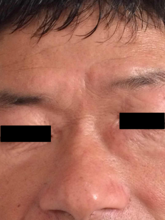
Figure 1 A 54-year-old male presented with a chief complaint of a 2-year history of the left lacrimal sac occupation that had grown rapidly for nearly half a year.
Physical examination touched a tough nodule in the left lacrimal sac.
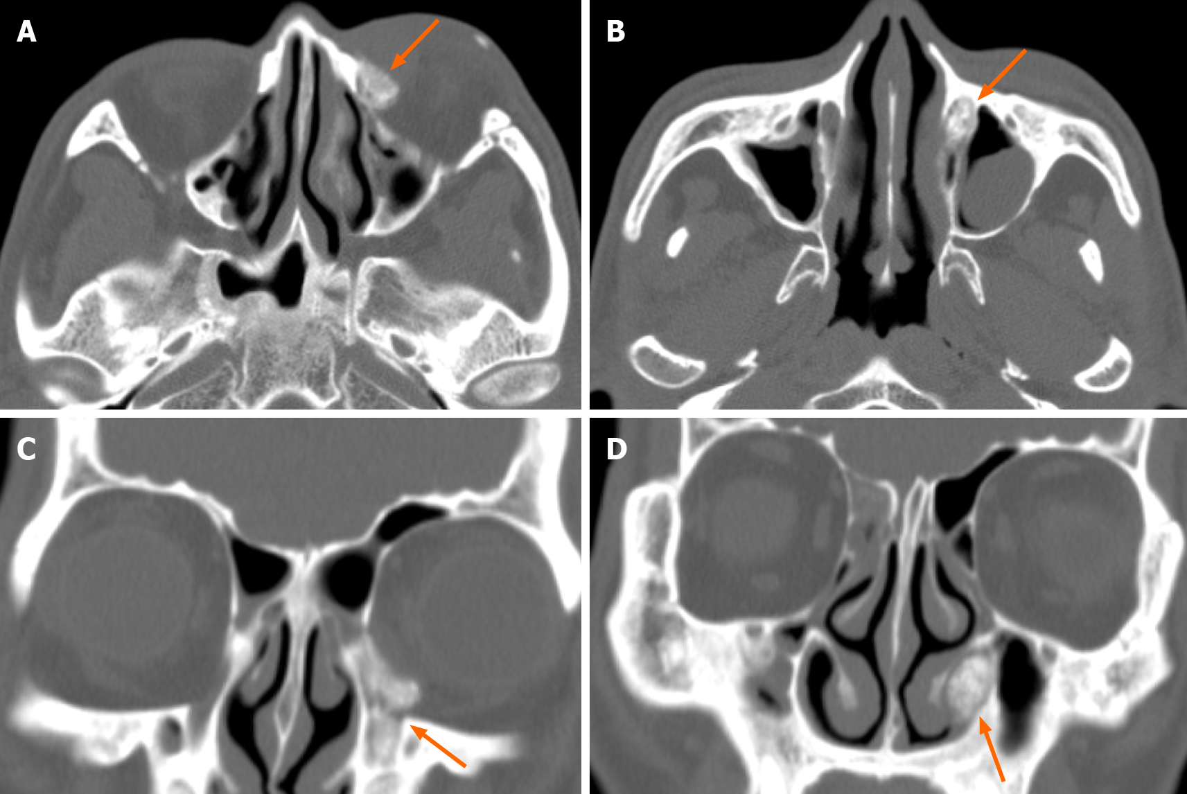
Figure 2 Axial and coronal images of non-contrast enhanced computed tomography scan showed the speckle high density in the left lacrimal sac and the dilated nasolacrimal duct.
A and B: Axial images of non-contrast enhanced computed tomography scan; C and D: Coronal images of non-contrast enhanced computed tomography scan.
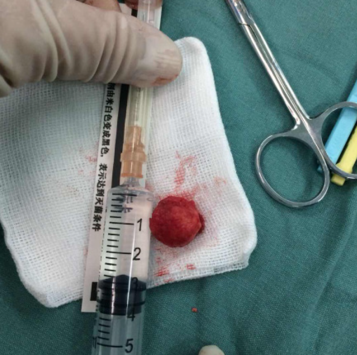
Figure 3 One larger stone removed in the operation.
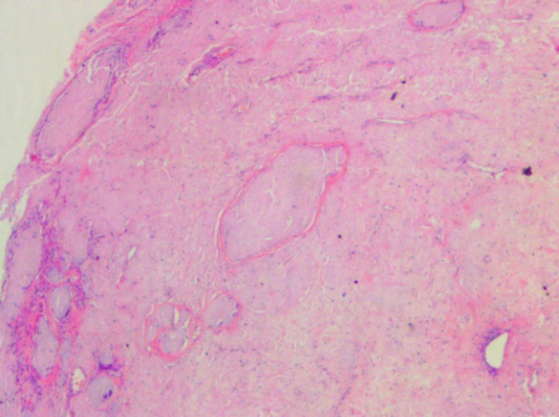
Figure 4 Orbital pathology (hematoxylin-eosin, × 100).
Amorphous pink material and multinucleated giant cell reaction were found in the fibrous tissue.
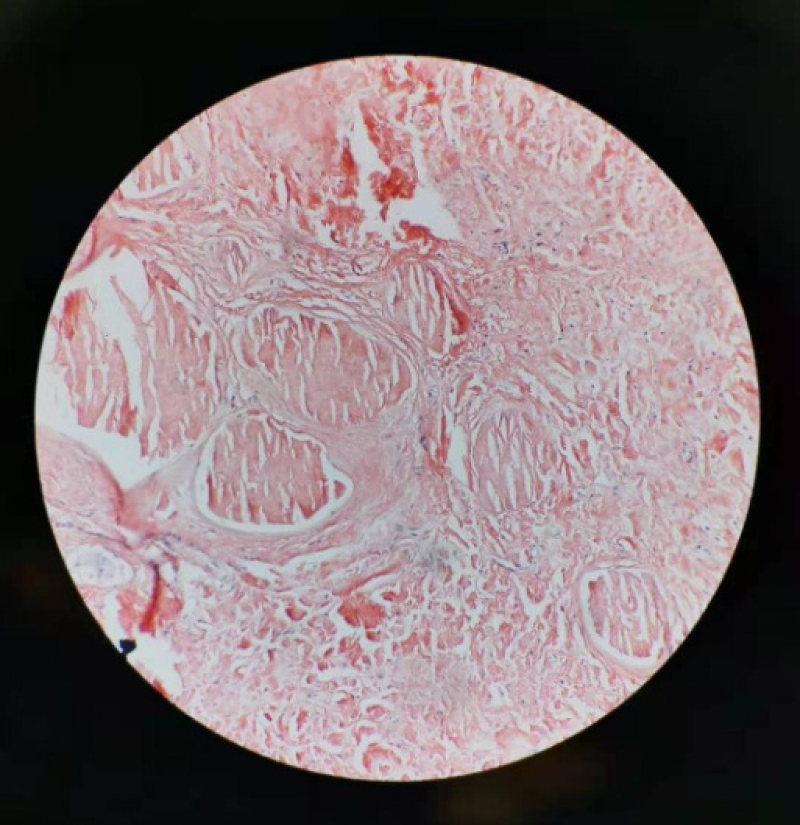
Figure 5 Photomicrograph (magnification, × 200) demonstrates homogeneous orange material without structure in coarse granular, massive, nodular forms on Congo red staining.
- Citation: Che ZG, Ni T, Wang ZC, Wang DW. Computed tomography imaging features for amyloid dacryolith in the nasolacrimal excretory system: A case report. World J Clin Cases 2021; 9(8): 1940-1945
- URL: https://www.wjgnet.com/2307-8960/full/v9/i8/1940.htm
- DOI: https://dx.doi.org/10.12998/wjcc.v9.i8.1940


