Copyright
©The Author(s) 2021.
World J Clin Cases. Mar 16, 2021; 9(8): 1931-1939
Published online Mar 16, 2021. doi: 10.12998/wjcc.v9.i8.1931
Published online Mar 16, 2021. doi: 10.12998/wjcc.v9.i8.1931
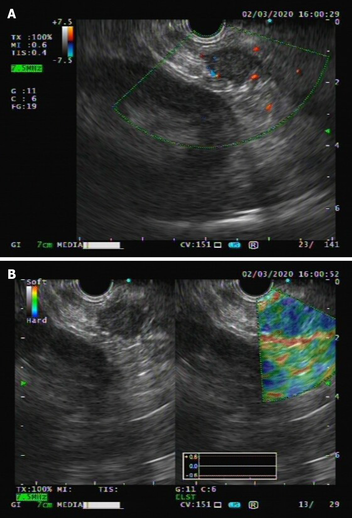
Figure 1 Endoscopic ultrasound images of the epithelioid angiomyolipoma.
A: Endoscopic ultrasound showed a hypoechogenic mass in the tail of the pancreas, measuring 1.9 cm, without obvious blood flow on color doppler flow imaging; B: Elastography showed that the elastic properties of the mass were a little harder than the surrounding tissue.
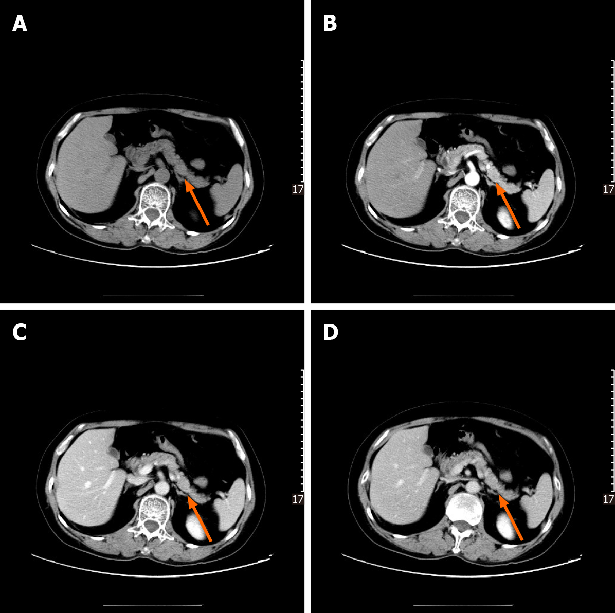
Figure 2 Contrast-enhanced computed tomography images of the epithelioid angiomyolipoma.
A-D: Axial contrast-enhanced multidetector computed tomography scan series failed to demonstrate any mass.
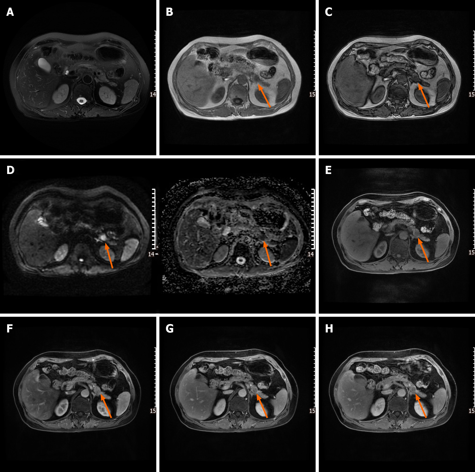
Figure 3 Gadolinium-enhanced magnetic resonance images of the epithelioid angiomyolipoma.
A: Magnetic resonance (MR) imaging showed an isointensity to pancreatic parenchyma on fs-T2-weighted imaging; B and C: Axial T1-weighted in and out phase MR images, respectively, showing a slight loss of signal intensity (SI) related to microscopic lipid component; D: Diffusion-weighted image revealing a high SI area in the pancreas with corresponding low SI on the ADC map consistent with restricted diffusion; E-H: Axial T1-weighted fat saturated MR image before and after gadolinium administration, showing a mass with slightly intake of more contrast than surrounding pancreatic tissue, followed by rapid washout in the portal and delayed phases.
Figure 4 Photomicrograh (hematoxylin and eosin staining; magnification, × 200).
Microscopically, the tumor was composed of large epithelioid cells, while adipose tissue was scarcely observed.
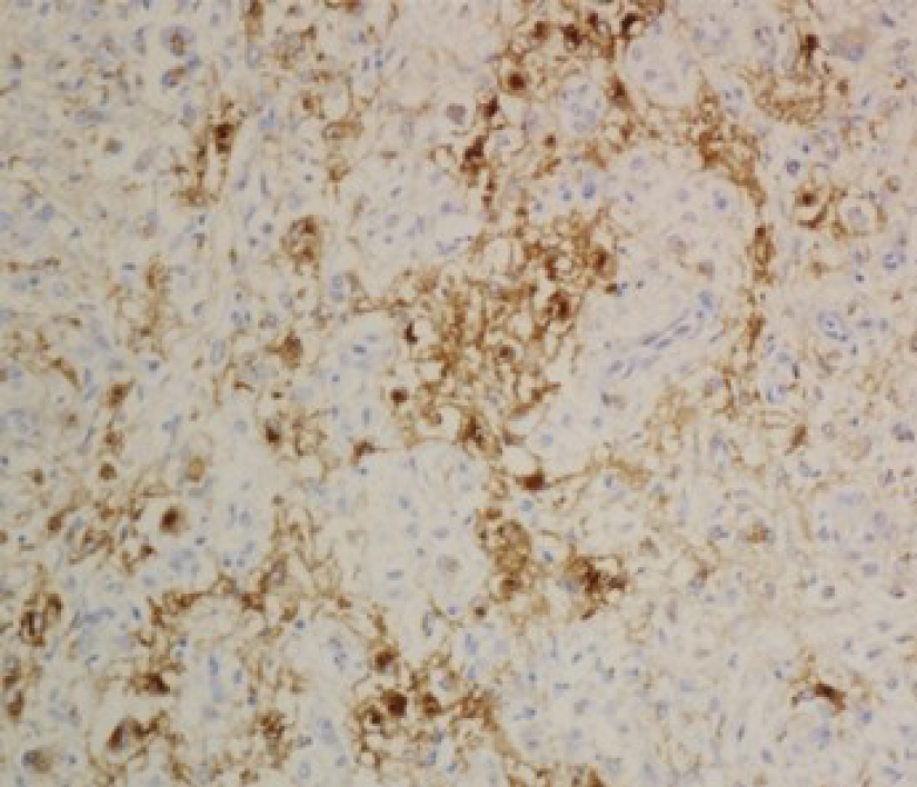
Figure 5 Immunohistochemical staining (magnification, × 200) demonstrated that the epithelioid cells and spindle smooth cells focally expressed human melanoma black-45.
- Citation: Zhu QQ, Niu ZF, Yu FD, Wu Y, Wang GB. Epithelioid angiomyolipoma of the pancreas: A case report and review of the literature. World J Clin Cases 2021; 9(8): 1931-1939
- URL: https://www.wjgnet.com/2307-8960/full/v9/i8/1931.htm
- DOI: https://dx.doi.org/10.12998/wjcc.v9.i8.1931


