Copyright
©The Author(s) 2021.
World J Clin Cases. Mar 16, 2021; 9(8): 1909-1915
Published online Mar 16, 2021. doi: 10.12998/wjcc.v9.i8.1909
Published online Mar 16, 2021. doi: 10.12998/wjcc.v9.i8.1909
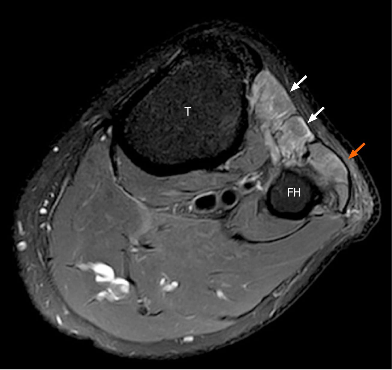
Figure 1 Magnetic resonance image study of the left knee.
The T2-weighted magnetic resonance images show volume loss and edema at the anterior (white arrow) and lateral (orange arrow) muscular compartments in the left lower leg, consistent with subacute to chronic common peroneal neuropathy. T: Tibia; FH: Fibular head.
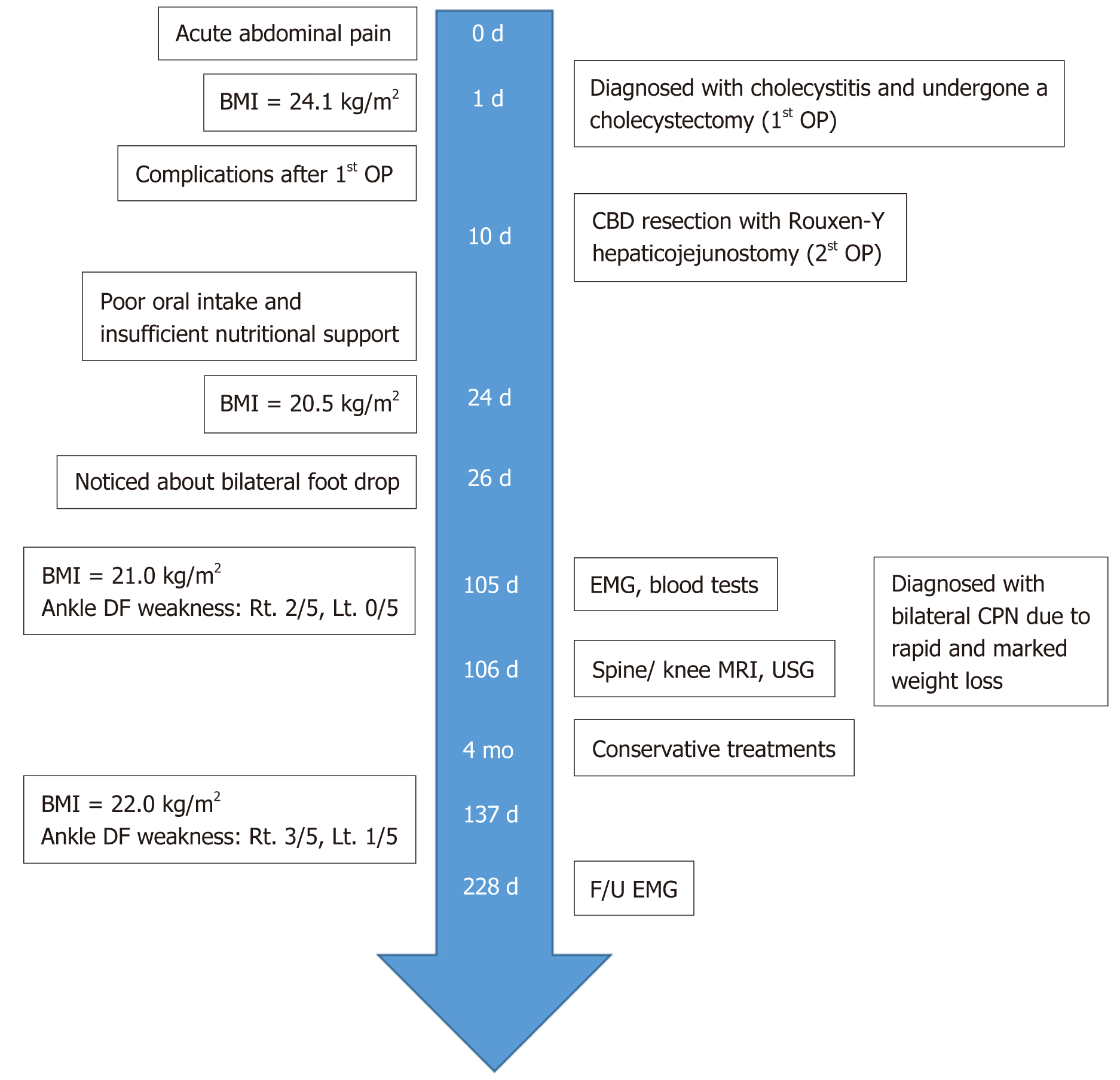
Figure 2 Case timeline.
BMI: Body mass index; OP: Operation; CBD: Common bile duct; DF: Dorsiflexor; EMG: Electromyography; MRI: Magnetic resonance imaging; USG: Ultrasonography; CPN: Common peroneal neuropathy.
- Citation: Oh MW, Gu MS, Kong HH. Bilateral common peroneal neuropathy due to rapid and marked weight loss after biliary surgery: A case report. World J Clin Cases 2021; 9(8): 1909-1915
- URL: https://www.wjgnet.com/2307-8960/full/v9/i8/1909.htm
- DOI: https://dx.doi.org/10.12998/wjcc.v9.i8.1909


