Copyright
©The Author(s) 2021.
World J Clin Cases. Mar 16, 2021; 9(8): 1893-1900
Published online Mar 16, 2021. doi: 10.12998/wjcc.v9.i8.1893
Published online Mar 16, 2021. doi: 10.12998/wjcc.v9.i8.1893
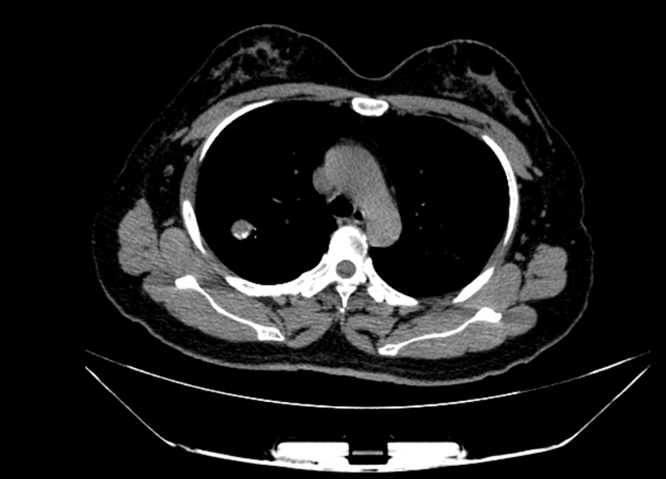
Figure 1 Computed tomography images of primary pulmonary synovial sarcoma.
The computed tomography images showed a dense soft tissue lesion in the posterior upper lobe of the right lung.
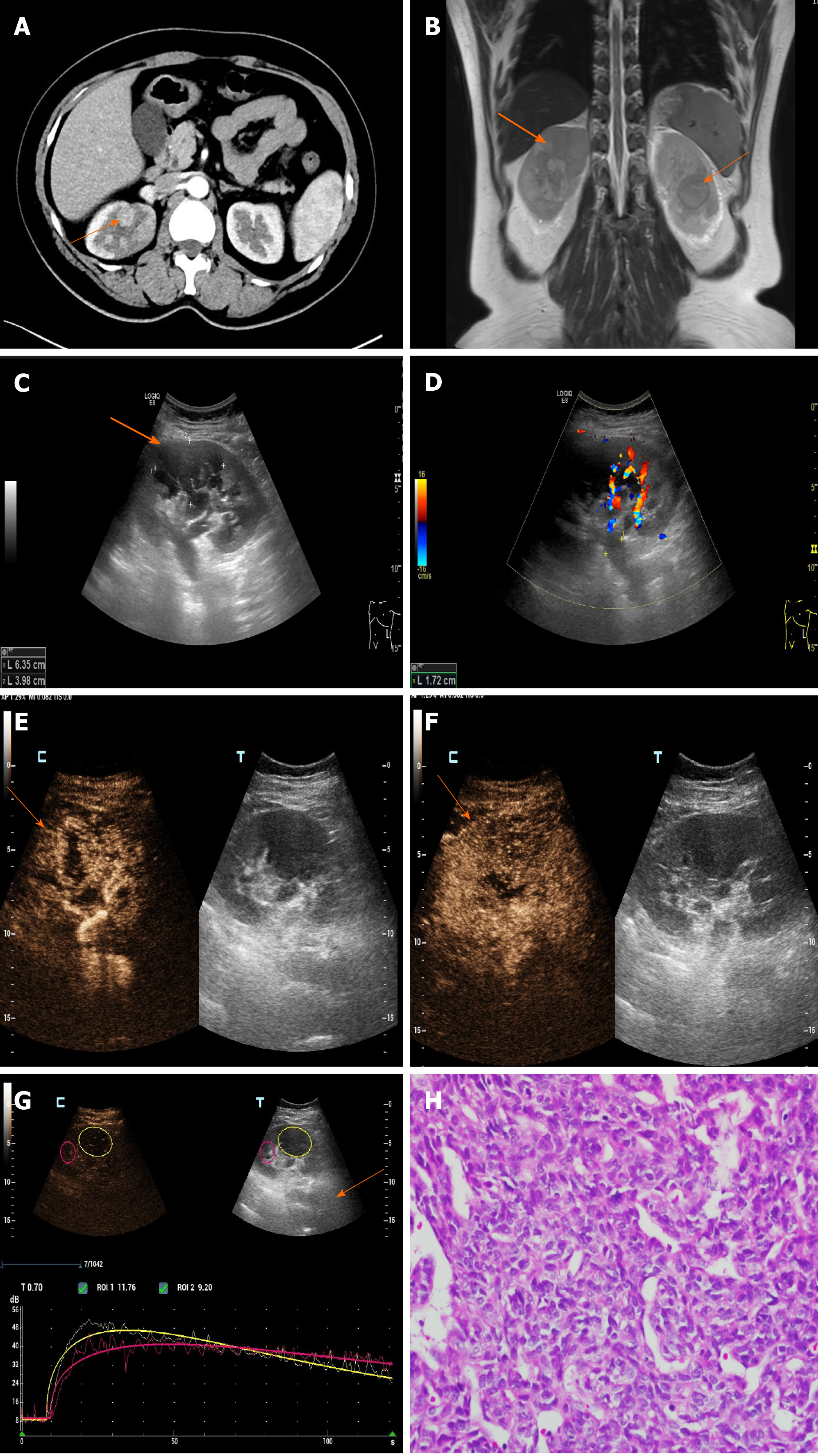
Figure 2 Renal metastases.
A: Enhanced computed tomography showed mild to moderate heterogeneous enhancement; B: Magnetic resonance imaging showed long T1 and short T2 signals; C and D: Color Doppler flow imaging: There were several hypoechoic lesions in the renal parenchyma, without obvious blood flow signals. The inner diameter of the renal vein in the renal hilum was widened and hypoechoic filling was seen inside; E-G: Contrast-enhanced ultrasonography: The whole lesion showed rapid high enhancement, and the contrast medium quickly withdrew, indicating the “fast forward and fast retreat” enhancement mode; H: Pathology image of renal metastatic synovial sarcoma (hematoxylin and eosin staining, × 400).
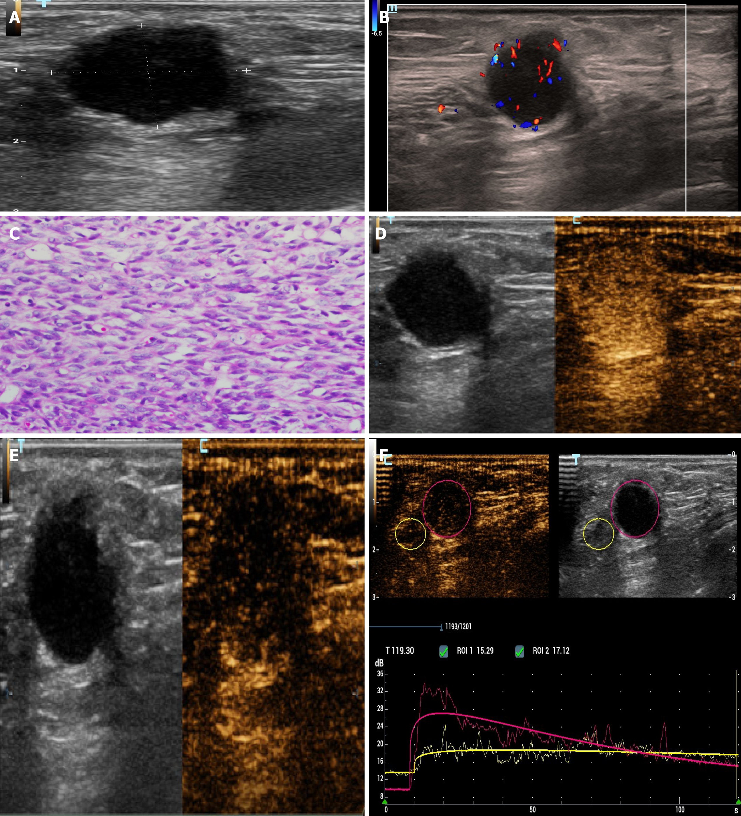
Figure 3 Chest wall metastasis.
A and B: The chest wall mass showed a hypoechoic mass in the superficial fascia layer on two-dimensional ultrasonography. Color Doppler flow imaging: Dot strip blood flow signal was seen in the mass; C: Pathological image of chest wall metastatic synovial sarcoma (hematoxylin and eosin staining, × 400); D-F: The ultrasound contrast agent quickly withdrew after rapidly high enhancement, showing the “fast forward and fast retreat” enhancement mode.
- Citation: Li R, Teng X, Han WH, Li Y, Liu QW. Imaging findings of primary pulmonary synovial sarcoma with secondary distant metastases: A case report. World J Clin Cases 2021; 9(8): 1893-1900
- URL: https://www.wjgnet.com/2307-8960/full/v9/i8/1893.htm
- DOI: https://dx.doi.org/10.12998/wjcc.v9.i8.1893


