Copyright
©The Author(s) 2021.
World J Clin Cases. Mar 16, 2021; 9(8): 1871-1876
Published online Mar 16, 2021. doi: 10.12998/wjcc.v9.i8.1871
Published online Mar 16, 2021. doi: 10.12998/wjcc.v9.i8.1871
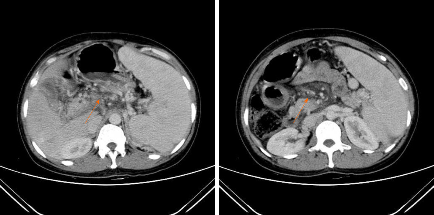
Figure 1
Spleen enlargement, and portal vein thrombosis as shown by the orange arrow and a dilatation of portal hypertension on abdominal computed tomography.
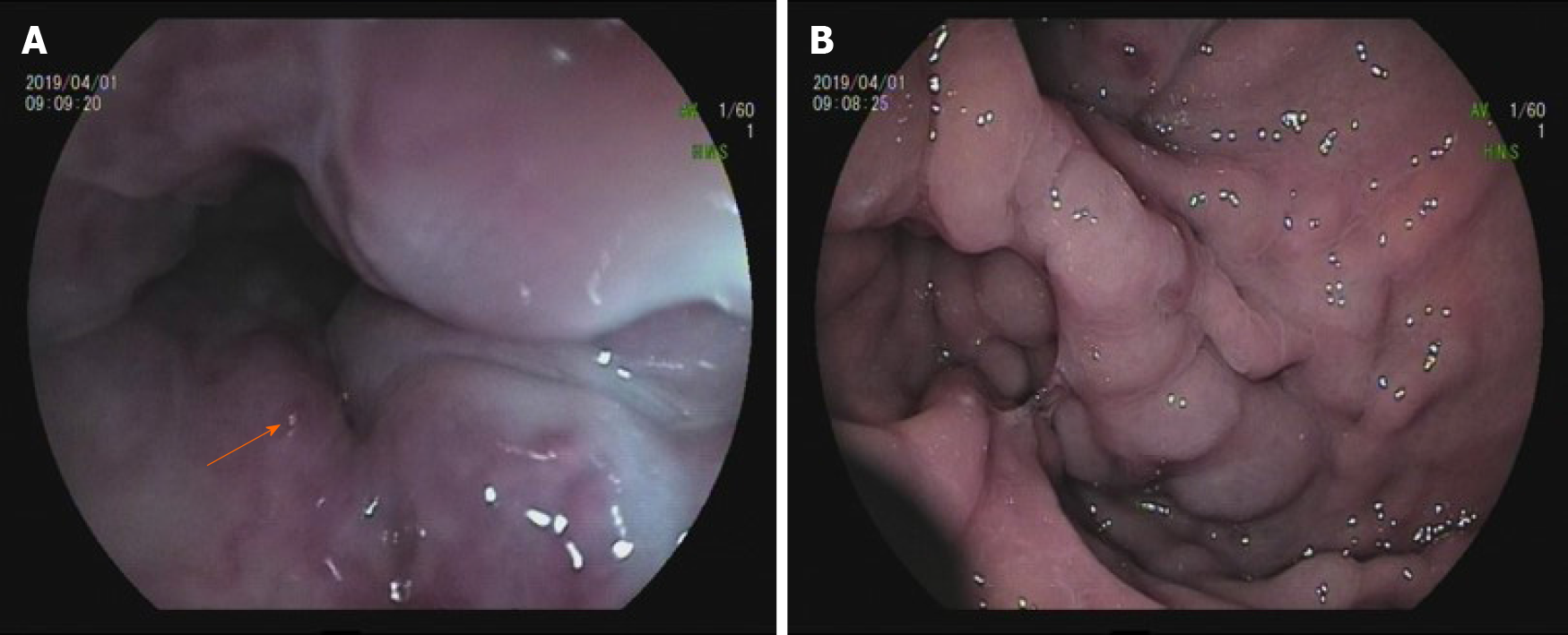
Figure 2 Endoscopic imaging of the esophagus and gastric fundus.
A: Shows a red color sign (i.e., a hematocystic spot) in the lower esophagus as indicated by the orange arrow; B: Gastric fundal varices.
- Citation: Wang JB, Gao Y, Liu JW, Dai MG, Yang SW, Ye B. Gastroesophageal varices in a patient presenting with essential thrombocythemia: A case report. World J Clin Cases 2021; 9(8): 1871-1876
- URL: https://www.wjgnet.com/2307-8960/full/v9/i8/1871.htm
- DOI: https://dx.doi.org/10.12998/wjcc.v9.i8.1871


