Copyright
©The Author(s) 2021.
World J Clin Cases. Mar 16, 2021; 9(8): 1863-1870
Published online Mar 16, 2021. doi: 10.12998/wjcc.v9.i8.1863
Published online Mar 16, 2021. doi: 10.12998/wjcc.v9.i8.1863
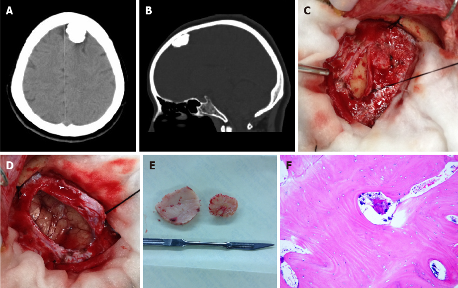
Figure 1 Images of Case 1.
A: Computed tomography showed a lesion, round in shape and with high density, in the left frontal area; B: In the sagittal bone window of computed tomography, a curvilinear lucent line was present between the inner table of the skull and the ossified mass; C: Gross intraoperative view right after opening the skull and retracting the dura. A bony, hard mass was found attached to the inner surface of the dura mater; D: Gross intraoperative view after lesion removal. The underlying arachnoid and compressed brain tissue were intact; E: Gross view of the resected intradural osteoma (right) and the normal inner plate of the skull (left); F: Hematoxylin-eosin-stained section (100 ×) showed mature lamella bone, comprised of the Haver’s system and normal osteocytes between osteoid layers, in pathology examination.
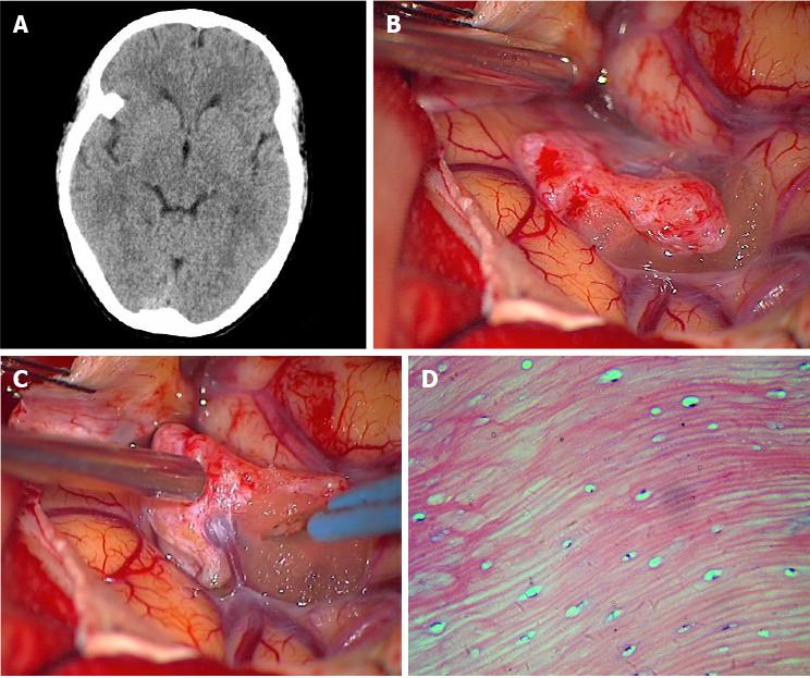
Figure 2 Images of Case 2.
A: Computed tomography showed a homogeneous high-density lesion isolated under the right greater wing of the sphenoid; B: Gross view of the arachnoid-covered bony tumor, located in the sylvian fissure; C: Gross view of a large superficial cortical vein passing through the mass; D: Hematoxylin-eosin-stained section (400 ×) showed mature bone, made up of the Haver's system and normal osteocytes between the layers of osteoid material, in pathology examination.
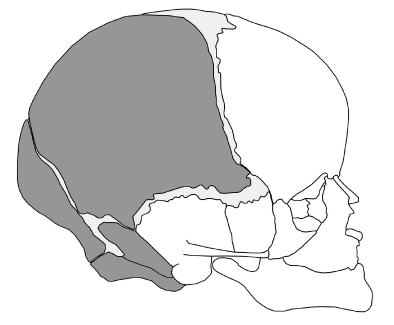
Figure 3 Schematic illustrating the skeletal structure of the skull.
The mesenchyme for these structures is derived from the neural crest (white) and mesoderm (gray).
- Citation: Li L, Ying GY, Tang YJ, Wu H. Intradural osteomas: Report of two cases. World J Clin Cases 2021; 9(8): 1863-1870
- URL: https://www.wjgnet.com/2307-8960/full/v9/i8/1863.htm
- DOI: https://dx.doi.org/10.12998/wjcc.v9.i8.1863


