Copyright
©The Author(s) 2021.
World J Clin Cases. Mar 16, 2021; 9(8): 1853-1862
Published online Mar 16, 2021. doi: 10.12998/wjcc.v9.i8.1853
Published online Mar 16, 2021. doi: 10.12998/wjcc.v9.i8.1853
Figure 1 Radiograph and facial appearance of the child aged 17 mo.
A and B: Cranial computed tomography (CT) scan shows significantly increased bone density and thickened bone plate of the skull. The sinus cavity was small without inflation, and the nasal cavity was obviously narrowed. The nasal bone was thickened with abnormal morphology; C: CT scan shows that the middle ear cavities were narrowed; the lumen of the labyrinth (vestibular, semicircular canal and cochlear) was sclerotic and the ossicular chain was thickened. The width of the left optic canal was 3.93 mm, the width of the right optic canal was 4.17 mm; D: CT scan shows sclerosis of the clavicles and ribs; E: X-ray image shows pronounced metaphyseal flaring in the distal femora and “Flask deformation” of the proximal metaphysis on both sides (Erlenmeyer flask configuration); F: Facial appearance of the patient shows a wide nasal bridge, paranasal bossing, widely spaced eyes with an increased bizygomatic width, and a prominent mandible.
Figure 2 Radiograph and appearance of the child aged 4 yr and 2 mo after low-calcium diet.
No significant changes on cranial and femoral radiographs were observed compared to Figure 1.
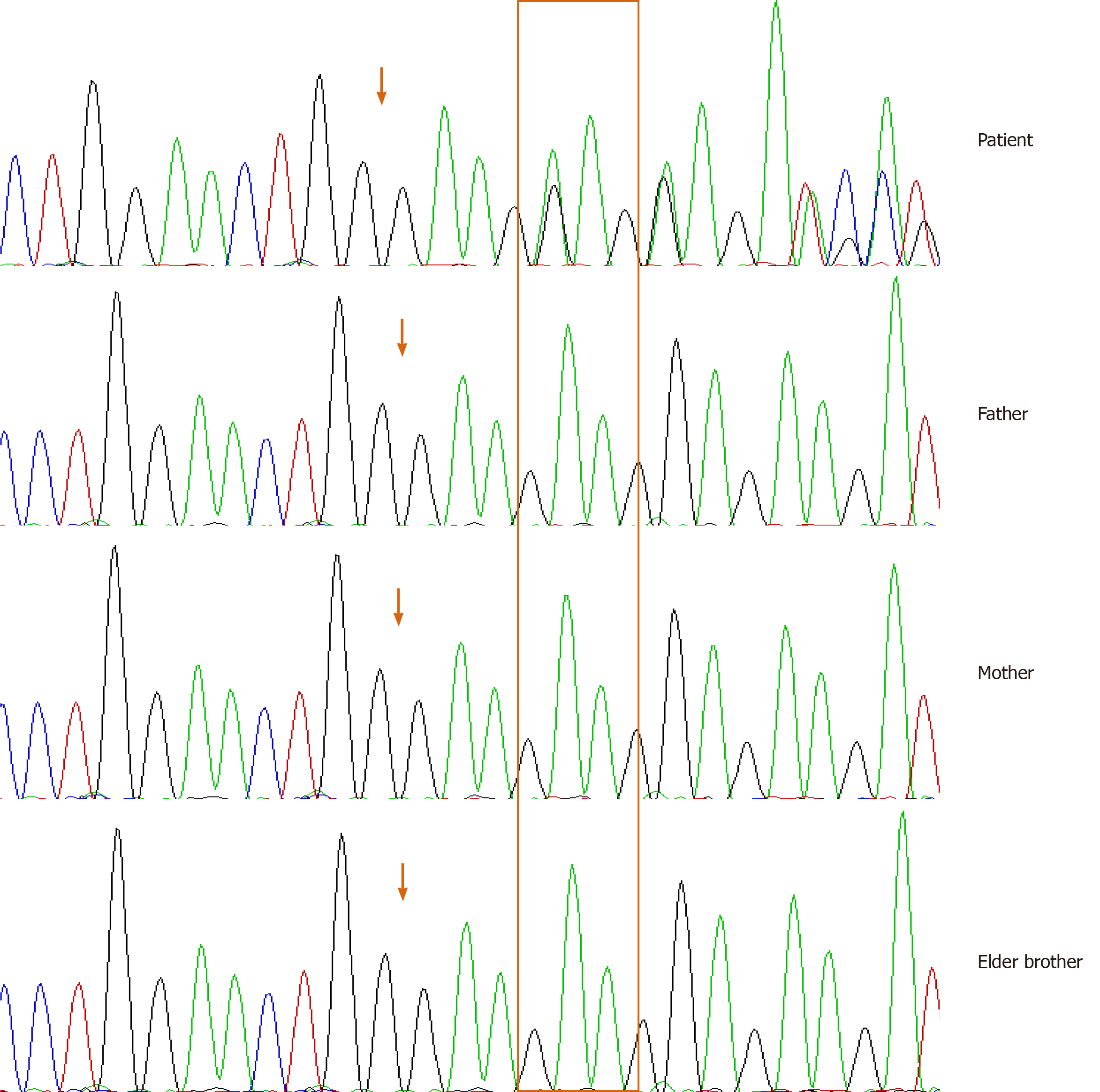
Figure 3 Sanger sequencing results of the ANKH gene.
ANKH gene test results of the patient, father, mother and his elder brother are shown from top to bottom, respectively: a heterozygous mutation c.1129_1131del on exon 9 of gene ANKH was identified in the patient.
- Citation: Wu JL, Li XL, Chen SM, Lan XP, Chen JJ, Li XY, Wang W. A three-year clinical investigation of a Chinese child with craniometaphyseal dysplasia caused by a mutated ANKH gene. World J Clin Cases 2021; 9(8): 1853-1862
- URL: https://www.wjgnet.com/2307-8960/full/v9/i8/1853.htm
- DOI: https://dx.doi.org/10.12998/wjcc.v9.i8.1853


