Copyright
©The Author(s) 2021.
World J Clin Cases. Mar 6, 2021; 9(7): 1748-1754
Published online Mar 6, 2021. doi: 10.12998/wjcc.v9.i7.1748
Published online Mar 6, 2021. doi: 10.12998/wjcc.v9.i7.1748
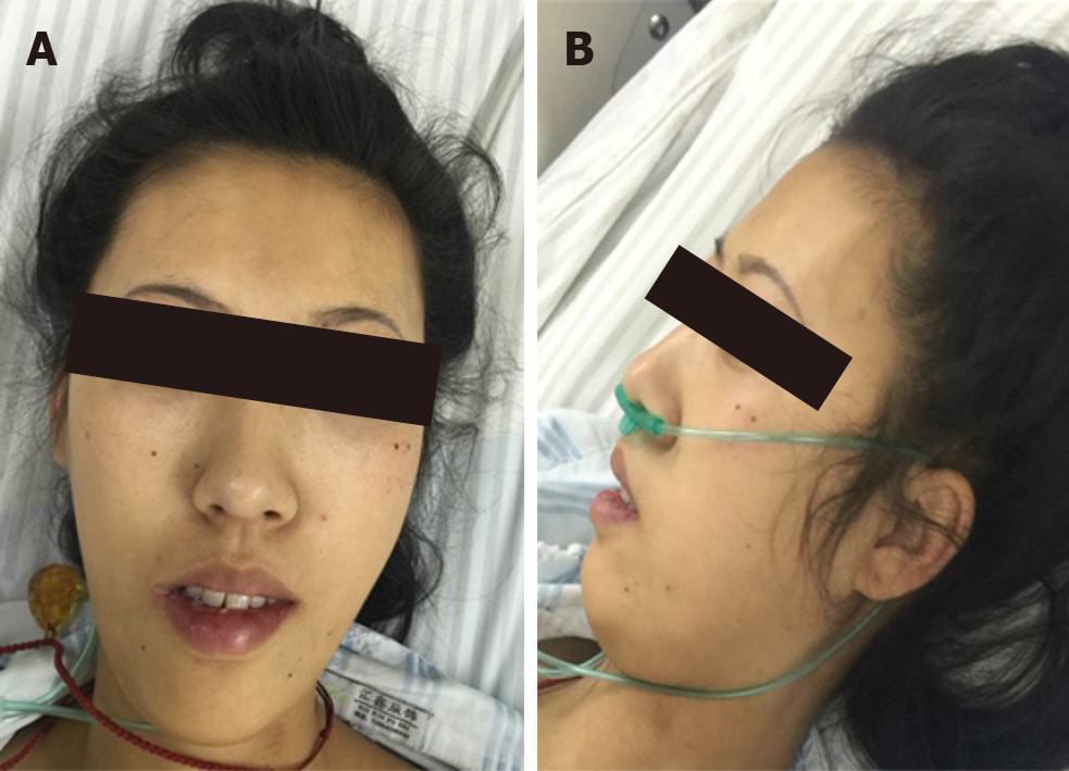
Figure 1 Facial features of the patient.
The patient has characteristic facial features of congenital fiber-type disproportion: thin, elongated face and a high arched palate.
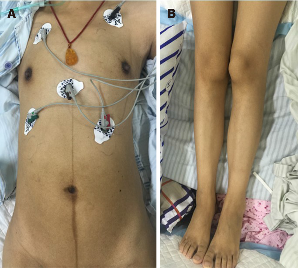
Figure 2 Low muscle volume.
Muscle volume was less in the limbs and trunk.
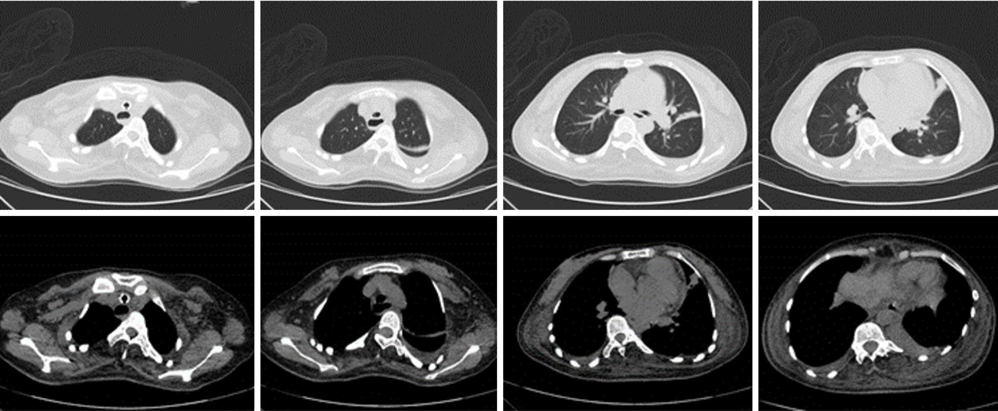
Figure 3 Chest computed tomography.
Scoliosis was observed on her chest computed tomography.
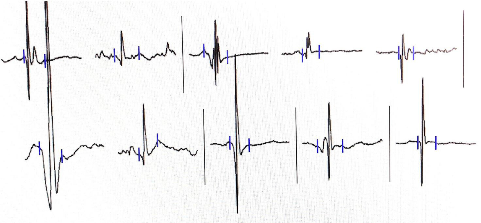
Figure 4 Electromyography suggesting a myopathic process.
Decrease in the duration and amplitude of the motor unit potential were observed. Duration and amplitude findings in the right quadriceps femoris were 7.7 ms and 500 µV (36% decrease), respectively.
Figure 5 Biopsy of the left biceps brachia (100 ×).
A: Hematoxylin and eosin staining: muscle fiber size differed with a dominant type I muscle fiber atrophy; B: Adenosine triphosphate staining (pH, 4.3): type I muscle fiber enzyme activity was well preserved and seen as dark stains; unstained Type II muscle fiber demonstrated a loss of enzyme activity; C: Adenosine triphosphate staining (pH, 10.4): Enzyme activity of type I muscle fiber was partially lost, which can be seen as light stains; type II muscle fiber enzyme activity was well preserved and can be seen as dark stains. Panel A, B, C revealed that the diameters of type I muscle fibers were < type II muscle fibers.
- Citation: Yang HM, Guo JX, Yang YM. Congenital fiber-type disproportion presenting with type II respiratory failure after delivery: A case report. World J Clin Cases 2021; 9(7): 1748-1754
- URL: https://www.wjgnet.com/2307-8960/full/v9/i7/1748.htm
- DOI: https://dx.doi.org/10.12998/wjcc.v9.i7.1748


