Copyright
©The Author(s) 2021.
World J Clin Cases. Feb 26, 2021; 9(6): 1455-1460
Published online Feb 26, 2021. doi: 10.12998/wjcc.v9.i6.1455
Published online Feb 26, 2021. doi: 10.12998/wjcc.v9.i6.1455
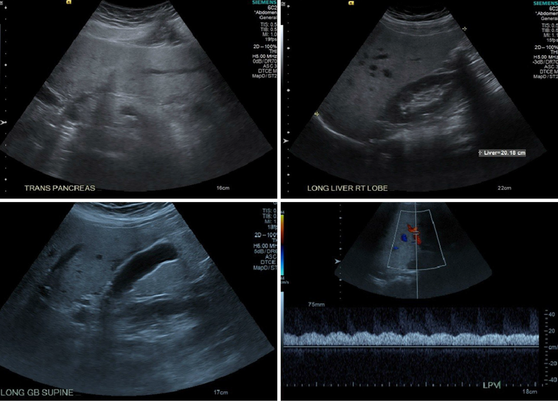
Figure 1 Intrahepatic vessels appear unremarkable including the hepatic veins.
Increased liver echogenicity with moderately enlarged liver most consistent with fatty infiltration. Mild heterogeneous liver around the gallbladder fossa likely related parenchymal sparing in an otherwise fatty liver.
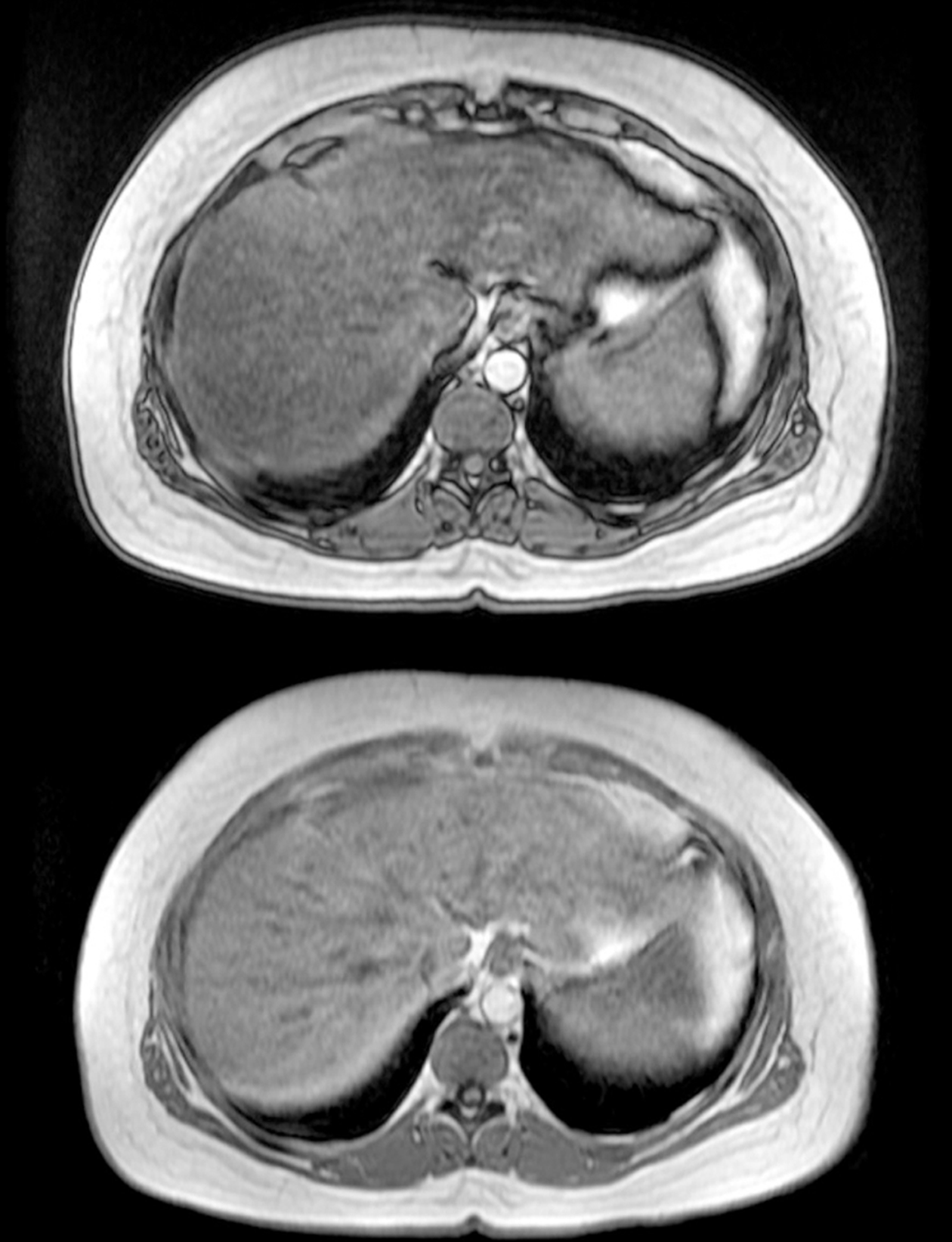
Figure 2 Magnetic resonance cholangiopancreatography results.
Mild hepatosplenomegaly and hepatic steatosis.
- Citation: Khiatah B, Nasrollah L, Covington S, Carlson D. Nonalcoholic fatty liver disease as a risk factor for cytomegalovirus hepatitis in an immunocompetent patient: A case report. World J Clin Cases 2021; 9(6): 1455-1460
- URL: https://www.wjgnet.com/2307-8960/full/v9/i6/1455.htm
- DOI: https://dx.doi.org/10.12998/wjcc.v9.i6.1455


