Copyright
©The Author(s) 2021.
World J Clin Cases. Feb 16, 2021; 9(5): 1175-1183
Published online Feb 16, 2021. doi: 10.12998/wjcc.v9.i5.1175
Published online Feb 16, 2021. doi: 10.12998/wjcc.v9.i5.1175
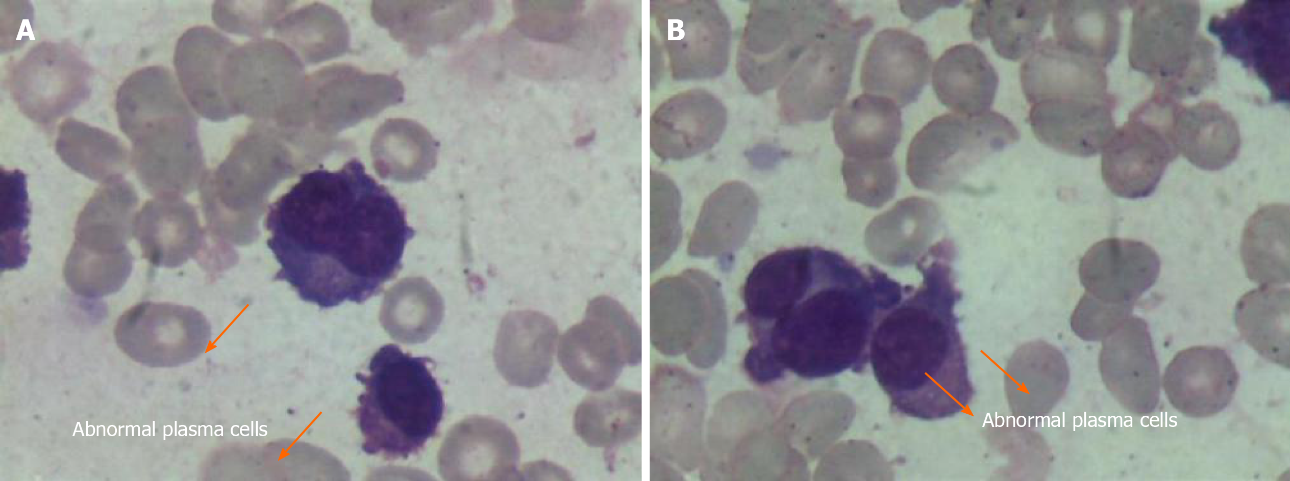
Figure 1 Circulating plasma cells as evident on the peripheral smear (1000 ×) in June 2015.
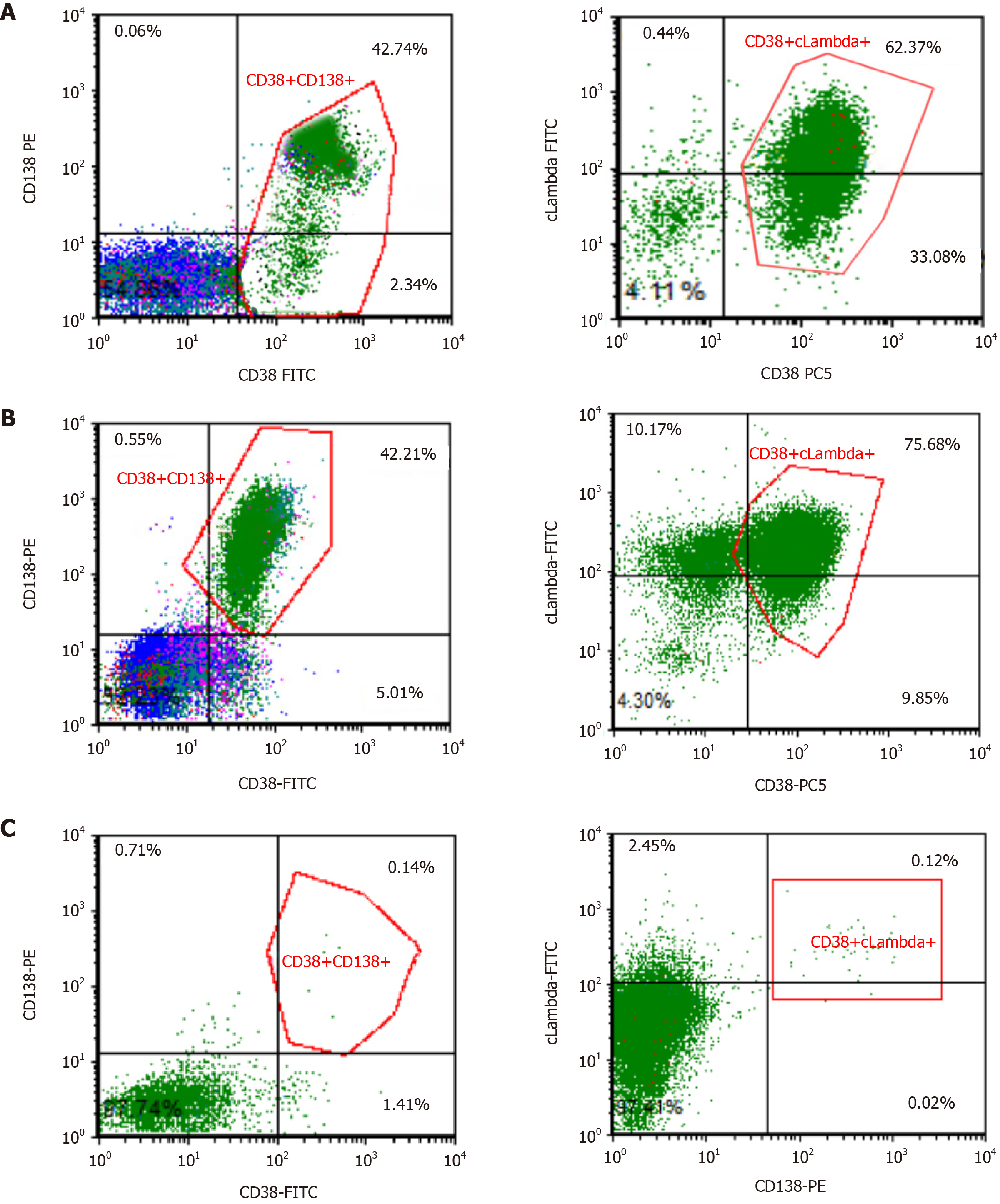
Figure 2 Flow-cytometry analysis.
A: Plasma cells (PCs) in the peripheral blood at the time of diagnosis (here exemplarily shown CD38 and CD138); B: PCs in the peripheral blood before venetoclax use; C: PCs in the bone marrow after two cycles of venetoclax.
Figure 3 Hematoxylin and eosin staining and immunohistostaining of bone marrow clot section (400 ×).
H/E: Hematoxylin and eosin staining.
Figure 4 Fluorescence in situ hybridization analysis.
A: RB1 (13q14) gene deletion in fluorescence in situ hybridization; B: CKS1B (1q21) gene amplification in fluorescence in situ hybridization.
- Citation: Yang Y, Fu LJ, Chen CM, Hu MW. Venetoclax in combination with chidamide and dexamethasone in relapsed/refractory primary plasma cell leukemia without t(11;14): A case report. World J Clin Cases 2021; 9(5): 1175-1183
- URL: https://www.wjgnet.com/2307-8960/full/v9/i5/1175.htm
- DOI: https://dx.doi.org/10.12998/wjcc.v9.i5.1175


