Copyright
©The Author(s) 2021.
World J Clin Cases. Feb 16, 2021; 9(5): 1103-1110
Published online Feb 16, 2021. doi: 10.12998/wjcc.v9.i5.1103
Published online Feb 16, 2021. doi: 10.12998/wjcc.v9.i5.1103
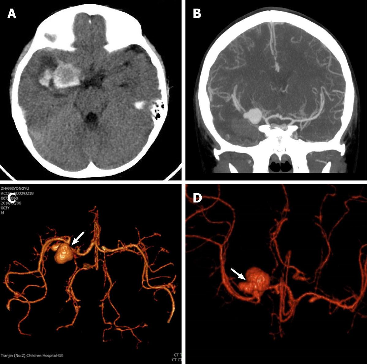
Figure 1 Preoperative imaging examination.
A: Subarachnoid hemorrhage caused by ruptured intracranial dissecting aneurysm (IDA); B: Computed tomography angiography shows intracranial aneurysm in the right medical council on alcohol; C and D: Pearl-and-string sign of IDAs (focal stenoses proximally and distally, which are noted by white arrows.
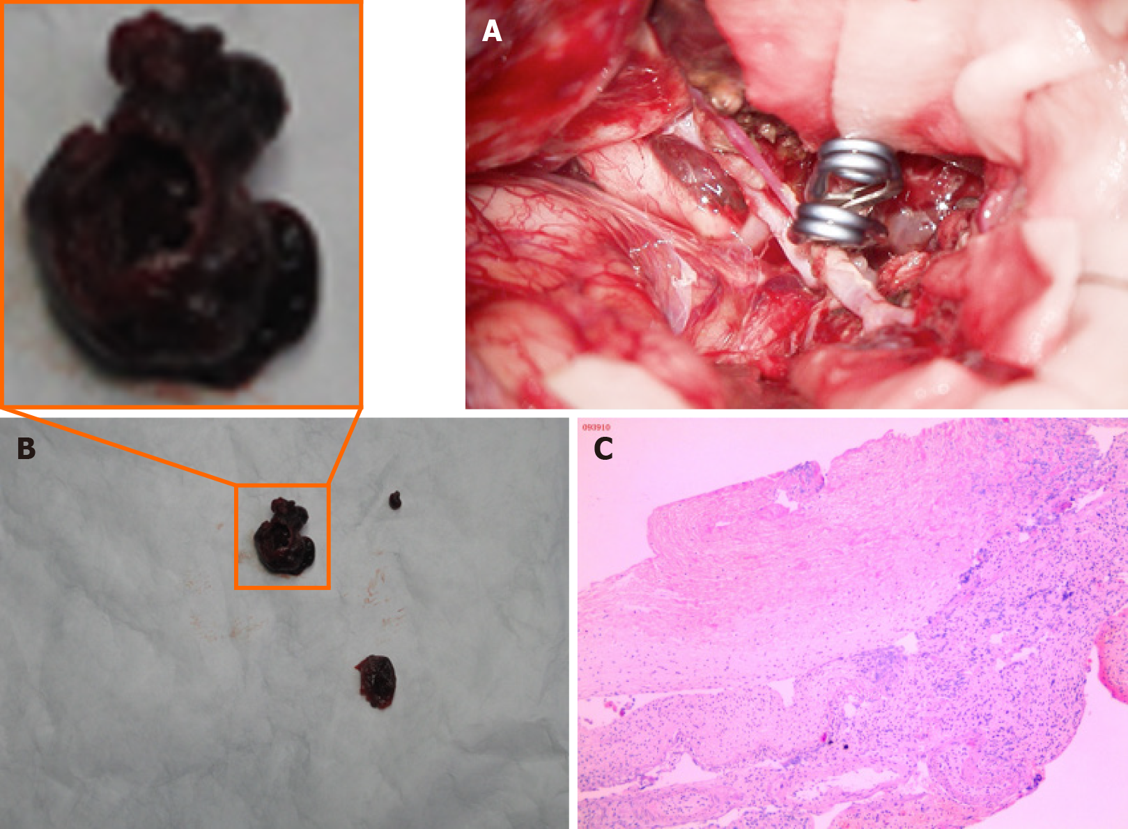
Figure 2 Clipping and angioplasty for intracranial dissecting aneurysms, and pathological examination.
A: The aneurysm was clipped; B: The wall of the intracranial dissecting aneurysm was very thin, and a thrombus was adhered to the wall; C: The intracranial dissecting aneurysm was resected and sent for pathological examination. Pathological examination indicated irregular and malformed vascular wall structure with inflammatory infiltration.
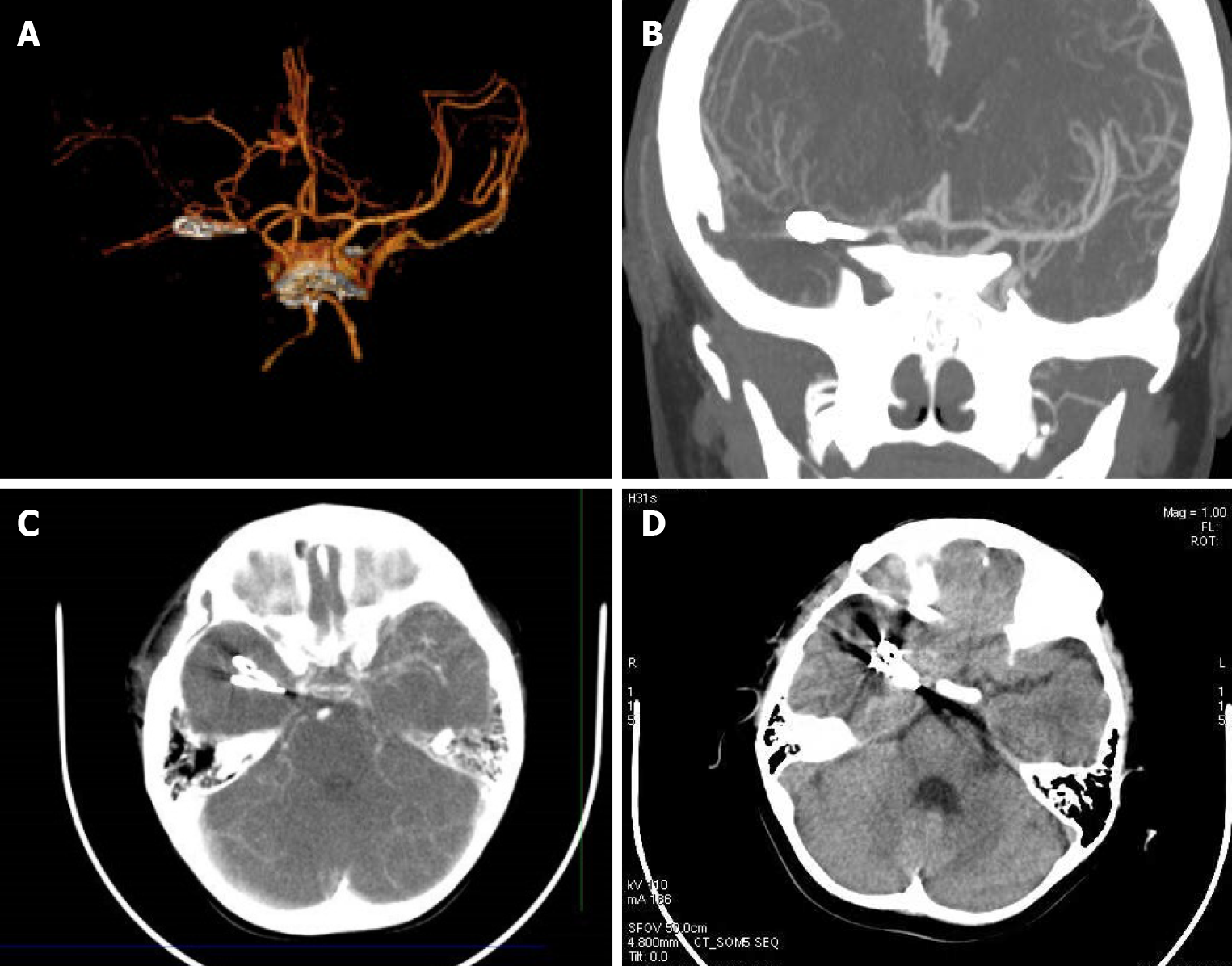
Figure 3 Postoperative computed tomography angiography examination and follow-up.
A-C: Postoperative computed tomography angiography examination indicated that the aneurysm had been resected, and the blood flow of the constructed medical council on alcohol was unobstructed; D: The 3-year follow-up showed no recurrence.
- Citation: Sun N, Yang XY, Zhao Y, Zhang QJ, Ma X, Wei ZN, Li MQ. Treatment of pediatric intracranial dissecting aneurysm with clipping and angioplasty, and next-generation sequencing analysis: A case report and literature review . World J Clin Cases 2021; 9(5): 1103-1110
- URL: https://www.wjgnet.com/2307-8960/full/v9/i5/1103.htm
- DOI: https://dx.doi.org/10.12998/wjcc.v9.i5.1103


