Copyright
©The Author(s) 2021.
World J Clin Cases. Feb 6, 2021; 9(4): 878-885
Published online Feb 6, 2021. doi: 10.12998/wjcc.v9.i4.878
Published online Feb 6, 2021. doi: 10.12998/wjcc.v9.i4.878
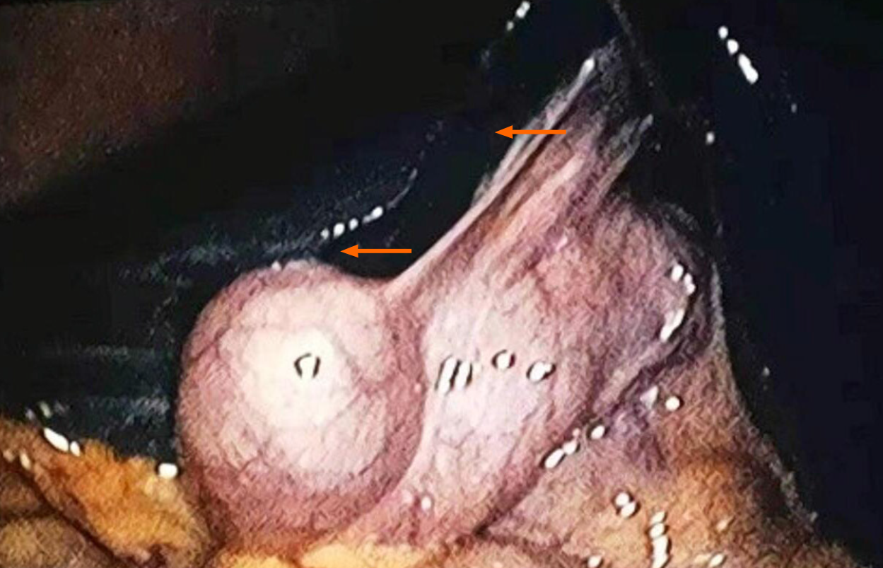
Figure 1 Laparoscopic images presenting a deep black-brown liver with a smooth surface and the slightly shorter edges, and a stone-filled gallbladder (orange arrows).
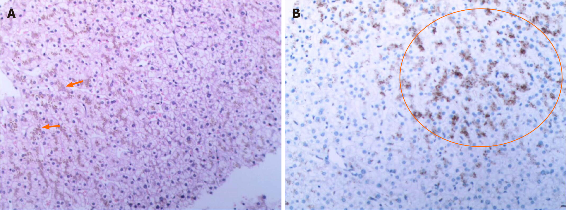
Figure 2 Imaging results.
A: Hematoxylin and eosin staining of liver cells showing a large number of pigment granule deposits (orange arrow) in a patient with suspected Dubin-Johnson syndrome (DJS); original magnification × 20; B: Hepatic tissue immunohistochemistry results showing that liquified or vacuolized liver cells (orange circle), which indicates DJS.
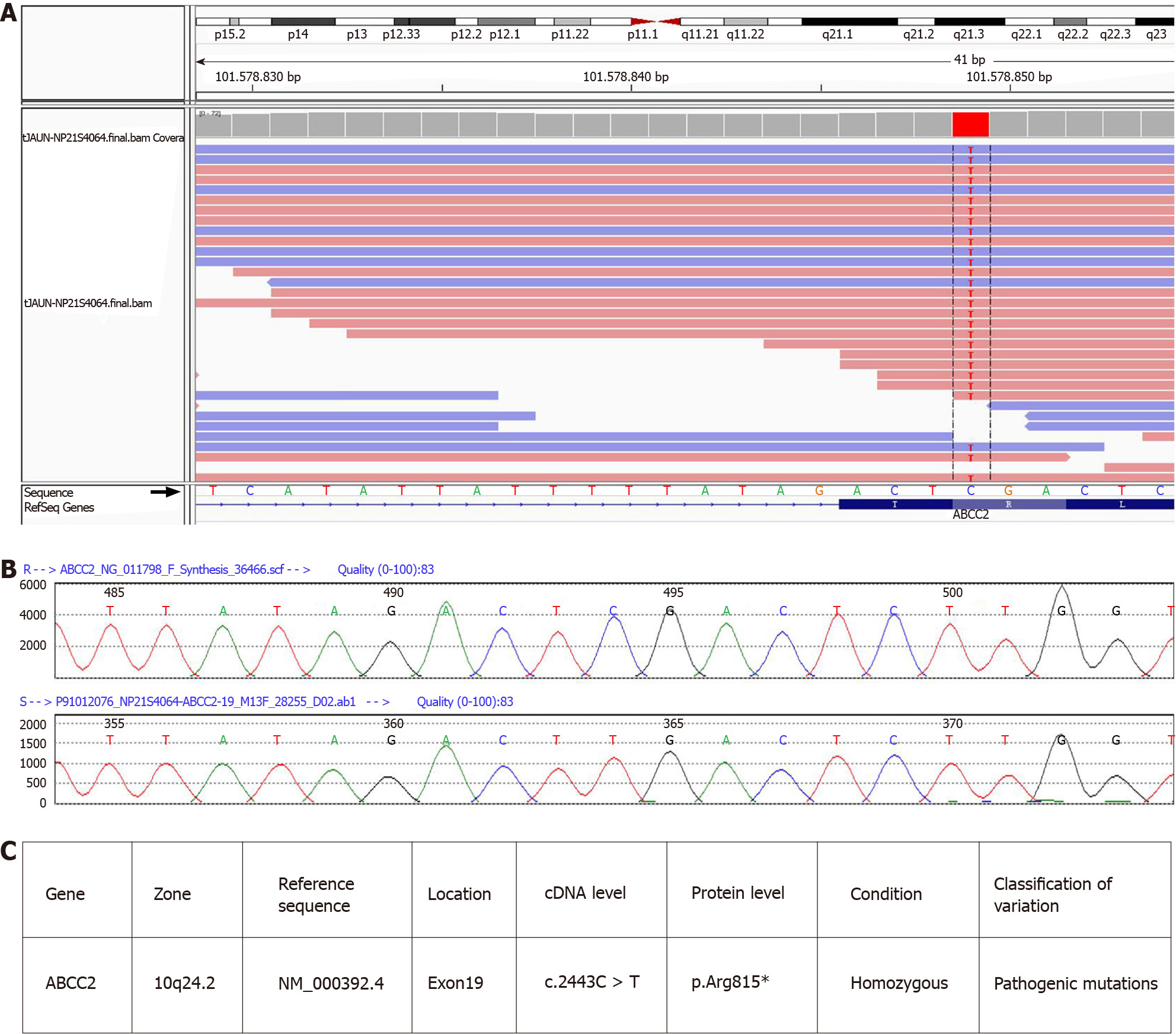
Figure 3 Next-generation sequencing results of ABCC2 in a Dubin-Johnson syndrome patient.
A: Next-generation sequencing chromatographic representation of ABCC2 mutation in the patient. The red arrow indicates that the cytosine (C) at position 2443 of the ABCC2 gene coding region is mutated to thymine (T); B: Sanger sequencing chromatogram. The lower panel shows the mutated sequence in the patient, and the upper panel shows the normal sequence in control subjects; C: Details of the mutation sites.
- Citation: Wu H, Zhao XK, Zhu JJ. Clinical characteristics and ABCC2 genotype in Dubin-Johnson syndrome: A case report and review of the literature. World J Clin Cases 2021; 9(4): 878-885
- URL: https://www.wjgnet.com/2307-8960/full/v9/i4/878.htm
- DOI: https://dx.doi.org/10.12998/wjcc.v9.i4.878


