Copyright
©The Author(s) 2021.
World J Clin Cases. Feb 6, 2021; 9(4): 854-863
Published online Feb 6, 2021. doi: 10.12998/wjcc.v9.i4.854
Published online Feb 6, 2021. doi: 10.12998/wjcc.v9.i4.854
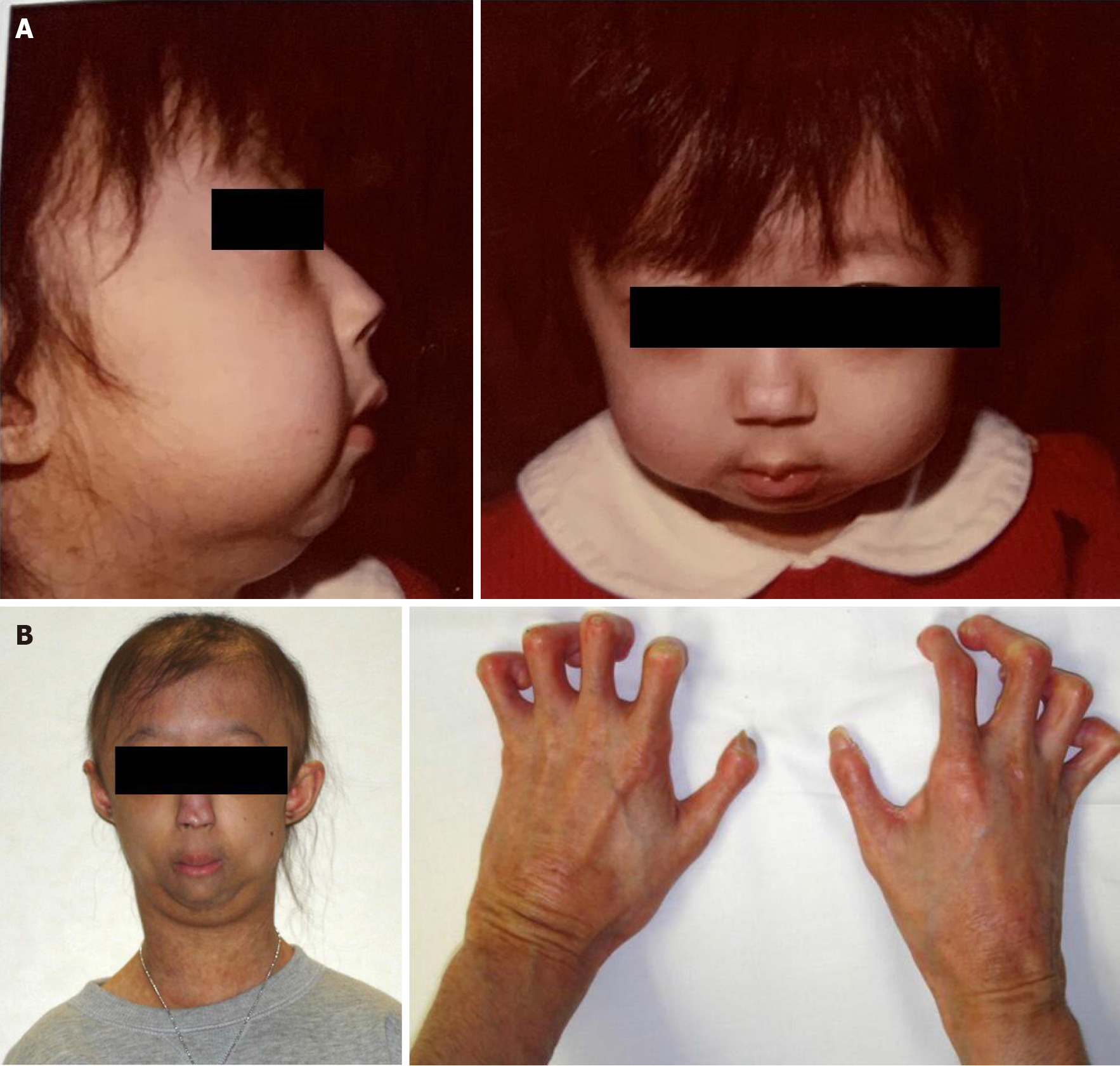
Figure 1 Clinical photos.
A: 3 years old; B: Thin nose, prominent eyes and occipital dysplasia are indicated. 18 years old when she presented at our hospital with osteosarcoma. She had ichthyosis-like skin, bird-like facies, lack of subcutaneous fat, hair loss, flexion contractures in the fingers, and sloping shoulders.
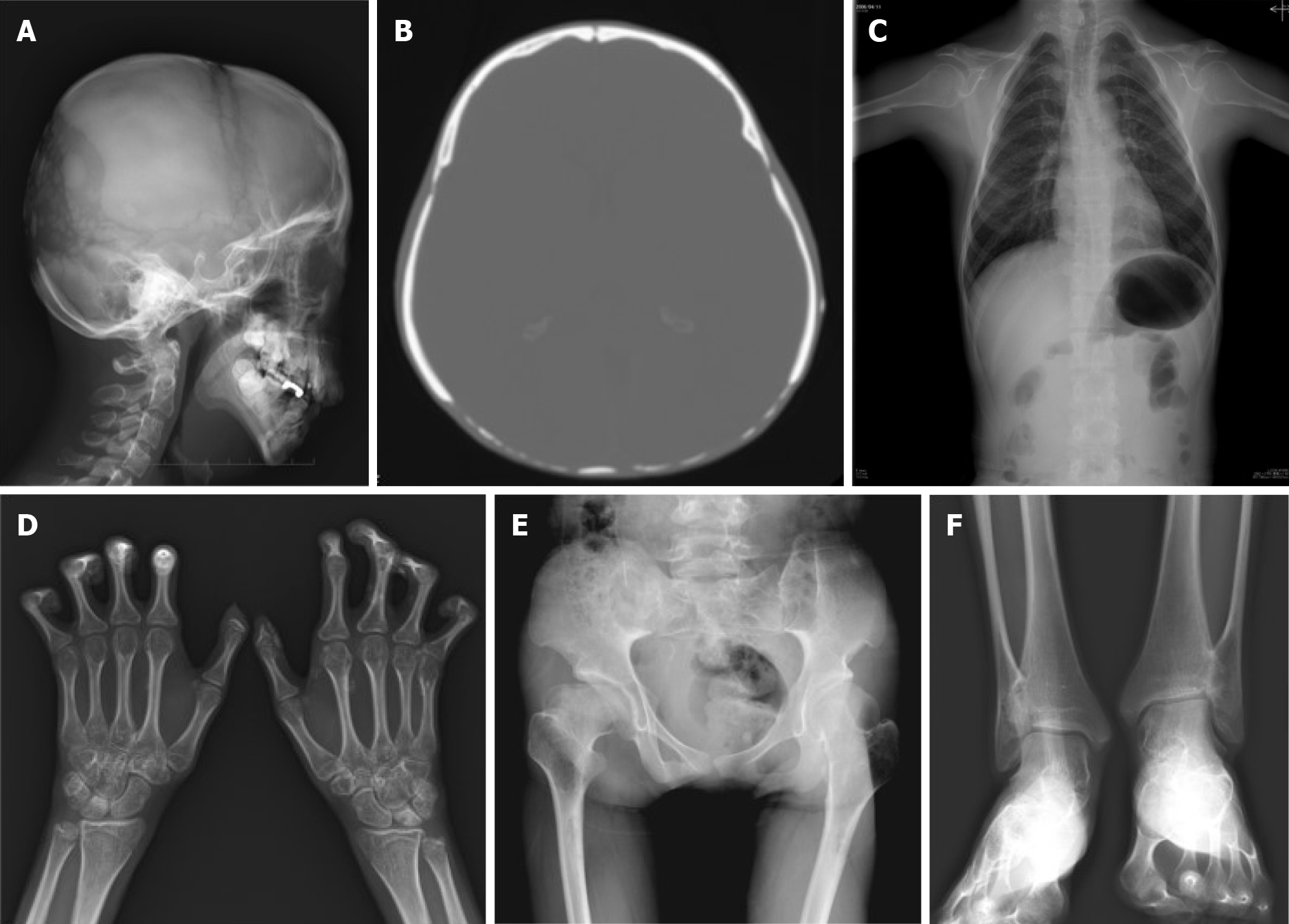
Figure 2 X-rays.
A and B: Occipital dysplasia; C: Short clavicles and pyriform thorax; D: DIP and PIP joints in the hands bilaterally had flexion contractures and heterotopic ossification was noted; E: Post-Perthes-disease deformity; F: Distal tibiofibular synostosis.
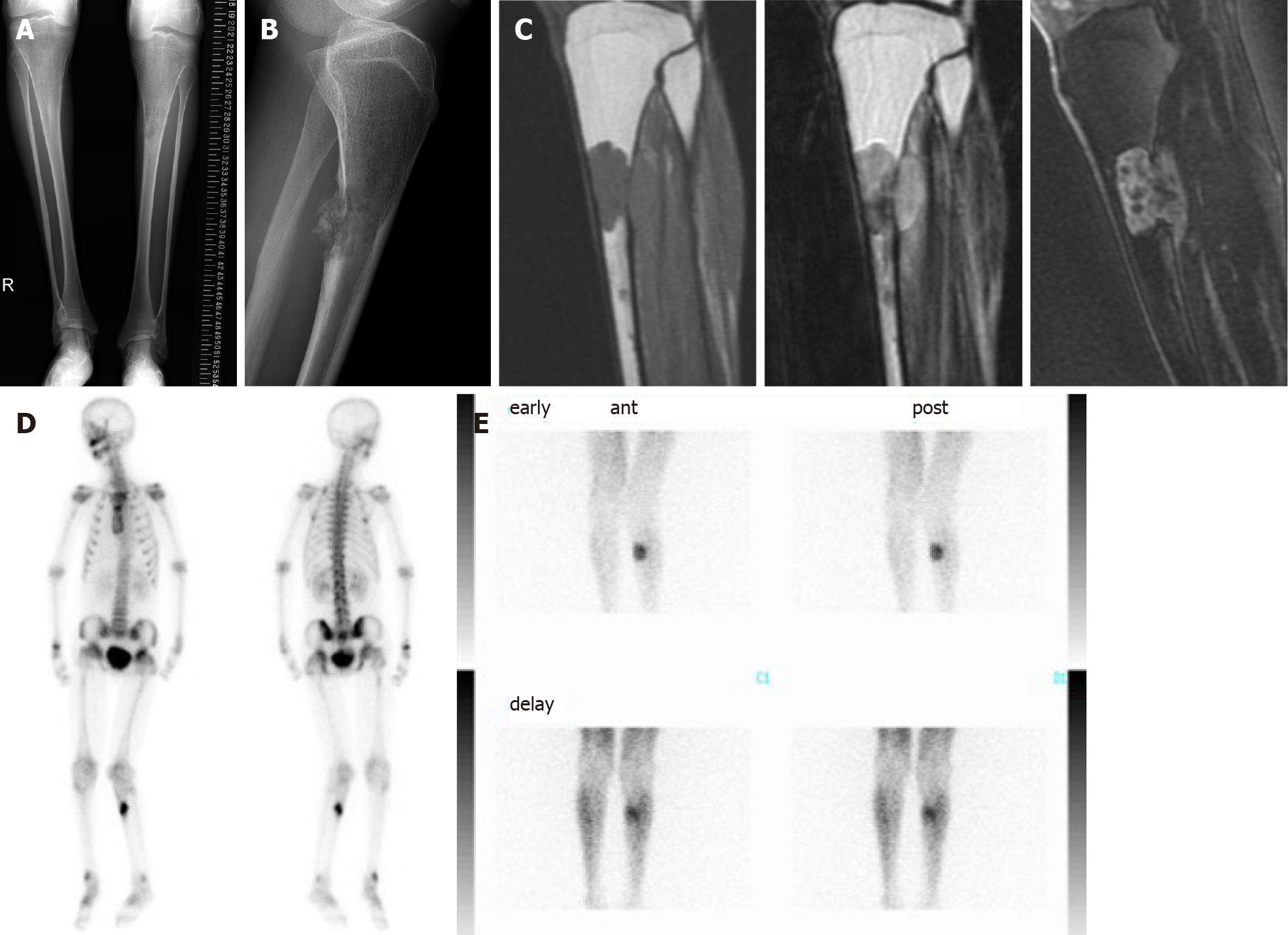
Figure 3 Image findings of bone tumor of tibia.
A and B: Bilateral tibial shafts were morphologically small in diameter and narrow in the medullary cavities. An irregular osteolytic lesion was observed in the anteroposterior view, and posterior cortical destruction and extraosseous calcified lesions were observed in the lateral view; C: Magnetic resonance imaging showed intra- and extra-osseous lesions with homogenous low intensity in T1-weighted images, low-to-high intensity in T2-weighted images, and enhanced inhomogeneity by gadolinium; D: Bone scintigraphy showed accumulation in the proximal tibia, but no other bone lesions were detected; E: Thallium scintigraphy showed high accumulation at the tumor site in the tibia.
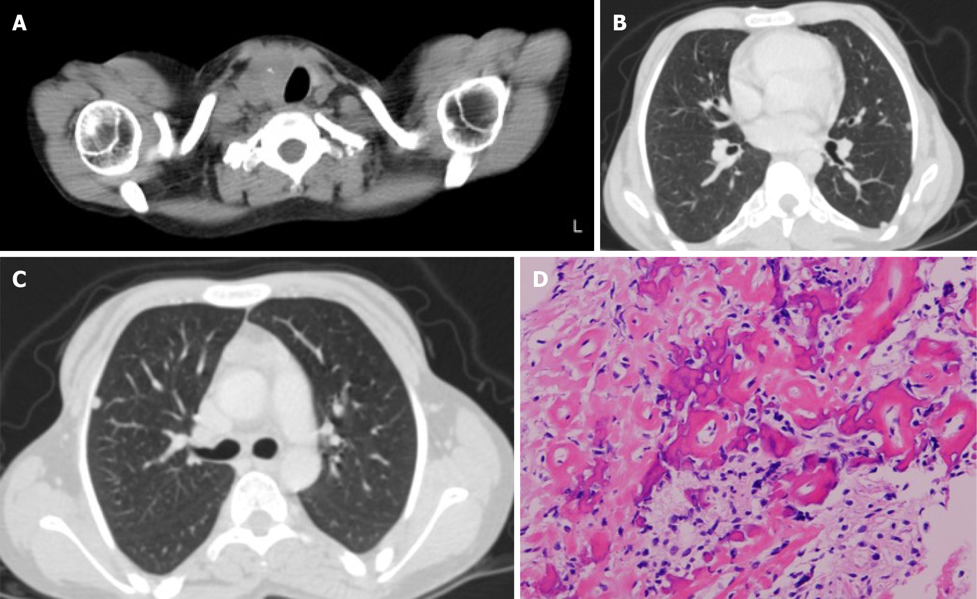
Figure 4 Thyroid tumor and biopsy result of bone tumor.
A-C: Computed tomography showed left tracheal deviation and thyroid tumor and multiple small nodules in the lungs were also detected; D: Open biopsy of the tibial bone tumor revealed that the tumor cells produced eosinophilic immature osteoids and were varied in size with nuclear atypia. Both low- and high-grade variants were included, but the final diagnosis was a conventional fibroblastic osteosarcoma.
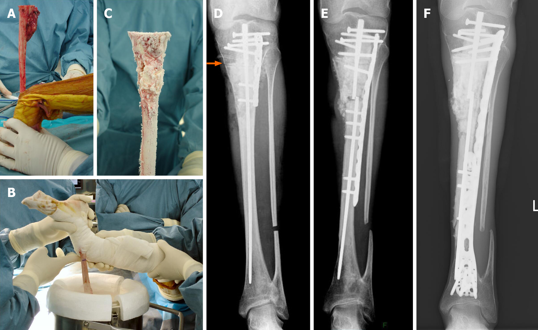
Figure 5 Frozen autografting of osteosarcoma and postoperative X-rays.
A: In the surgery, tumor bone was exposed with wide margins and a proximal tibial osteotomy was performed 3 cm below the knee joint; B: The tibia was rotated and put in the container filled with liquid nitrogen for 20 min; C: Thawed at room temperature for 15 min and in distilled water for 15 min; D: Post-operative X-ray. Frozen autograft was replaced with a small intramedullary nail, plate, and polymethylmethacrylate. Arrow is osteotomy line; E: Three years after surgery, an X-ray revealed fracture of the frozen bone at the end plate and additional plate fixation with autologous bone grafting was performed; F: 13 years post-operatively. Although multiple surgeries were needed for non-union and infection, limb salvage was successful and no recurrences have been detected.
- Citation: Hayashi K, Yamamoto N, Takeuchi A, Miwa S, Igarashi K, Araki Y, Yonezawa H, Morinaga S, Asano Y, Tsuchiya H. Long-term survival in a patient with Hutchinson-Gilford progeria syndrome and osteosarcoma: A case report. World J Clin Cases 2021; 9(4): 854-863
- URL: https://www.wjgnet.com/2307-8960/full/v9/i4/854.htm
- DOI: https://dx.doi.org/10.12998/wjcc.v9.i4.854


