Copyright
©The Author(s) 2021.
World J Clin Cases. Dec 26, 2021; 9(36): 11475-11481
Published online Dec 26, 2021. doi: 10.12998/wjcc.v9.i36.11475
Published online Dec 26, 2021. doi: 10.12998/wjcc.v9.i36.11475
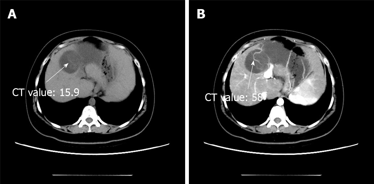
Figure 1 Computed tomography scan.
A: Non-contrast-enhanced axial computed tomography scan showing a hypodense multilocular cystic lesion; B: In the arterial phases, the cyst wall and intracystic separations are hyperdense. CT: Computed tomography.
Figure 2 Conventional magnetic resonance imaging scan.
A and B: Conventional magnetic resonance imaging scan showing that the lesion is hypointense on T1-weighted imaging (A) and hyperintense on T2-weighted imaging (B); C: The cyst wall is hyperdense on enhanced fat-suppressed T1-weighted imaging.
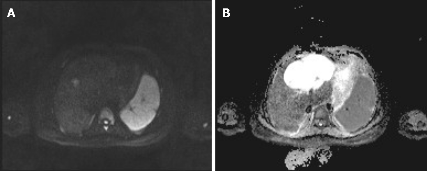
Figure 3 The diffusion of the cystic wall nodules is restricted on diffusion-weighted imaging and apparent diffusion coefficient imaging.
A: Diffusion-weighted imaging; B: Apparent diffusion coefficient imaging.
Figure 4 Pathological material and histopathological examination.
A: Grossly, the tumor was multilocular, and the cavities contained fluid of variable consistency; B and C: Histopathological examination showing a fibrous cyst wall tissue covered with tall columnar epithelium. Most segments contain ovarian-like stroma.
- Citation: Yu TY, Zhang JS, Chen K, Yu AJ. Mucinous cystic neoplasm of the liver: A case report. World J Clin Cases 2021; 9(36): 11475-11481
- URL: https://www.wjgnet.com/2307-8960/full/v9/i36/11475.htm
- DOI: https://dx.doi.org/10.12998/wjcc.v9.i36.11475


