Copyright
©The Author(s) 2021.
World J Clin Cases. Dec 26, 2021; 9(36): 11457-11466
Published online Dec 26, 2021. doi: 10.12998/wjcc.v9.i36.11457
Published online Dec 26, 2021. doi: 10.12998/wjcc.v9.i36.11457
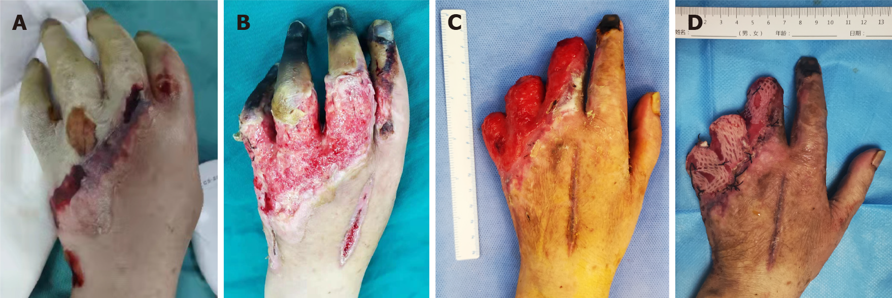
Figure 1 Appearance of the patient's hand during hospitalization.
A: Pale skin of the second to fifth fingers in the left hand mimicking Raynaud’s phenomenon upon admission; B: The dorsal skin of the hand was necrotic and exfoliated in 1 wk, and dry gangrene occured in the fingertips; C: The wound reached the standard for skin grafting about 3 wk after digital amputation; D: Appearance 1 wk after autologous skin grafting shows successful performance.
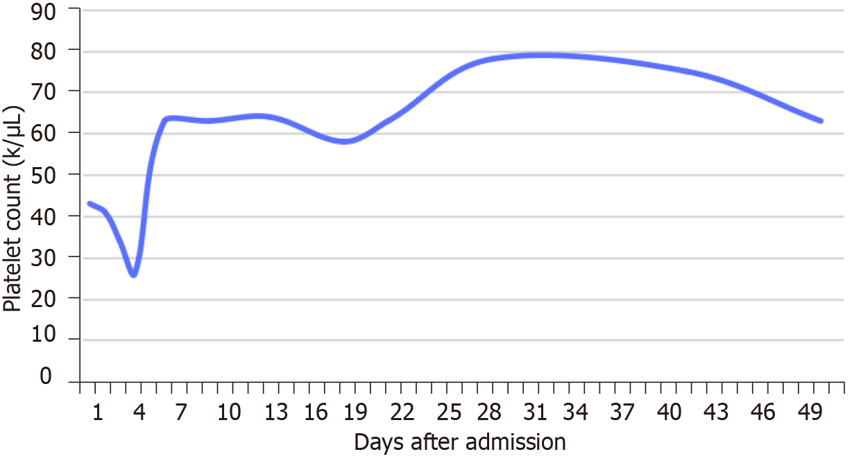
Figure 2 The patient’s platelet count changed during hospitalization.
The patient’s platelet count continued to decrease after admission, and gradually increased after hormonal therapy and platelet transfusion (1 U), but it did not reach the normal value (150-450 k/µL) during hospitalization.
Figure 3 Computed tomography and ultrasound imaging of the patient.
A: Abdominal computed tomography showed normal dimensions of the liver but irregular contours. There were several mass lesions that took up mixed contrast in the arterial phase (white arrows); B: The mass lesions lost their contrast in the portal venous phase; C: Color Doppler ultrasound in the abdomen showed cirrhosis with multiple solid masses in the liver, and high enhancement in the arterial phase.
- Citation: Chen JL, Yu X, Luo R, Liu M. Severe digital ischemia coexists with thrombocytopenia in malignancy-associated antiphospholipid syndrome: A case report and review of literature. World J Clin Cases 2021; 9(36): 11457-11466
- URL: https://www.wjgnet.com/2307-8960/full/v9/i36/11457.htm
- DOI: https://dx.doi.org/10.12998/wjcc.v9.i36.11457


