Copyright
©The Author(s) 2021.
World J Clin Cases. Dec 26, 2021; 9(36): 11362-11368
Published online Dec 26, 2021. doi: 10.12998/wjcc.v9.i36.11362
Published online Dec 26, 2021. doi: 10.12998/wjcc.v9.i36.11362
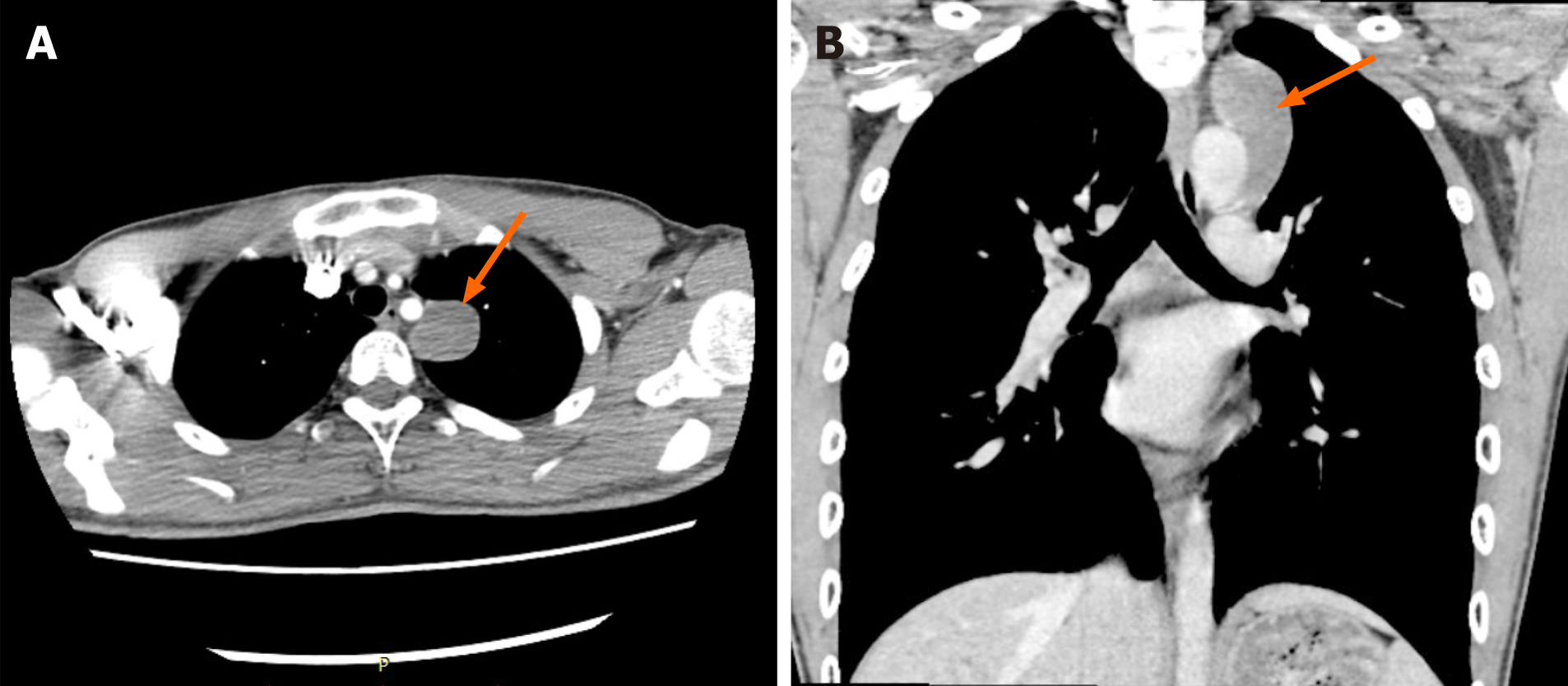
Figure 1 Chest computed tomography scan before surgery suggested a mediastinal mass above the left aortic arch.
A: Cross-section of chest computed tomography (CT); B: Coronal section of chest CT. The orange arrows refer to the mediastinal mass.
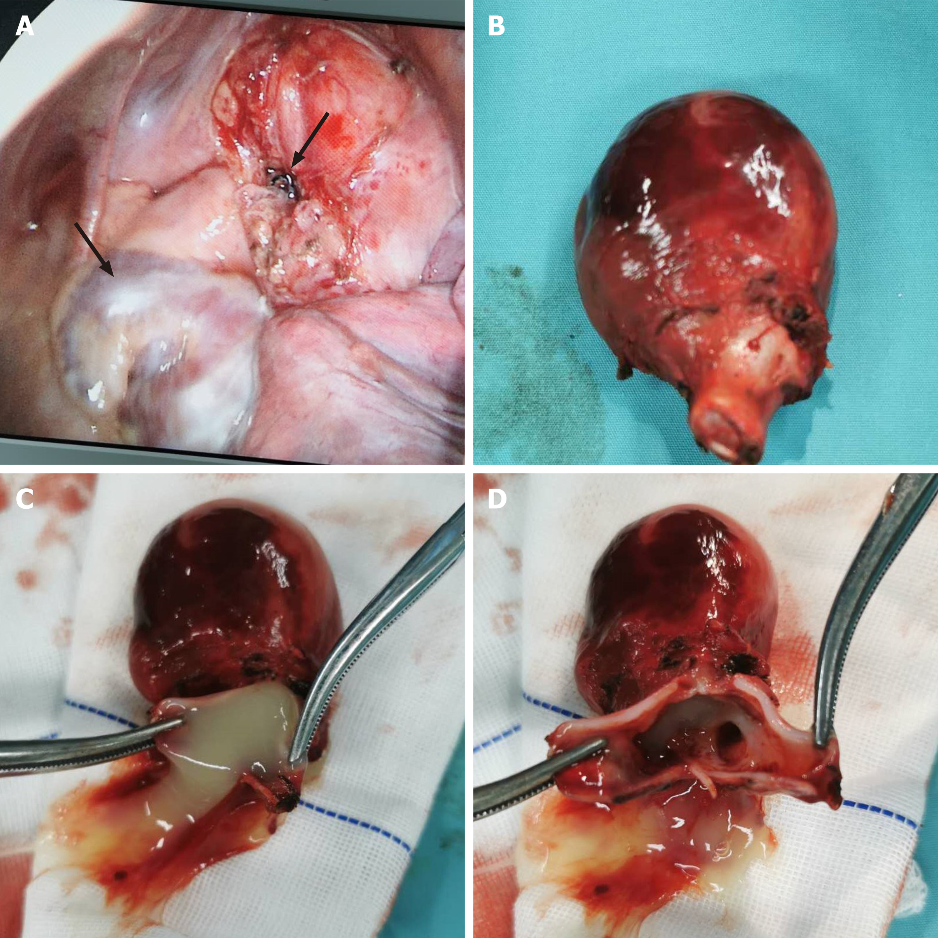
Figure 2 Cysts removed during surgery, and visual field under thoracoscopy.
A: The right arrow refers to the broken end of the cyst, the left arrow refers to the left atrium and left atrial appendage, and the lower right of the field of vision is the lung tissue. B: The cyst pedicle is solid tissue, and cartilage fragments can be seen from the pedicle; C: The fluid inside the cyst; D: Bronchial bifurcation-like structure in the cysts.
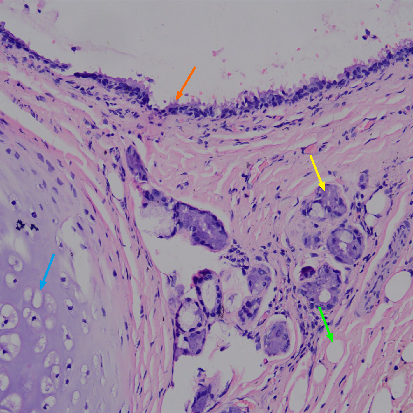
Figure 3 The pathological diagnosis of the tumor was a bronchial cyst.
The orange arrow refers to the pseudolayer ciliar epithelium, the yellow arrow refers to the bronchial glands, the blue arrow refers to the cartilage tissue, and the green arrow refers to the fat tissue. Tumor tissues were fixed with 10% neutral formaldehyde, embedded in paraffin, and then stained with hematoxylin and eosin (200 ×).
- Citation: Zhu X, Zhang L, Tang Z, Xing FB, Gao X, Chen WB. Mature mediastinal bronchogenic cyst with left pericardial defect: A case report. World J Clin Cases 2021; 9(36): 11362-11368
- URL: https://www.wjgnet.com/2307-8960/full/v9/i36/11362.htm
- DOI: https://dx.doi.org/10.12998/wjcc.v9.i36.11362


