Copyright
©The Author(s) 2021.
World J Clin Cases. Dec 16, 2021; 9(35): 11078-11084
Published online Dec 16, 2021. doi: 10.12998/wjcc.v9.i35.11078
Published online Dec 16, 2021. doi: 10.12998/wjcc.v9.i35.11078
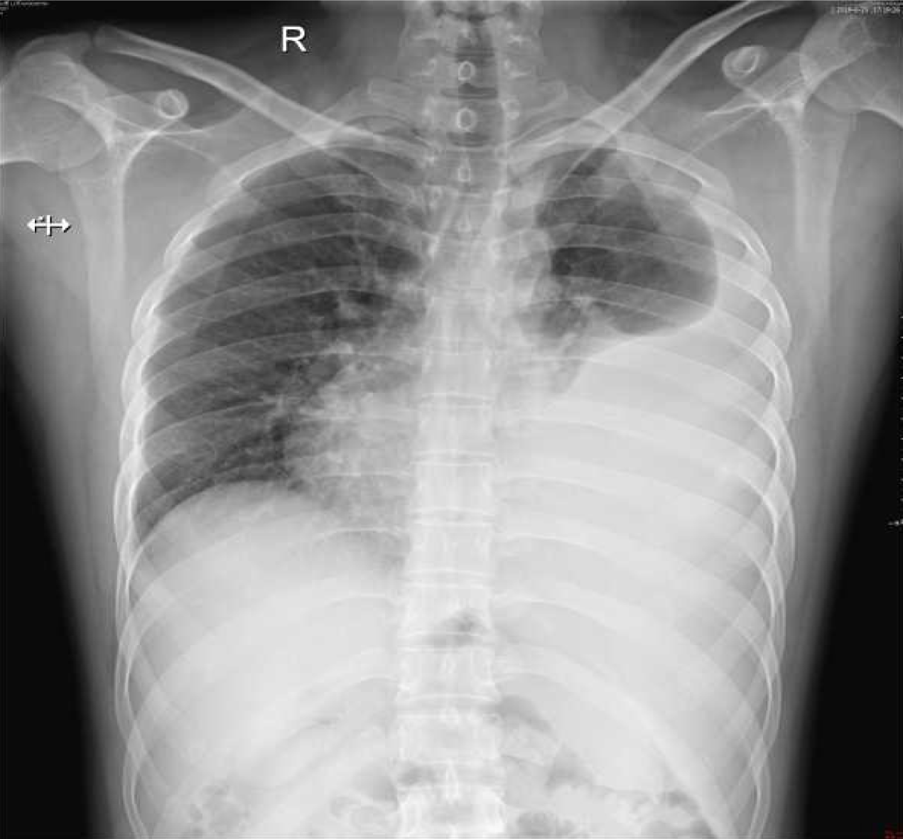
Figure 1 A chest radiograph showed severe left hydrothorax.
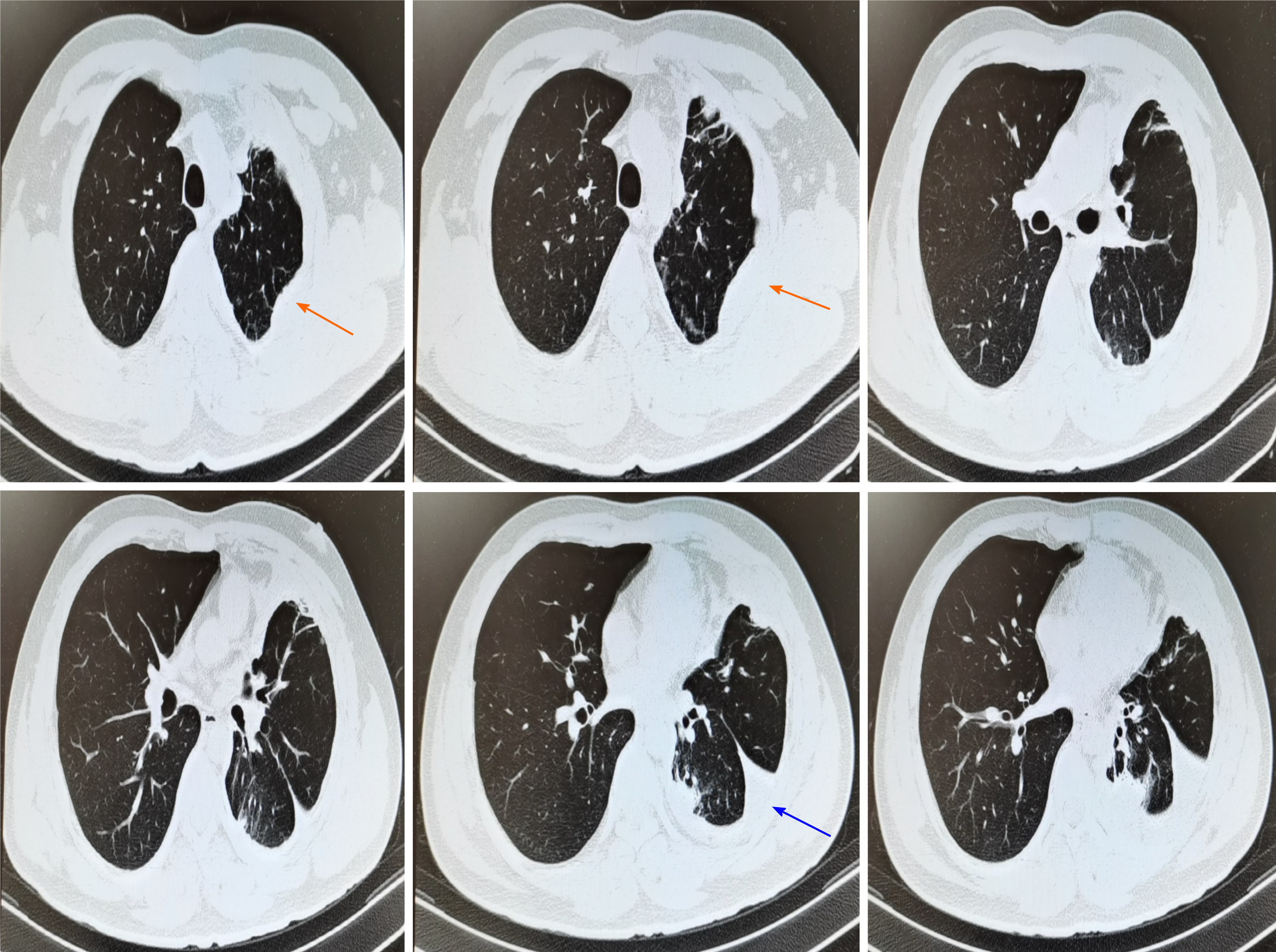
Figure 2 Chest computed tomography demonstrated multiple nodules on left visceral pleura (indicated by orange arrows) and part of atelectasis in the left lower lung (showed by the blue arrow).
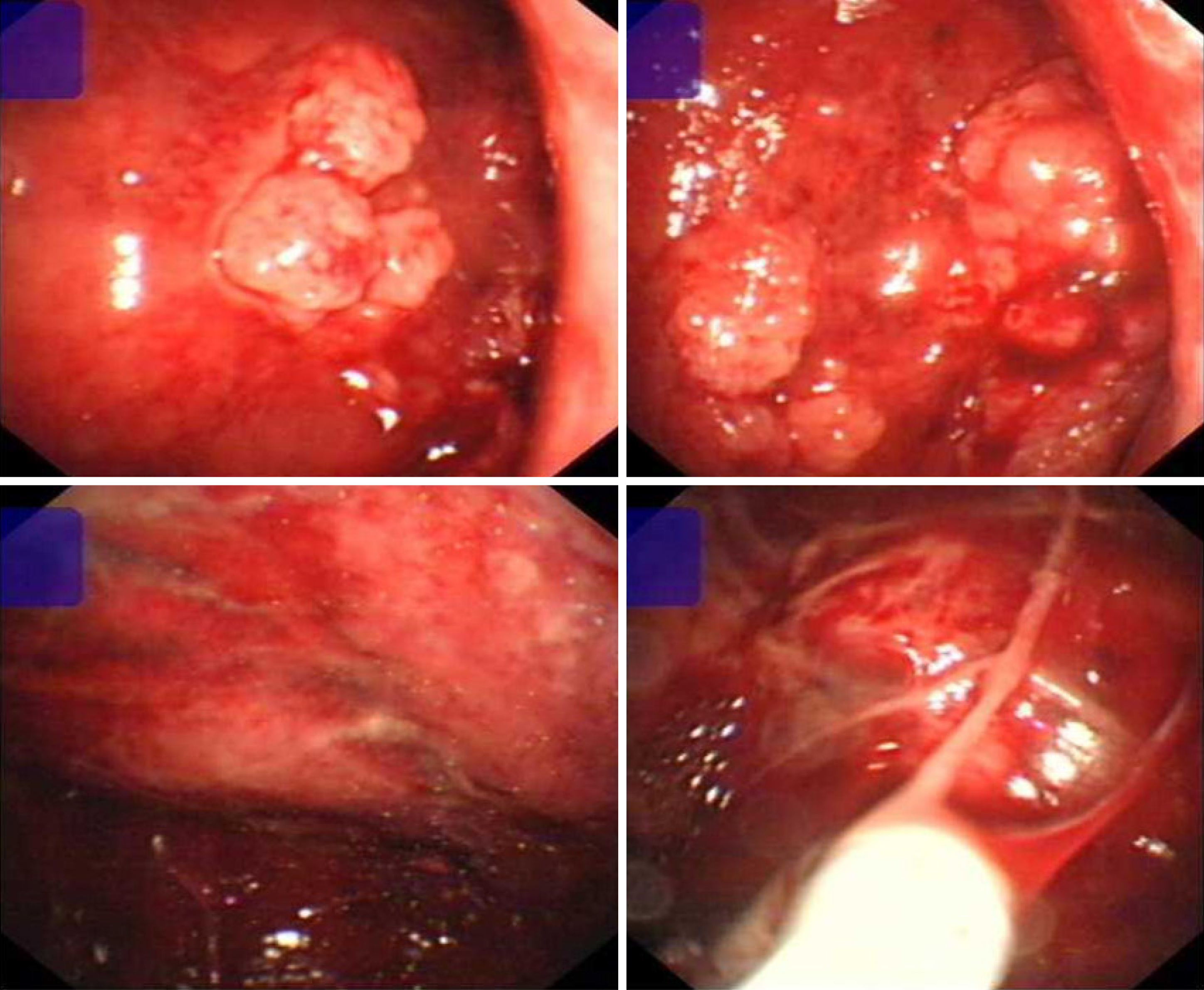
Figure 3 Thoracoscopy revealed visceral pleura almost full of nodular, cauliflower-like protrusions of various sizes.
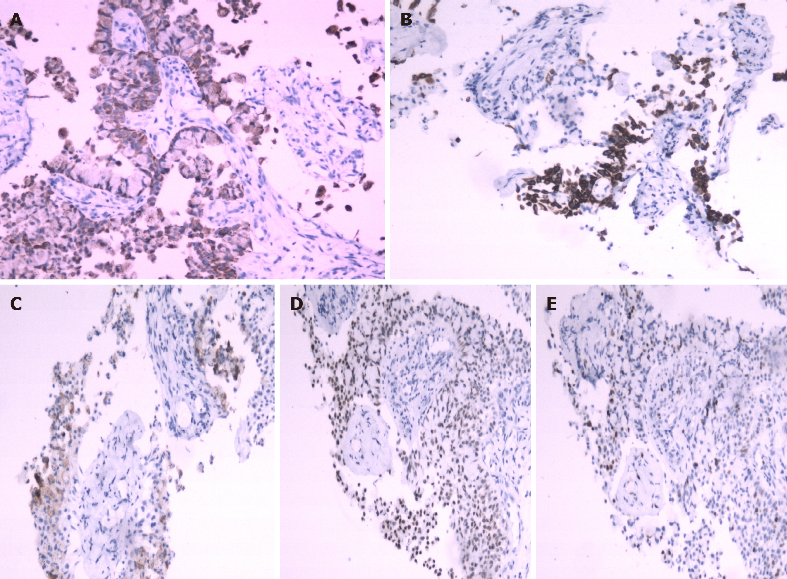
Figure 4 Immunohistochemical analysis showed that the tumor cells were positive for cytokeratin, cytokeratin 5/6, carcinoembryonic antigen, thyroid transcription factor-1 and Ki-67.
A: Cytokeratin; B: Cytokeratin 5/6, C: Carcinoembryonic antigen; D: Thyroid transcription factor-1; E: Ki-67.
- Citation: Wang T. Double-mutant invasive mucinous adenocarcinoma of the lung in a 32-year-old male patient: A case report. World J Clin Cases 2021; 9(35): 11078-11084
- URL: https://www.wjgnet.com/2307-8960/full/v9/i35/11078.htm
- DOI: https://dx.doi.org/10.12998/wjcc.v9.i35.11078


