Copyright
©The Author(s) 2021.
World J Clin Cases. Dec 16, 2021; 9(35): 11071-11077
Published online Dec 16, 2021. doi: 10.12998/wjcc.v9.i35.11071
Published online Dec 16, 2021. doi: 10.12998/wjcc.v9.i35.11071
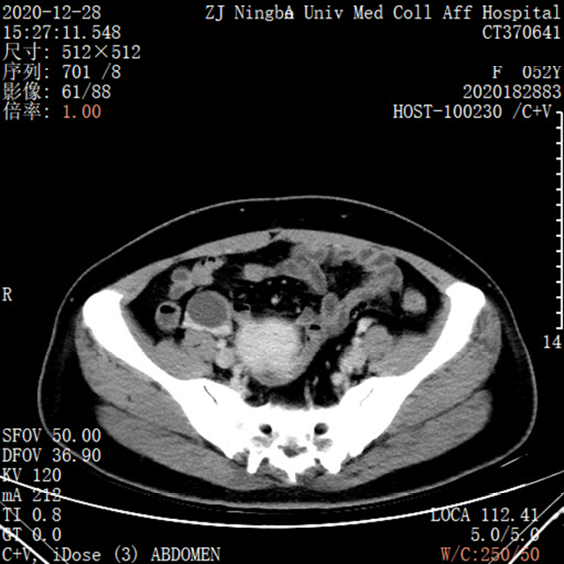
Figure 1 Contrast-enhanced computed tomography scans of the abdomen showed no specific abnormalities in the left colon.
Figure 2 Multiangle photographs of the pedunculated polyp under colonoscopy.
A-C: The pedunculated polyp shows the characteristic features of adenoma with unclear surface pattern; D: The pedunculated polyp was resected under colonoscopy.
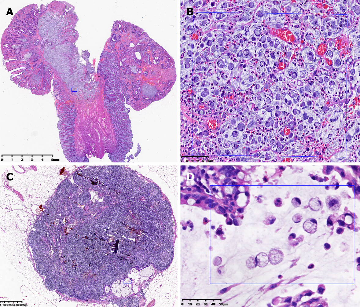
Figure 3 The pathologic results for pedunculated polyps and lymph nodes.
A: The tumor was composed of signet ring cell carcinoma (dark rectangle, hematoxylin and eosin: 0.52 ×); B: The pathologic result in the rectangle clearly showed that the signet ring cells infiltrated the submucosa (hematoxylin and eosin: 40 ×); C: The lymph node was invaded by signet ring cell adenocarcinoma (blue rectangle, hematoxylin and eosin: 1.32 ×); D: The pathologic result in the rectangle clearly showed that the signet ring cells invaded a lymph node (hematoxylin and eosin: 40 ×).
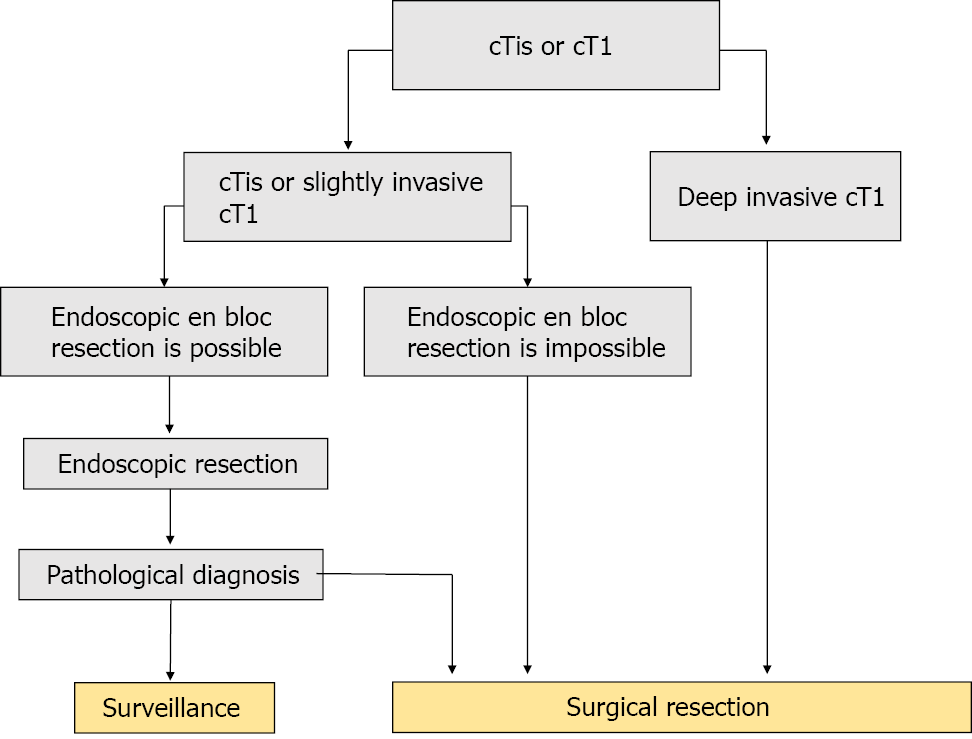
Figure 4 The treatment strategies for cTis and cT1 colorectal cancer from the Japanese Society for Cancer of the Colon and Rectum Guidelines.
- Citation: Yan JN, Shao YF, Ye GL, Ding Y. Signet ring cell carcinoma hidden beneath large pedunculated colorectal polyp: A case report. World J Clin Cases 2021; 9(35): 11071-11077
- URL: https://www.wjgnet.com/2307-8960/full/v9/i35/11071.htm
- DOI: https://dx.doi.org/10.12998/wjcc.v9.i35.11071


