Copyright
©The Author(s) 2021.
World J Clin Cases. Dec 16, 2021; 9(35): 10828-10837
Published online Dec 16, 2021. doi: 10.12998/wjcc.v9.i35.10828
Published online Dec 16, 2021. doi: 10.12998/wjcc.v9.i35.10828
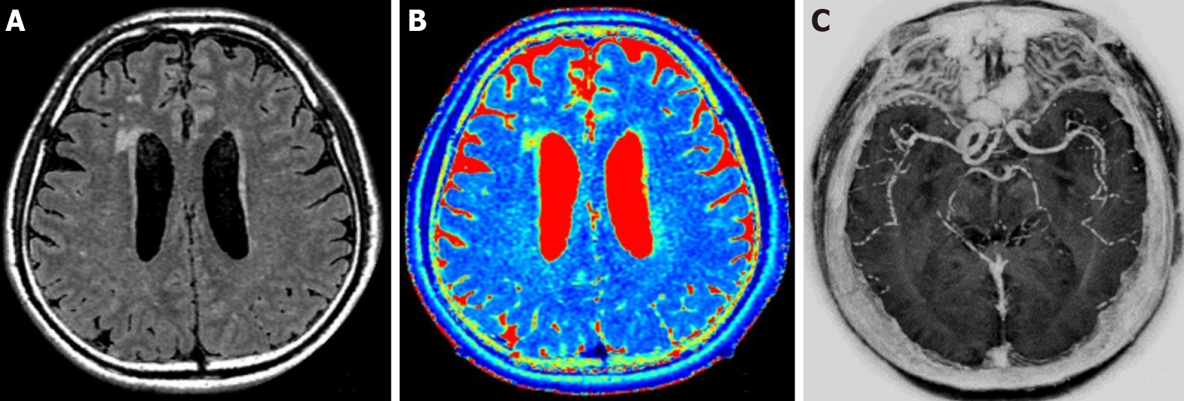
Figure 1 A 61-year-old man with transient ischemic attack.
Automatic image reconstruction from MAGnetic resonance imaging compilation (MAGiC) raw data. A: MAGiC T2-fluid-attenuated inversion recovery (left); B: MAGiC T2 mapping (middle); C: MAGiC phase sensitive inversion recovery Vessel (right).
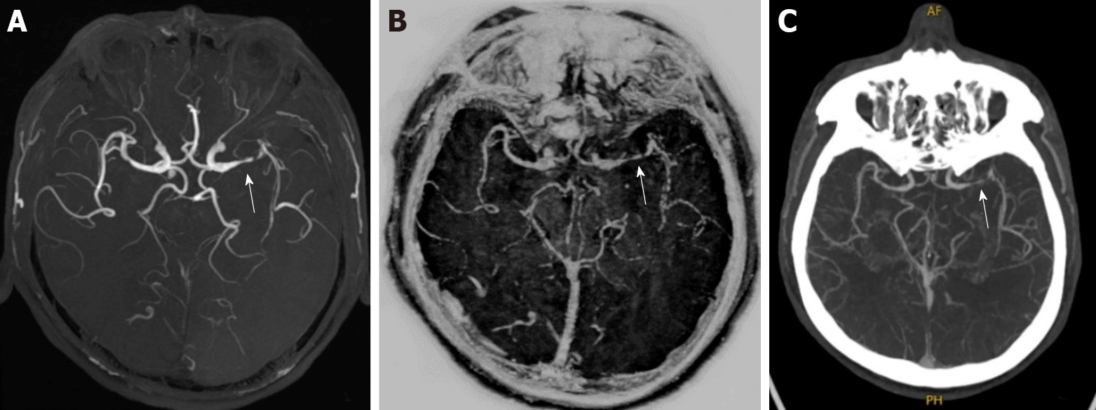
Figure 2 A 49-year-old man with bilateral lacunar cerebral infarction.
A: Three-dimensional time-of-flight magnetic resonance angiography (left); B: MAGnetic resonance imaging compilation phase sensitive inversion recovery Vessel (middle); C: Computed tomography angiography (right). All three examination methods showed severe local stenosis of the left middle cerebral artery (arrows).
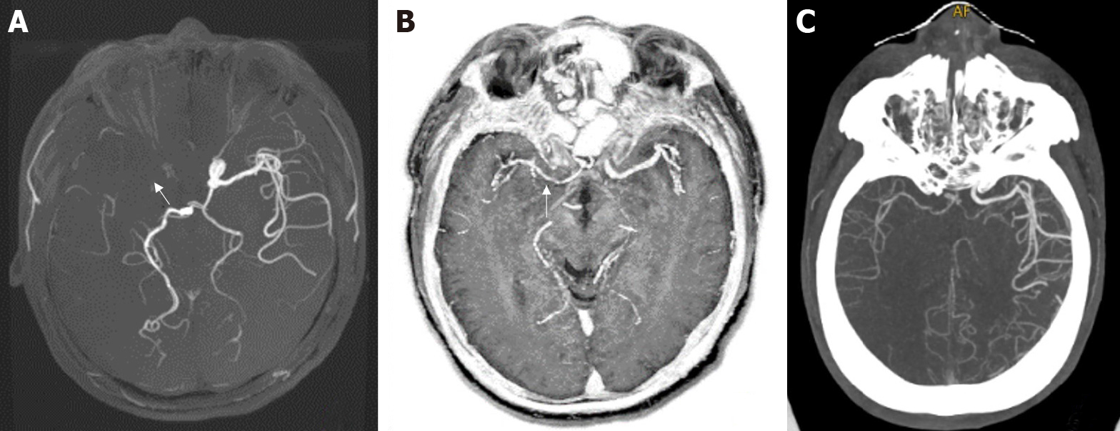
Figure 3 A 53-year-old man with right cerebral infarction.
A: The right middle cerebral artery was not shown in three-dimensional time-of-flight magnetic resonance angiography (left); B: MAGnetic resonance imaging compilation phase sensitive inversion recovery Vessel (middle); and C: Computed tomography angiography (right). Both the latter two showed mild and moderate stenosis of the local lumen of the right middle cerebral artery (arrows).
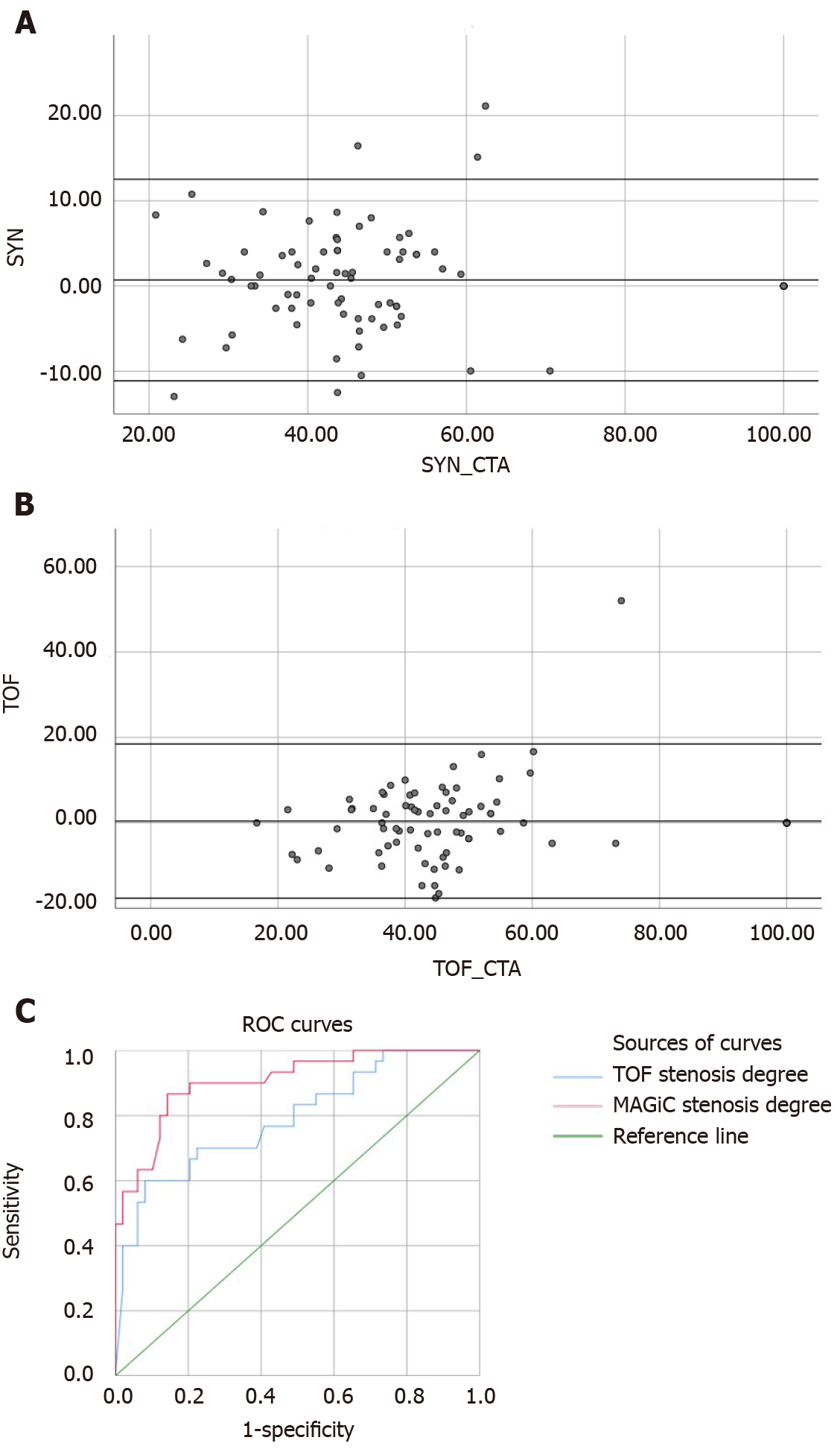
Figure 4 Bland-Altman and receiver operating characteristic curve evaluation of vascular stenosis degrees obtained by MAGnetic resonance imaging compilation phase-sensitive inversion recovery Vessel and time-of-flight magnetic resonance angiography.
A: MAGnetic resonance imaging compilation-computed tomography angiography; B: Time-of-flight magnetic resonance angiography; C: Receiver operating characteristic curves. TOF: Time-of-flight; CTA: Computed tomography angiography; ROC: Receiver operating characteristic.
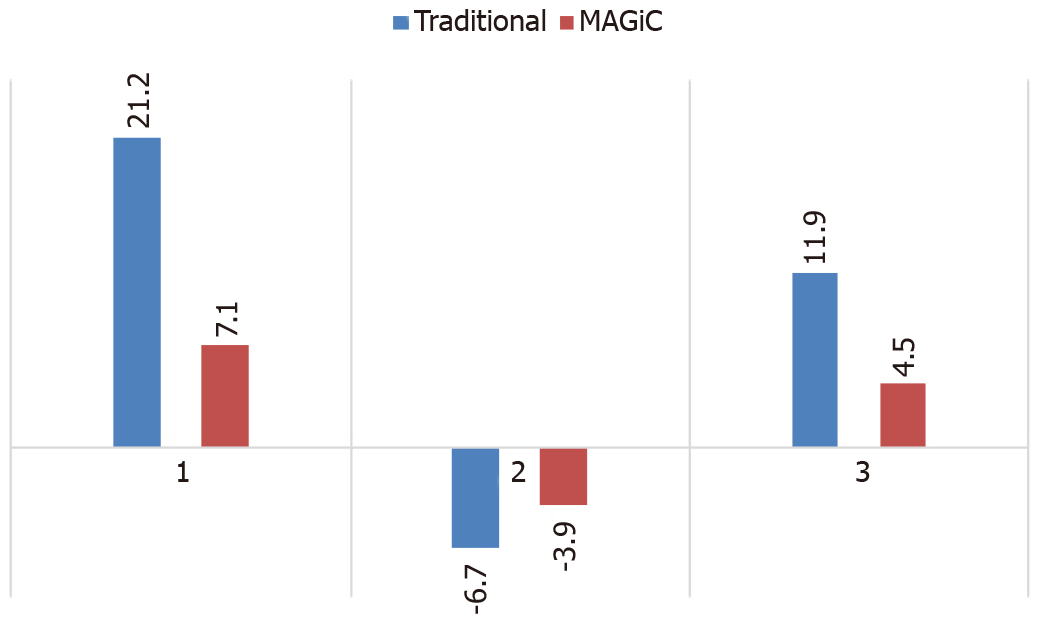
Figure 5 Contrast-to-noise ratio values of diffusion-weighted imaging diffusion restriction areas of MAGnetic resonance tmaging compilation-reconstructed multi-contrast images and traditional multi-contrast images.
1: T2-weighted image; 2: T1-weighted image; 3: T2-fluid-attenuated inversion recovery-weighted image; MAGiC: MAGnetic resonance imaging compilation.
- Citation: Wang Q, Wang G, Sun Q, Sun DH. Application of MAGnetic resonance imaging compilation in acute ischemic stroke. World J Clin Cases 2021; 9(35): 10828-10837
- URL: https://www.wjgnet.com/2307-8960/full/v9/i35/10828.htm
- DOI: https://dx.doi.org/10.12998/wjcc.v9.i35.10828


