Copyright
©The Author(s) 2021.
World J Clin Cases. Dec 6, 2021; 9(34): 10702-10707
Published online Dec 6, 2021. doi: 10.12998/wjcc.v9.i34.10702
Published online Dec 6, 2021. doi: 10.12998/wjcc.v9.i34.10702
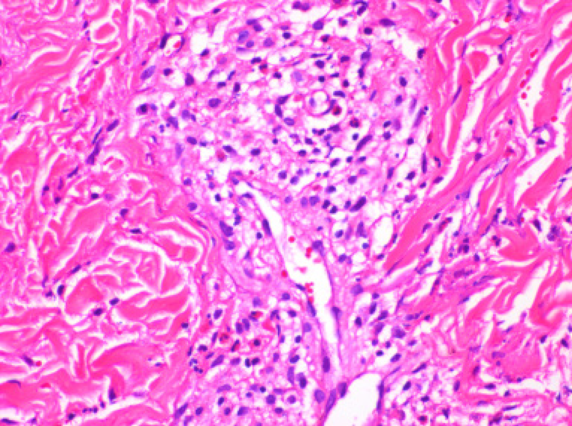
Figure 1 Pathological examination of skin biopsy specimens of the lower limbs showed eosinophil infiltration in the peripheral blood vessels.
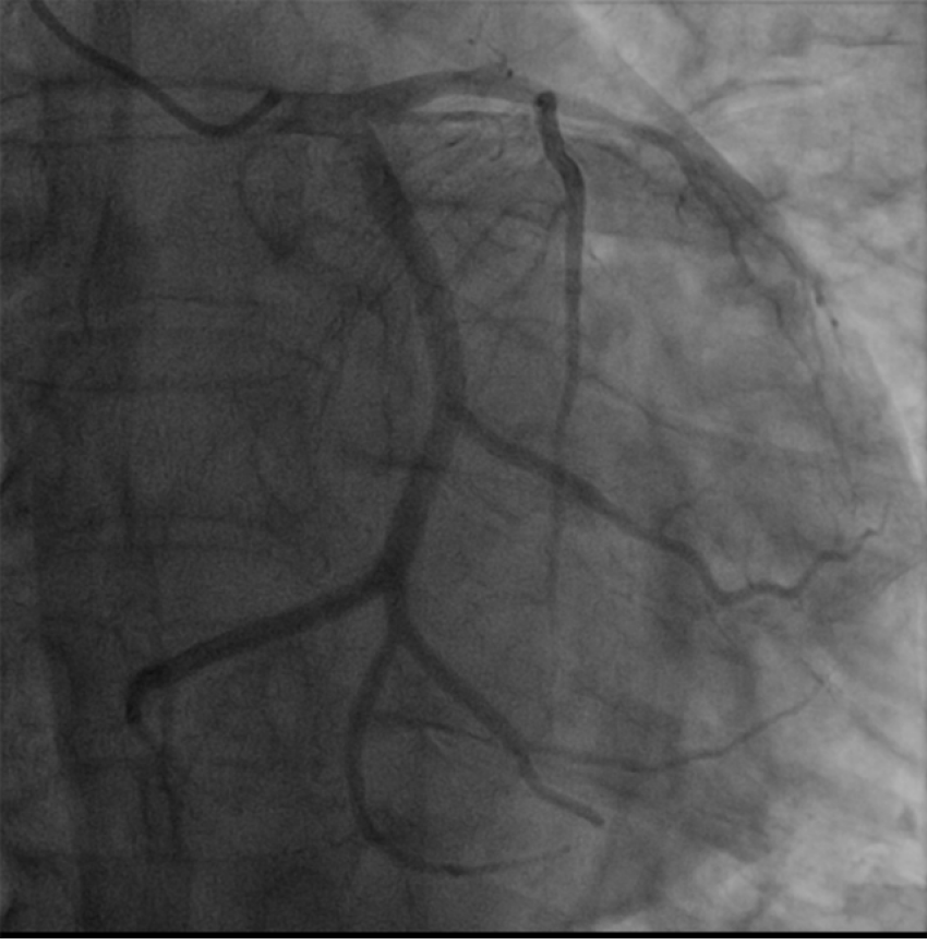
Figure 2 Coronary angiogram demonstrated occluded left circumflex arteries, the middle segment of the left anterior descending coronary artery was 30% narrowed, and the distal segment was occluded.
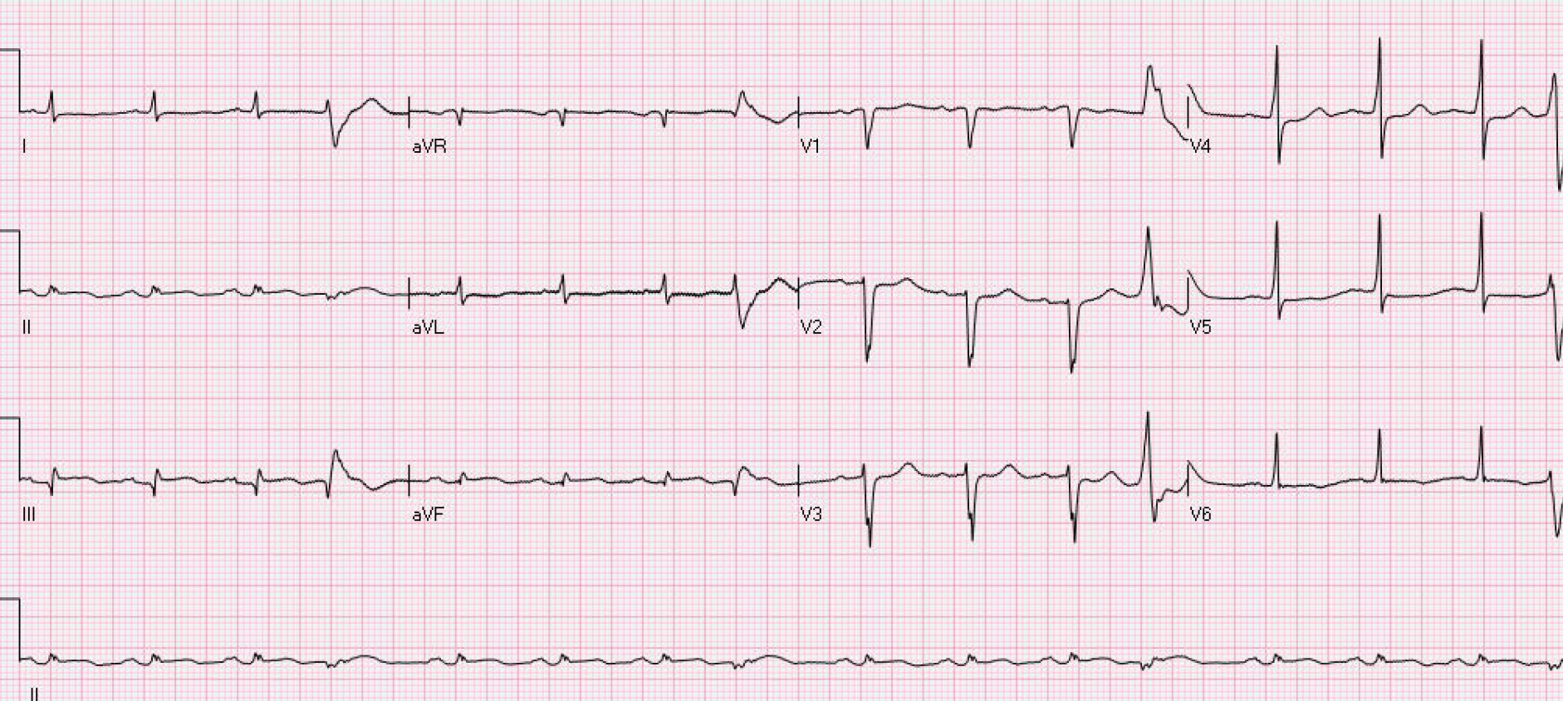
Figure 3 Electrocardiography showed inferior wall myocardial infarction, sinus rhythm, ventricular premature beats, poor progressive increase of the R wave in the anterior septum, low voltage in the limb leads, and ST-T changes.
- Citation: Jiang XD, Guo S, Zhang WM. Acute myocardial infarction induced by eosinophilic granulomatosis with polyangiitis: A case report. World J Clin Cases 2021; 9(34): 10702-10707
- URL: https://www.wjgnet.com/2307-8960/full/v9/i34/10702.htm
- DOI: https://dx.doi.org/10.12998/wjcc.v9.i34.10702


