Copyright
©The Author(s) 2021.
World J Clin Cases. Dec 6, 2021; 9(34): 10518-10529
Published online Dec 6, 2021. doi: 10.12998/wjcc.v9.i34.10518
Published online Dec 6, 2021. doi: 10.12998/wjcc.v9.i34.10518
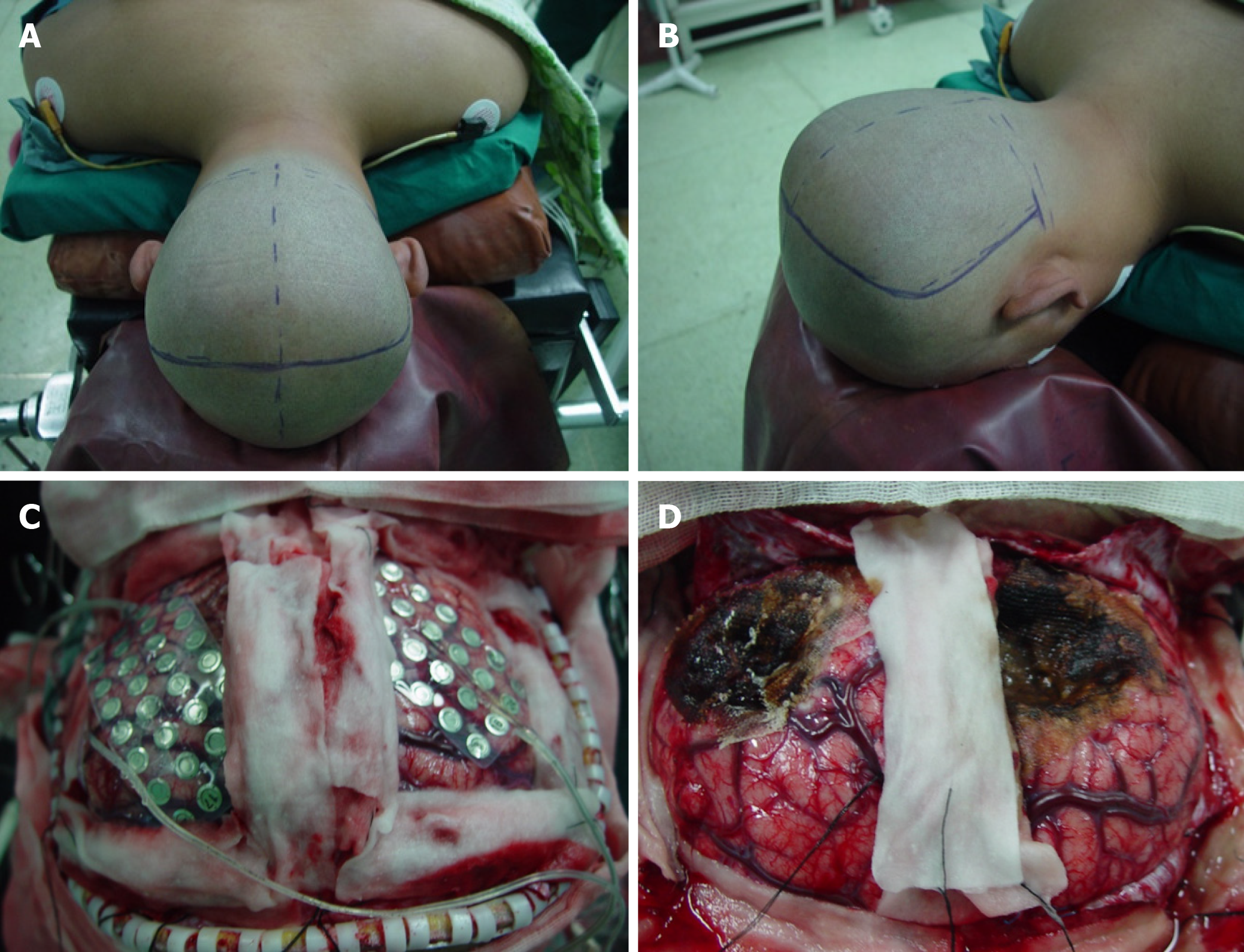
Figure 1 Surgical resection of bilateral occipital lesions.
A and B: The extent of the bilateral occipital craniotomy; C: Intracranial cortical electrodes were placed on the surface of the bilateral occipital lobe during surgery to enable monitoring of the electroencephalography; D: Photograph taken after resection of the lesions in the bilateral occipital lobe.
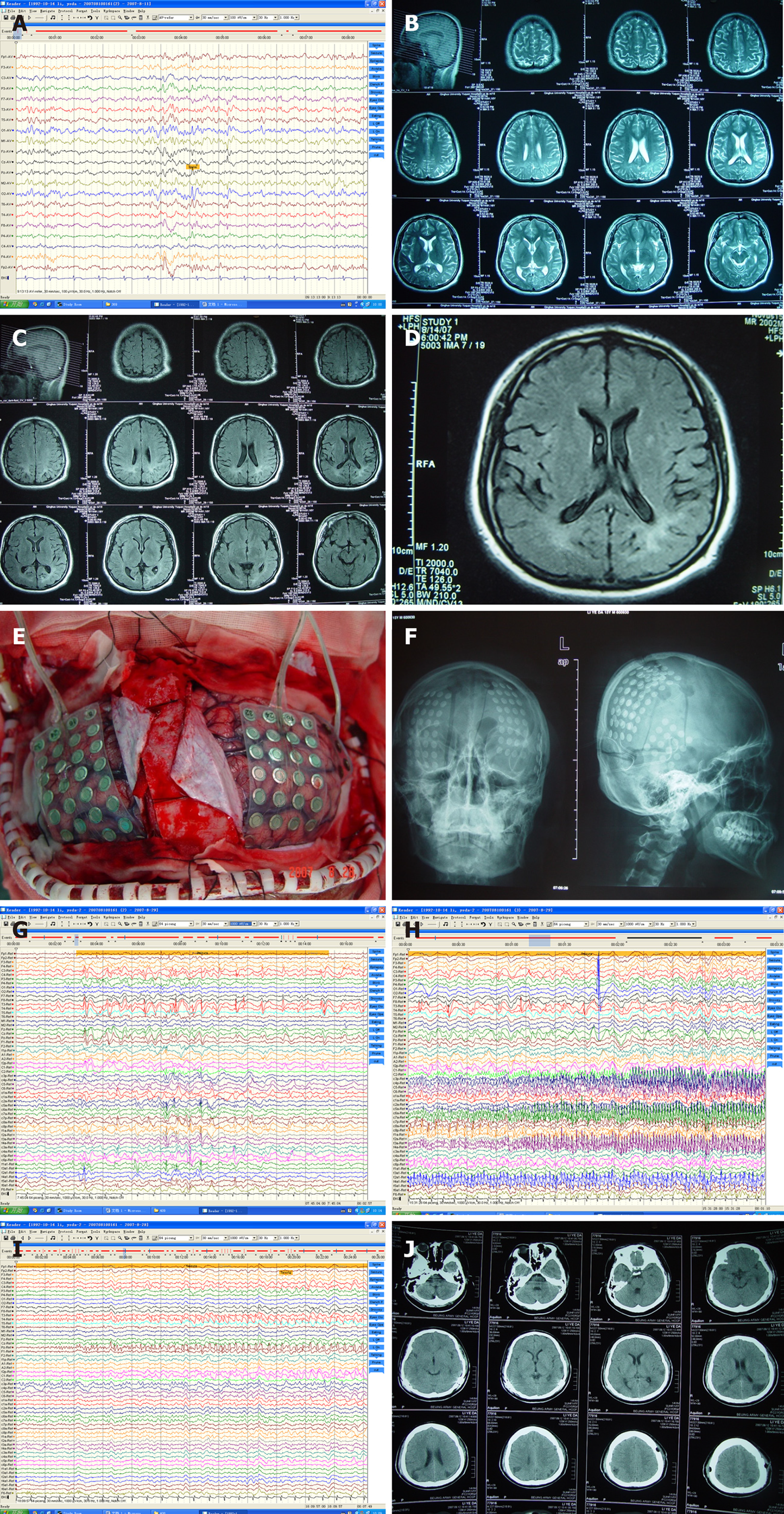
Figure 2 Clinical findings in a 15-year-old male patient with bilateral occipital lobe epilepsy.
A: Scalp video-electroencephalography (EEG) recordings demonstrated abnormal discharges in the right occipital region during the interictal period; B and C: magnetic resonance imaging (MRI) revealed abnormal signals in the bilateral occipital lobe; D: T2-FLAIR MRI showed irregular high signals in the bilateral occipital lobe that suggested ischemic changes; E: A subdural grid electrode was placed during surgery under general anesthesia; F: Anteroposterior and lateral head X-rays (taken after closure of the craniotomy) showing the position of the subdural grid electrode; G-I: Representative EEG recordings made using the subdural grid electrode showing abnormal discharges arising from both sides of the occipital lobe; The upper half of each trace shows recordings obtained from the left occipital lobe, and the lower half of each trace shows recordings obtained from the right occipital lobe; J: Postoperative cranial computed tomography.
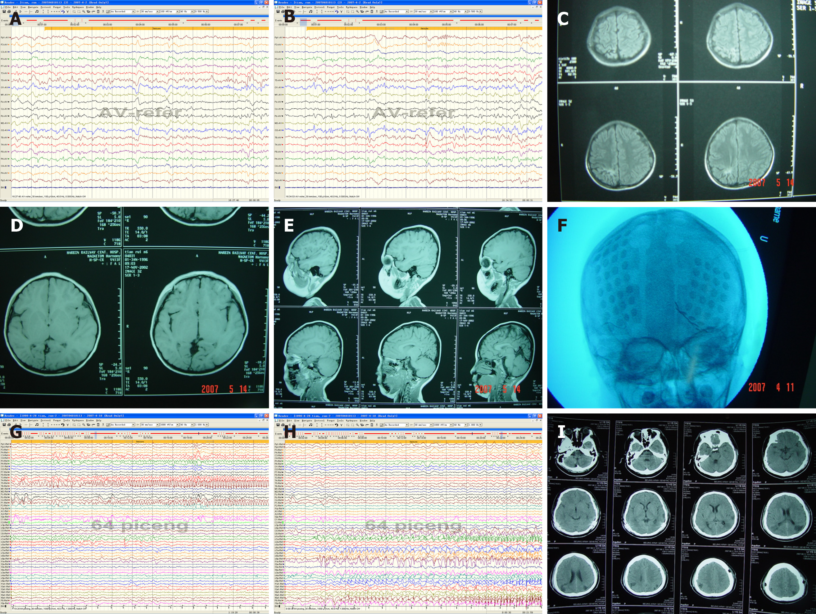
Figure 3 Clinical findings in an 11-year-old male patient with bilateral occipital lobe epilepsy.
A: Representative scalp video-electroencephalography (EEG) recording demonstrating abnormal discharges originating in the left occipital region during the interictal period; B: Representative scalp video-EEG recording demonstrating abnormal discharges originating in the right occipital region during the interictal period; C-E: magnetic resonance imaging showing bilateral occipital dysplasia and a high signal on T2-FLAIR imaging that was obvious on the right side; F: Anteroposterior X-ray illustrating the position of the subdural grid electrode; G and H: Representative EEG recordings made using the subdural grid electrode showing abnormal discharges arising from both the left (G) and right (H) sides of the occipital lobe; I: Postoperative cranial computed tomography.
- Citation: Lyu YE, Xu XF, Dai S, Feng M, Shen SP, Zhang GZ, Ju HY, Wang Y, Dong XB, Xu B. Resection of bilateral occipital lobe lesions during a single operation as a treatment for bilateral occipital lobe epilepsy. World J Clin Cases 2021; 9(34): 10518-10529
- URL: https://www.wjgnet.com/2307-8960/full/v9/i34/10518.htm
- DOI: https://dx.doi.org/10.12998/wjcc.v9.i34.10518


