Copyright
©The Author(s) 2021.
World J Clin Cases. Dec 6, 2021; 9(34): 10418-10429
Published online Dec 6, 2021. doi: 10.12998/wjcc.v9.i34.10418
Published online Dec 6, 2021. doi: 10.12998/wjcc.v9.i34.10418
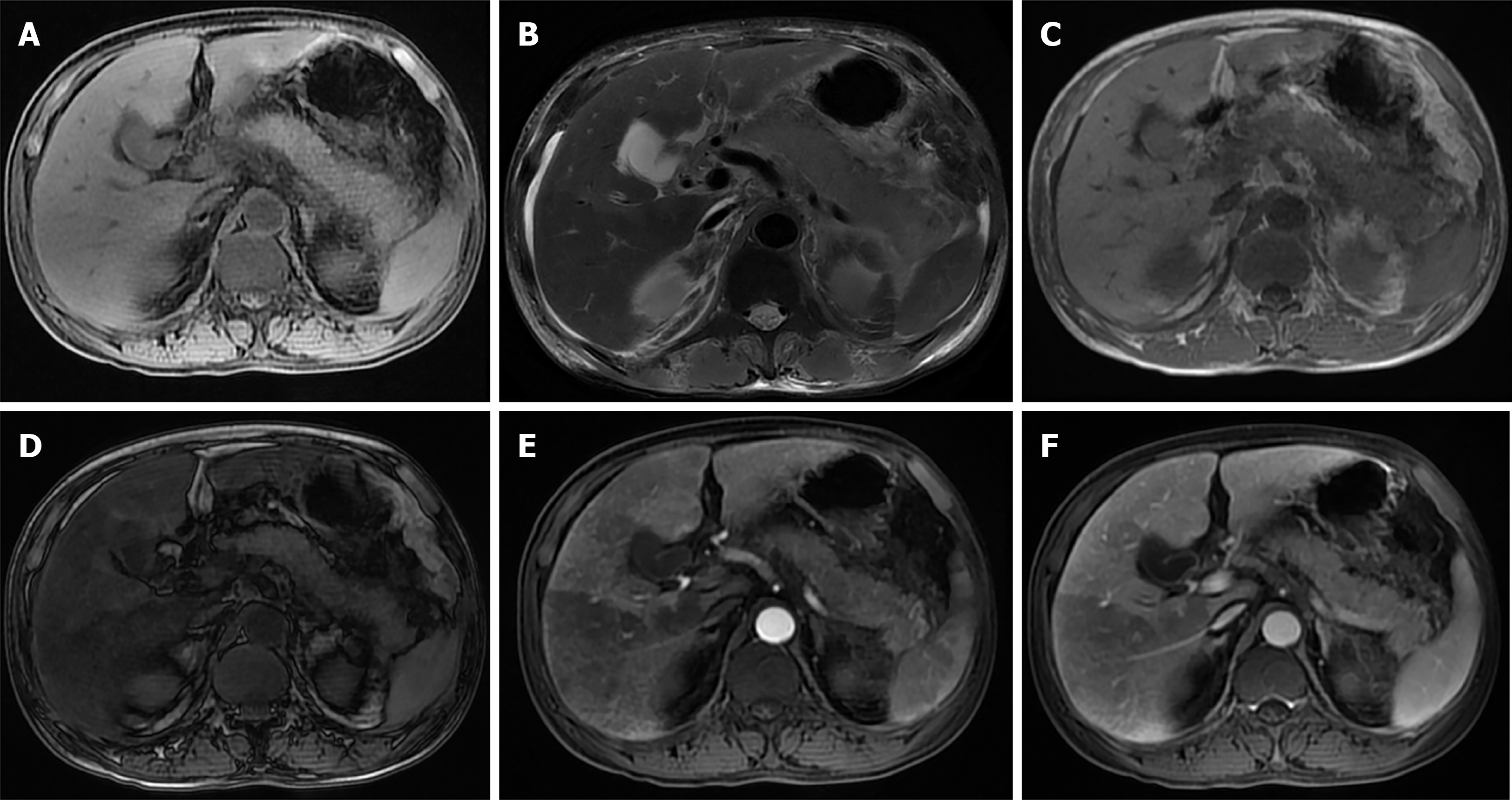
Figure 1 A 49-year-old acute pancreatitis male patient with fatty liver, whose liver perfusion is abnormal.
A and B: On the third day after onset, the pancreas parenchyma shows swelling on the T1-weighted imaging (A) and the T2-weighted imaging (B); C and D: The liver shows hypo-/hyper-intensity on the in-phase (C) and out-phase (D); E and F: The liver presents heterogeneous enhancement after contrast agent administration.
- Citation: Liu W, Du JJ, Li ZH, Zhang XY, Zuo HD. Liver injury associated with acute pancreatitis: The current status of clinical evaluation and involved mechanisms. World J Clin Cases 2021; 9(34): 10418-10429
- URL: https://www.wjgnet.com/2307-8960/full/v9/i34/10418.htm
- DOI: https://dx.doi.org/10.12998/wjcc.v9.i34.10418


