Copyright
©The Author(s) 2021.
World J Clin Cases. Nov 26, 2021; 9(33): 10369-10373
Published online Nov 26, 2021. doi: 10.12998/wjcc.v9.i33.10369
Published online Nov 26, 2021. doi: 10.12998/wjcc.v9.i33.10369
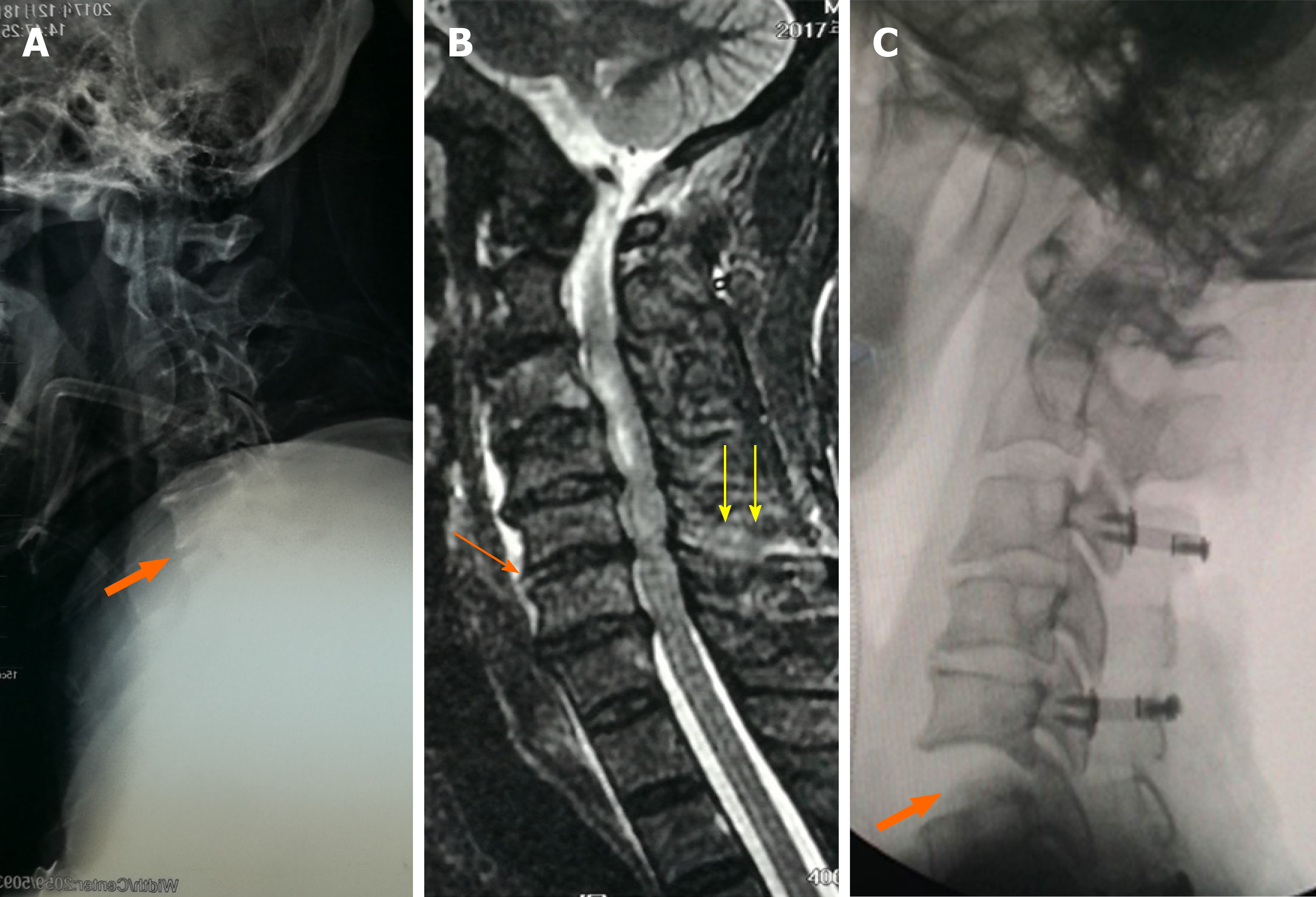
Figure 1 A case of occult cervical spine instability.
A: The standard lateral X-ray at admission showed no obvious instability of the cervical spine; B: The magnetic resonance imaging at admission showed injuries involving the disc (orange arrow) and posterior interspinous ligament (yellow arrows) at the C5-6 level; C: The intraoperative C-arm fluoroscopic X-ray after anesthesia and muscle relaxation revealed significantly increased intervertebral space at C5-6, indicating instability at this level.
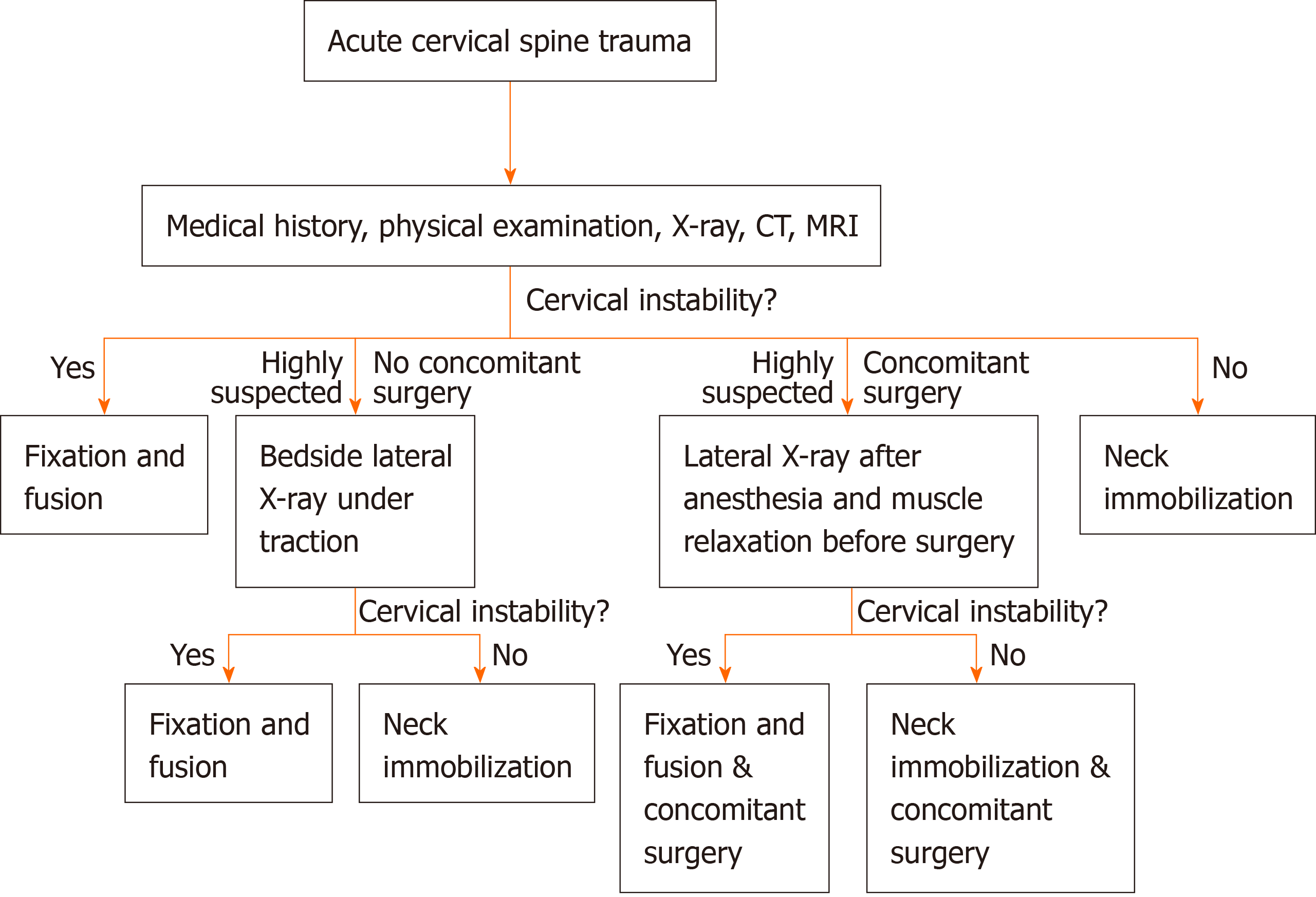
Figure 2 Clinical algorithm for diagnosing occult cervical spine instability.
CT: Computed tomography; MRI: Magnetic resonance imaging.
- Citation: Zhu C, Yang HL, Im GH, Liu LM, Zhou CG, Song YM. Clinical algorithm for preventing missed diagnoses of occult cervical spine instability after acute trauma: A case report. World J Clin Cases 2021; 9(33): 10369-10373
- URL: https://www.wjgnet.com/2307-8960/full/v9/i33/10369.htm
- DOI: https://dx.doi.org/10.12998/wjcc.v9.i33.10369


