Copyright
©The Author(s) 2021.
World J Clin Cases. Nov 26, 2021; 9(33): 10328-10336
Published online Nov 26, 2021. doi: 10.12998/wjcc.v9.i33.10328
Published online Nov 26, 2021. doi: 10.12998/wjcc.v9.i33.10328
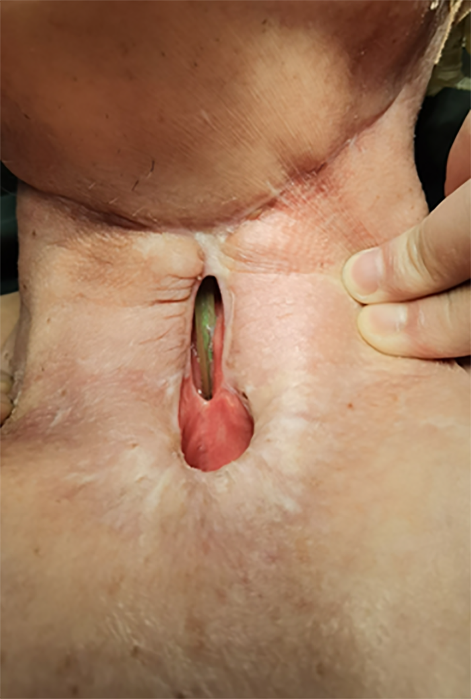
Figure 1 The pharyngeal fistula and tracheoesophageal fistula before reconstruction.
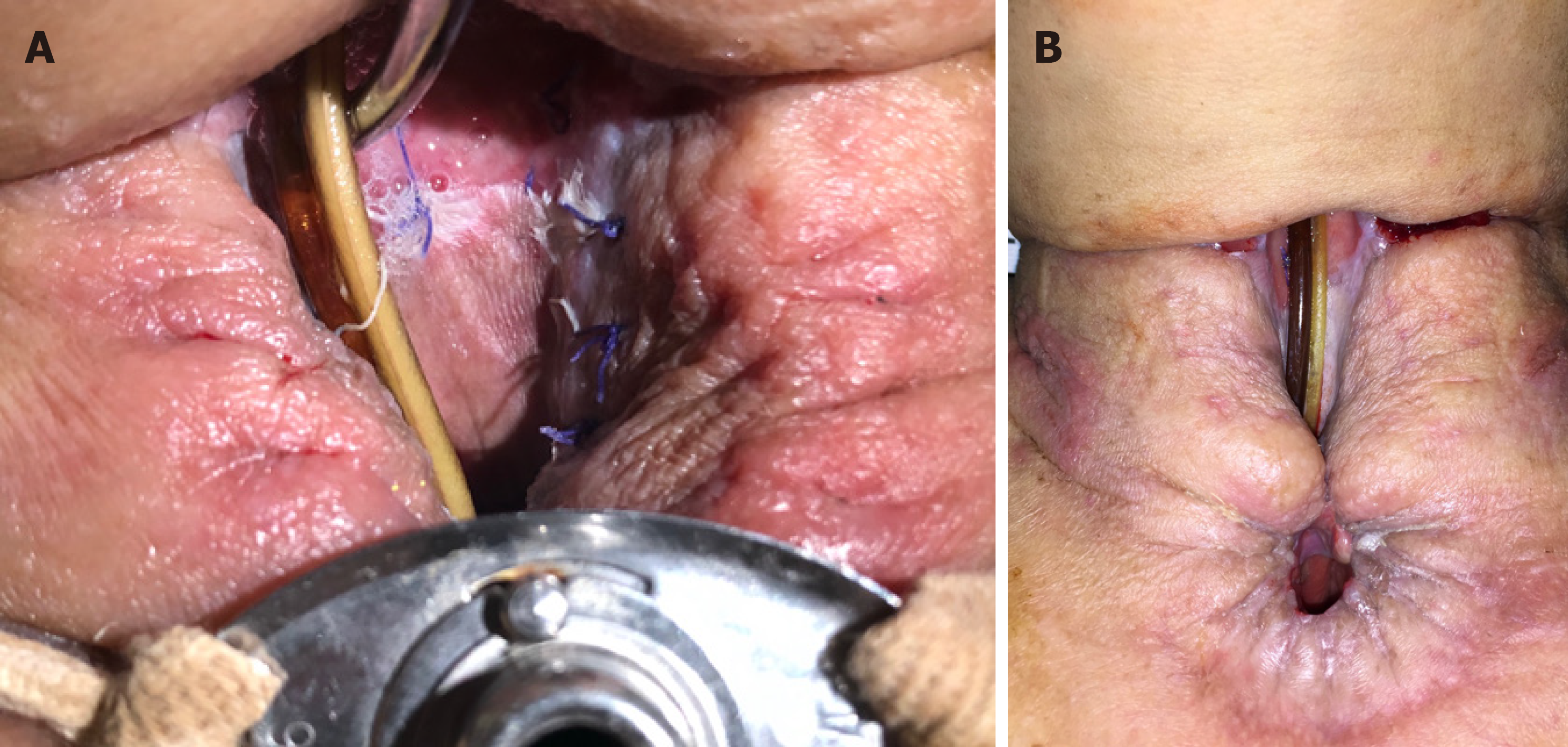
Figure 2 Split thickness skin grafting.
A: The 4th day after the split thickness skin grafting; B: The skin graft stably healed with the surrounding granulation and the cervical skin.
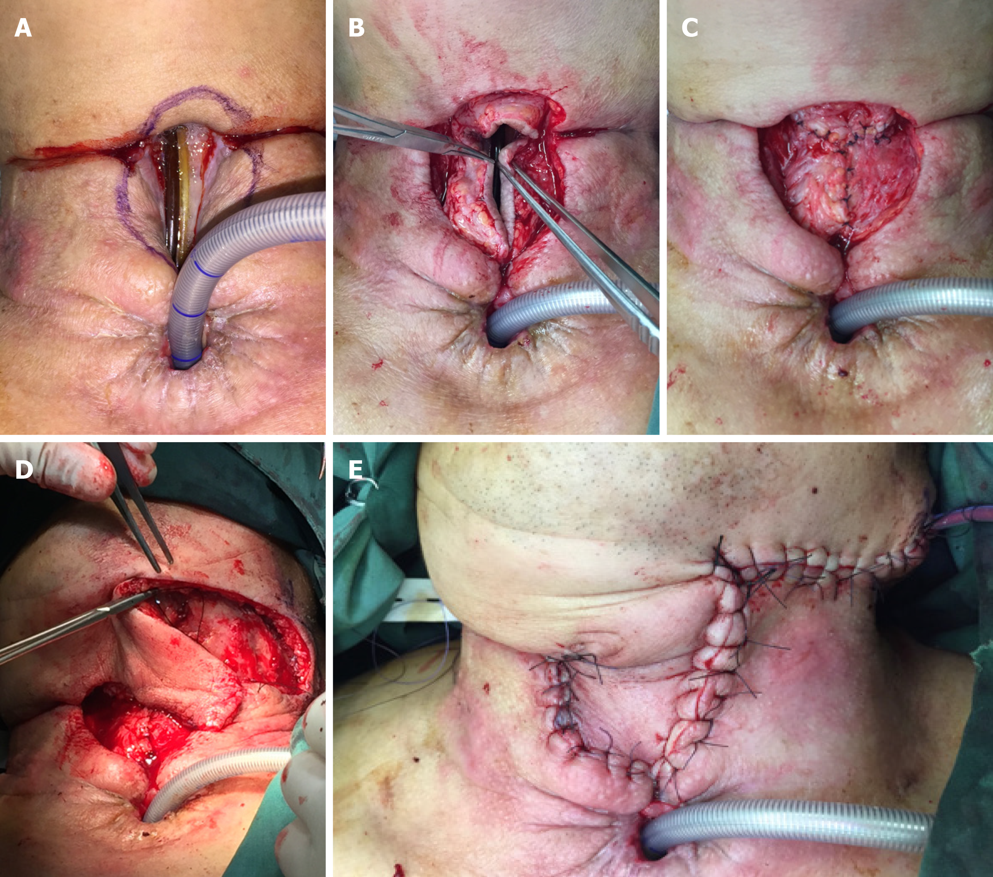
Figure 3 Intraoperative images of case 1.
A: The flap design; B: Using the surrounding tissue including the skin to become the anterior wall of the pharynx from three directions; C: The reconstructed anterior wall of the pharynx; D: Taking a random flap from the submandibular part; E: The skin defect of the neck was reconstructed.
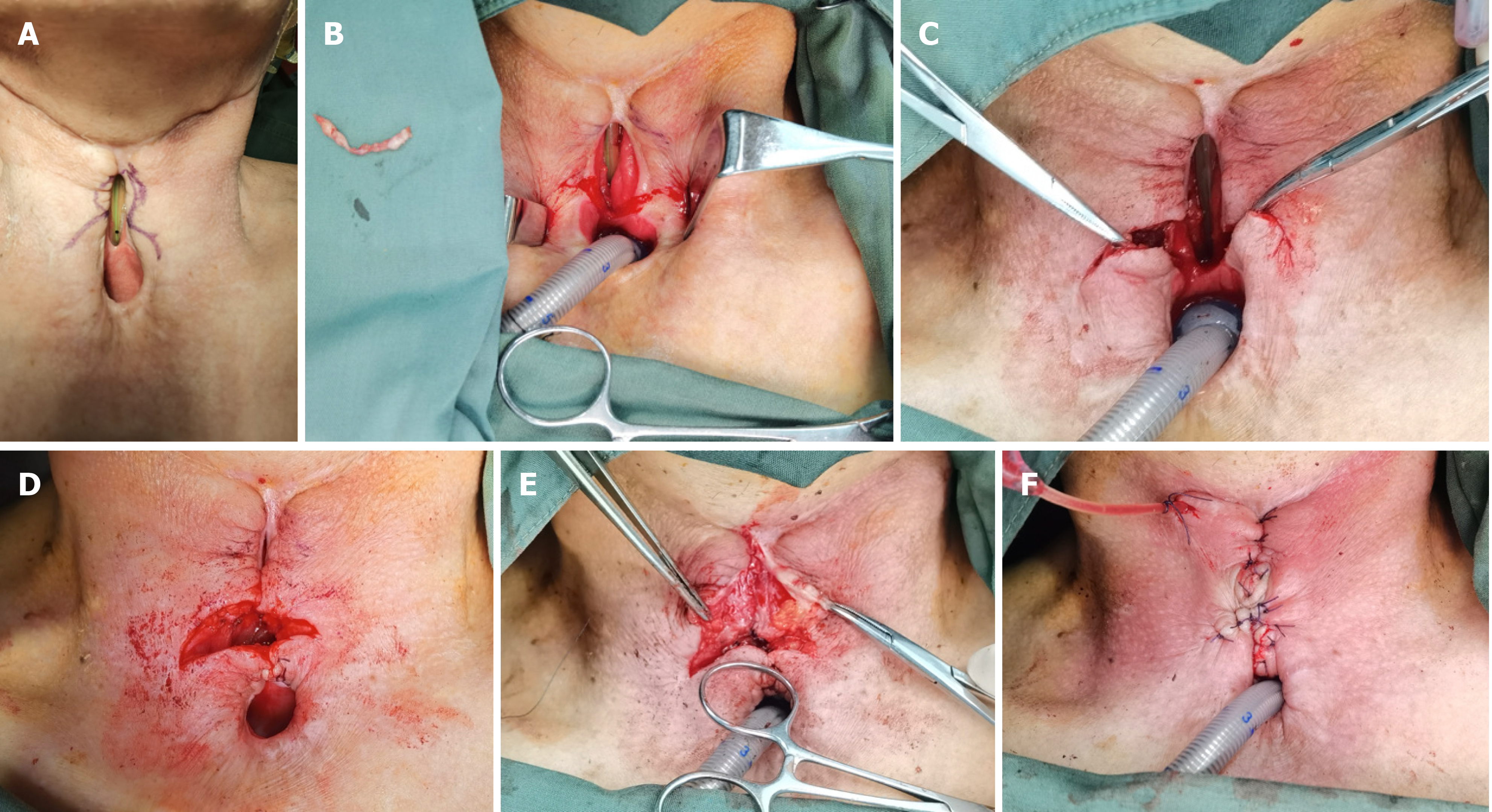
Figure 4 Intraoperative images of case 2.
A: The flap design; B: Cutting off the skin as designed to bare the subcutaneous tissue for further sewing; C: Using the two-side lower part of the surrounding tissue including the skin to become the upper wall of the tracheostoma; D: The reconstructed upper wall of the tracheostoma; E: Dissociating the side walls of the pharyngeal fistula and sewing them to become the anterior wall of the reconstructed pharynx; F: Sewing the local random flaps from three directions in a “inverted-T” fashion to close the skin defect.
- Citation: Zhang Y, Liu Y, Sun Y, Xu M, Wang XL. Local random flaps for cervical circumferential defect or tracheoesophageal fistula reconstruction after failed gastric pull-up: Two case reports. World J Clin Cases 2021; 9(33): 10328-10336
- URL: https://www.wjgnet.com/2307-8960/full/v9/i33/10328.htm
- DOI: https://dx.doi.org/10.12998/wjcc.v9.i33.10328


