Copyright
©The Author(s) 2021.
World J Clin Cases. Nov 26, 2021; 9(33): 10300-10307
Published online Nov 26, 2021. doi: 10.12998/wjcc.v9.i33.10300
Published online Nov 26, 2021. doi: 10.12998/wjcc.v9.i33.10300
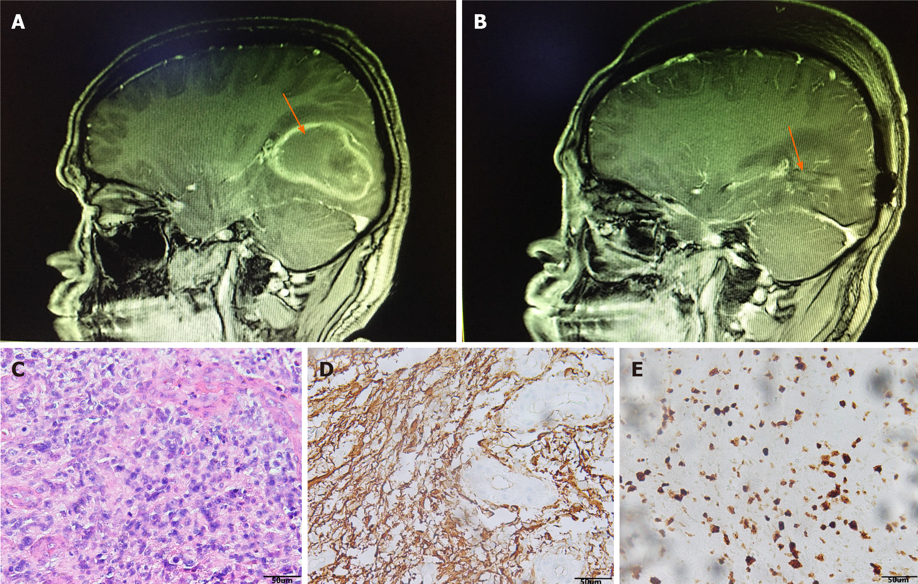
Figure 1 Images and pathology before and after the first operation.
A: Preoperative brain contrast-enhanced magnetic resonance imaging (MRI) indicated right temporal lobe lesions and the tumor border is located next to the lateral ventricle; B: Postoperative brain contrast-enhanced MRI indicated complete resection of the tumor; C: Hematoxylin-eosin staining showed a large amount of necrosis and scattered heteromorphic cells, considering high-grade glioblastoma (magnification, × 200); D: Glial fibrillary acidic protein (+) (magnification, × 200); E: Ki67 (+20%) (magnification, × 200).
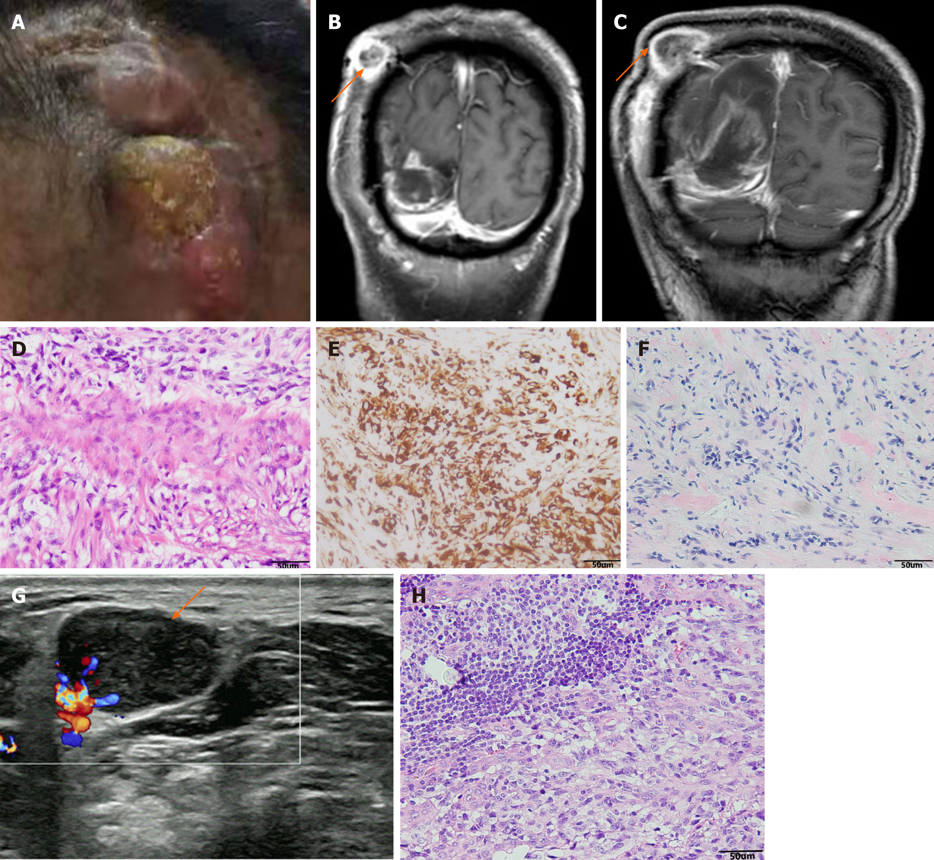
Figure 2 Imaging and pathology before and after the second operation.
A: The patient has a lump on the scalp at the first surgical incision in the right temple; B: 6 mo after first surgery, brain contrast-enhanced magnetic resonance imaging (MRI): intracranial recurrence and scalp transfer; C: Brain contrast-enhanced MRI was re-examined 9 mo after first surgery: both the intracranial recurrent tumors and the scalp metastases have grown up; D: Hematoxylin-eosin staining suggested fibrous tissue proliferation and necrosis, and scattered abnormal cells, which was considered as intracranial GBM metastasis (magnification, × 200); E: GFAP (+) (magnification, × 200); F: Vim (+) (magnification, × 200); G: Ultrasonography revealed enlarged lymph nodes in the right neck (2.1 cm × 1.5 cm); H: Pathological examination of enlarged lymph nodes in the right neck was diagnosed as glioblastoma, hematoxylin-eosin staining (magnification, × 200).
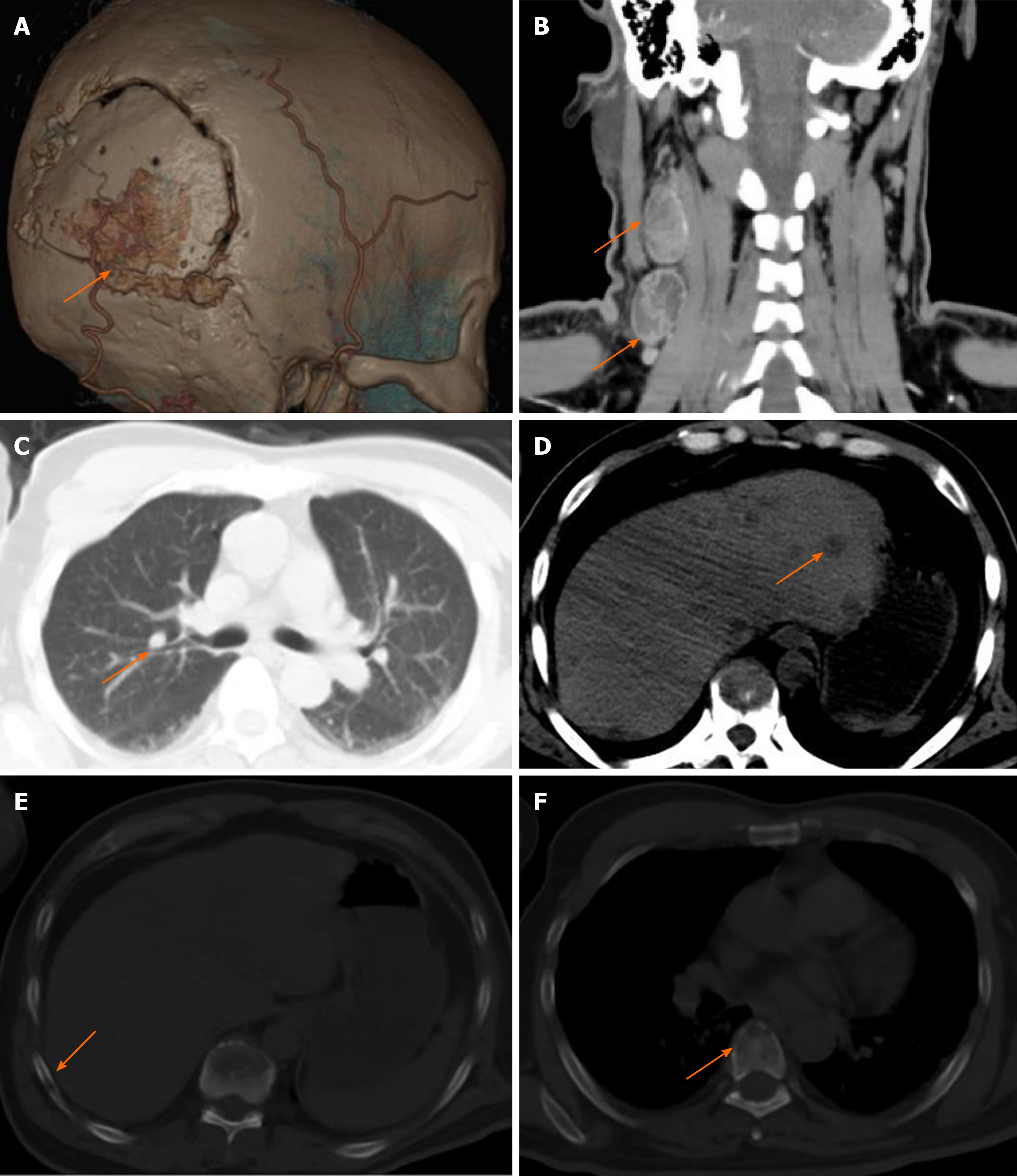
Figure 3 Follow-up after secondary surgery revealed multiple extracranial metastases.
A: Three-dimensional reconstruction of the skull suggested that the skull was infiltrated by the tumor; B: Cervical computed tomography (CT) suggested enlargement of cervical lymph nodes; C-F: Chest CT showed multiple nodules in the liver (C) and lungs (D), infiltration in the ribs (E), and infiltration in the spine (F), and all the above lesions are considered for multiple extracranial metastases of intracranial glioblastoma.
- Citation: Luan XZ, Wang HR, Xiang W, Li SJ, He H, Chen LG, Wang JM, Zhou J. Extracranial multiorgan metastasis from primary glioblastoma: A case report. World J Clin Cases 2021; 9(33): 10300-10307
- URL: https://www.wjgnet.com/2307-8960/full/v9/i33/10300.htm
- DOI: https://dx.doi.org/10.12998/wjcc.v9.i33.10300


