Copyright
©The Author(s) 2021.
World J Clin Cases. Nov 26, 2021; 9(33): 10265-10272
Published online Nov 26, 2021. doi: 10.12998/wjcc.v9.i33.10265
Published online Nov 26, 2021. doi: 10.12998/wjcc.v9.i33.10265
Figure 1 Radiological images at the time of diagnosis.
A: Abdominal magnetic resonance imaging showed a hypovascular mass present in the tail of the pancreas 6.4 cm × 4.2 cm in size; B: Multiple hypovascular nodules of no more than 7.1 cm × 5.5 cm scattered in the liver parenchyma; C: Colonoscopy image showing a 2.2 cm × 1.6 cm mass with surface congestive erosions, which was 33 cm from the anus.
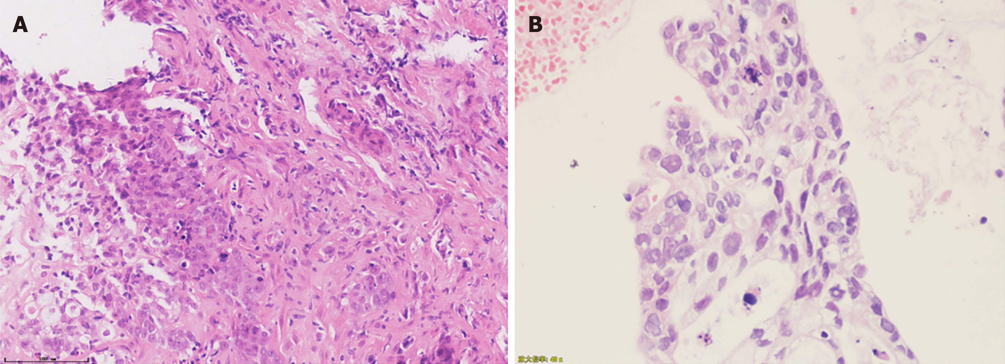
Figure 2 Results of pathologic diagnosis.
A: Fine-needle aspiration biopsy of the left lobe of the liver; B: Biopsy from colonoscopy. Magnification: 40 ×; scale bar: 100 μm.
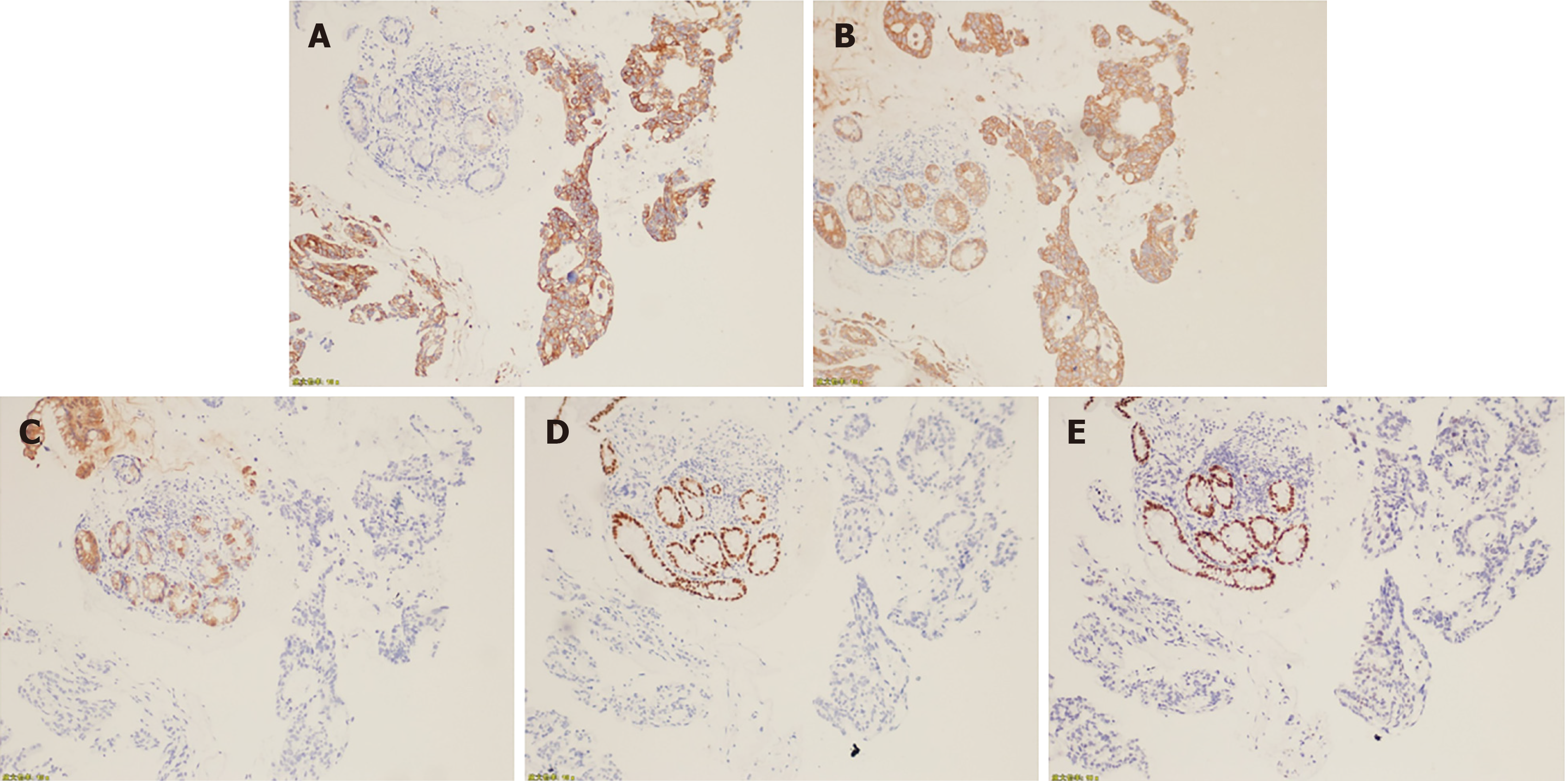
Figure 3 Histopathological analysis of biopsy from the colon lesion.
A: CK 7 positive; B: CK positive; C: CK 20 negative; D: CDX2 negative; E: SATB2 negative. Magnification: 10 ×.
Figure 4 Radiological images of response assessment to FOLFIRINOX treatment.
A: Abdominal magnetic resonance imaging after 12 cycles of FOLFIRINOX treatment showing the size of the hypovascular mass in the tail of the pancreas had shrunk to 4.2 cm × 2.0 cm; B: Multiple hypovascular nodules in the liver parenchyma were also reduced to less than 3.5 cm; C: A colonoscopy after 9 cycles of FOLFIRINOX showing that the mucosa of the original lesion site was slightly rough and red, and that no protuberant or new mucosal lesions were found.
- Citation: Dong YM, Sun HN, Sun DC, Deng MH, Peng YG, Zhu YY. Pancreatic cancer with synchronous liver and colon metastases: A case report. World J Clin Cases 2021; 9(33): 10265-10272
- URL: https://www.wjgnet.com/2307-8960/full/v9/i33/10265.htm
- DOI: https://dx.doi.org/10.12998/wjcc.v9.i33.10265


